 AACHARYA KOTA CLASSES
What is living?
- Books Name
- AACHARYA KOTA CLASSES Biology Book
- Publication
- AACHARYA KOTA CLASSES
- Course
- CBSE Class 11
- Subject
- Biology
ggyyuyuyu
What is living?
- Books Name
- ACME SMART COACHING Biology Book
- Publication
- ACME SMART PUBLICATION
- Course
- CBSE Class 11
- Subject
- Biology
WHAT IS LIVING?
Life is a unique, complex organisation of molecules expressing itself through chemical reactions (metabolism) which lead to growth, development, responsiveness, adaptation and reproduction. Hence, "the object by itself exhibiting the growth, development, death, consciousness, reproduction etc. is designated as living being."
A. Characteristics of Living Beings
All the living beings share certain unique and basic characteristics which set them apart from non-living objects. These characteristics are listed below :
1. Growth
2. Reproduction
3. Metabolism
4. Cellular structure
5. Consciousness
1. Growth:
Increase in mass and increase in number of cells are twin characters of growth. Growth refers to irreversible increase in mass or overall size of a tissue, an organism or its parts.
Growth is the result of difference between anabolism (building up reactions) and catabolism (breakdown reactions).
Growth occurs when anabolism or synthetic processes exceeds catabolism.
Degrowth or negative growth will occur when catabolism exceeds anabolism. It will decrease the mass of body.
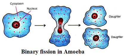
Unicellular organisms also grow by cell division. In majority of higher animals and plants, growth and reproduction are mutually exclusive events, but in unicelled organisms like Amoeba, reproduction is synonymous with growth, i.e., increase in number of cells.
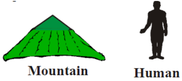
Non-living objects also grow if we take increase in body mass as a criterion of growth. But in these objects extrinsic growth is present i.e. increase in the mass of body from outside e.g., mountains, boulders and sand mounds. Growth, therefore, cannot be taken as a defining property of living organisms.
2. Reproduction:
Reproduction is the formation of new individuals of similar kind. It is, however, required for survival of the population as it compensates for the loss of life due to death.
Reproduction is of two types, asexual and sexual. Asexual reproduction is uniparental multiplication that occurs through binary fission, multiple fission, spore formation, fragmentation and vegetative multiplication.
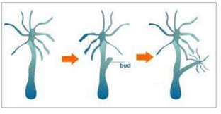
Fungi multiply by asexual spores, yeast and Hydra show budding, Planaria exhibits true regeneration. Fungi, filamentous algae, protonema of mosses easily multiplies by fragmentation. But, when we notice single-celled organisms like bacteria, unicellular alga and Amoeba, we are not clear about the usage of these two terms -growth and reproduction, i.e., increase in number of cells.
Hence, reproduction also cannot be an all inclusive property of living organisms. Still, no non-living object is capable of reproducing or replicating by itself. Further, there are some organisms which do not reproduce at all, e.g., worker bees, mules etc.
3. Metabolism:
Life is a never ending flow of energy and materials.
The energy are required by all living cells for building and functioning of their living matter.
Metabolism is the sum total of all chemical reactions occurring in an organism due to specific interactions amongst different types of molecules within the interior of cells.
Metabolism involves exchange of matter and energy between an organism and its environment and transformation of matter and energy within an organism.
Despite vast differences occurring in structure and functioning of cells in different organisms, metabolic reactions are unusually similar.
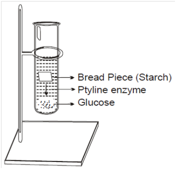
Metabolic reactions can be demonstrated outside the body in cell free systems.
An isolated metabolic reaction(s) in a test-tube is neither living nor non-living. Hence, while metabolism is a defining feature of all living organisms without exception , isolated metabolic reactions in-vitro are not living things but surely living reactions.
An isolated metabolic reaction(s) in a test-tube is neither living nor non-living. Hence, while metabolism is a defining feature of all living organisms without exception , isolated metabolic reactions in-vitro are not living things but surely living reactions.
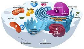
4. Cellular structure:
Body of all living organisms consists of cells and their products. Hence, cellular organisation of the body is the defining feature of life forms.
5. Consciousness:
All living beings, plants and animals, are aware of their environment. All organisms sense their surroundings and respond to sudden or smooth changes in it.
All organisms from prokaryota to eukaryota can sense and respond.
Humans also have "self-consciousness" hence, this becomes the defining property of living beings.
It is most obvious and technically complicated feature of all living organisms.
The foregoing discussion, nevertheless concludes that living beings are organized, self-replicating, evolving and self-regulating interactive systems, with consciousness at the head of all.
A question may arise whether a man lying in coma on the life support systems is living or non-living? The answer lies in the quantitative presence of consciousness in the living being in that particular state. Infact, the person in coma has lost the requisite quantity of consciousness to exhibit the features of living being, but still exhibits life. If the requisite consciousness is restored, the person may again start to be as living being or die otherwise.
Other characteristics of living beings are adaptation, life span, homeostasis, healing and repair, movement and variation.
Biodiversity
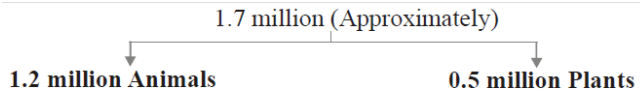
From a current estimation, approximately 1.7 million species have been scientifically named and classified. These include nearly 1.2 million animals and over 0.5 million species of plants.
Amongst animals, insects form the largest group, i.e., over 10,25,000 species. It is estimated that majority of species diversity is confined to tropical rain forests and huge diversity exist in under water reef formations in tropical oceans.
Existing living species are the outcome of about 3.5 billion years of evolutionary process on this earth. Nearly 15,000 new organisms are discovered every year.
A clearer understanding of this huge variety of organisms can be studied by dividing these into smaller groups or sub-groups (categories) and each group or sub-group comprising of organisms with more or less similar characters.
Method of placing organisms into groups or sub-groups depending upon extent of similarities and differences is called classification.
The division of organisms into different groups follows certain rules, that is why the term called taxonomy is used for classification of organisms following certain rules or principles.
Need for classification
There are millions of plants and animals varying greatly in their form, structure and complexity.
It is impossible to study all of them individually. To make the study of organisms possible and easier, scientists have divided organisms into different ranks or categories on the basis of similarities and differences.
Classification is just like systematically arranged library where we can easily find out the required book, in the same way, if the organisms are arranged according to a system, it makes their study easy.
Some important needs are
It is essential to understand the inter-relationship among different groups of organisms.
It serves as a base for the development of other biological sciences like biogeography.
Various applied biology fields also depend upon exact identification and classification.
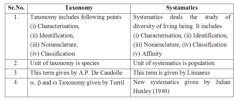
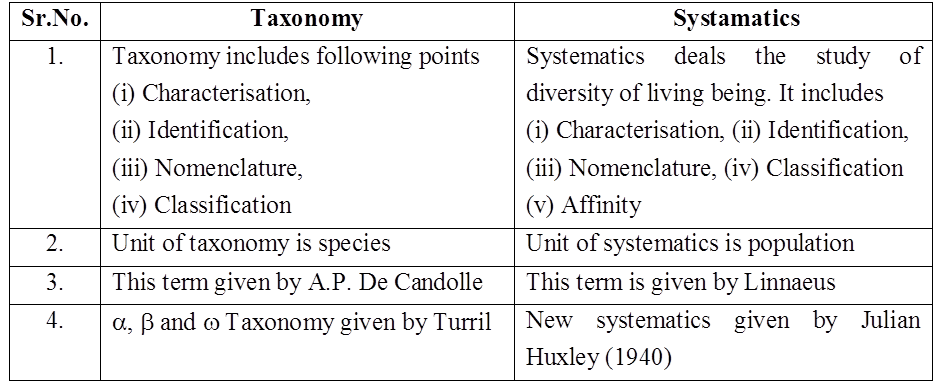
Taxonomy and Systematics
Taxonomy: The branch of science dealing with the study of principles and procedures of classification is called taxonomy. The term taxonomy was coined by A.P. de Candolle. Linnaeus is considered as Father of Taxonomy. Santapau is considered as Father of Indian Taxonomy. The fundamental elements of taxonomy are as follows :
- Characterisation and identification : It is the determination of the similarities of an organism with an already known organism, based upon specific characters.
- Nomenclature: It is the determination of correct name of an organism according to established universal rules.
- Classification: It is grouping of organisms into convenient categories on the basis of easily observable characters.
-
The classical taxonomy is based on observable morphological characters, however the modern taxonomy deals with several characters for the classification of organisms like
(a) External and internal structure alongwith the structure of cell.
(b) Development process.
(c) Ecological information of organisms
Systematics : The word "systematics" is derived from Latin word systema which means systematic arrangement of organisms. It was first used by Carolus Linnaeus. According to him, "systematics is the discipline of biology which deals with the kind and diversity of all organisms and the existing relationships amongst them."
Generally, the terms such as classification, systematics and taxonomy are used interchangeably but some taxonomists like Simpson (1961) relate them with a separate field. He defined systematics as
"The study of diversity of organisms and all their comparative and evolutionary relationships based on comparative anatomy, comparative ecology, comparative physiology and comparative biochemistry."
The main uses of systematics are as given below :
(i) It helps in providing knowledge of great diversity of animals and plants. It provides information regarding evolution which took place among plants and animals by knowing the distinction, relationship, habitat and habits. It thus, gives a vivid picture of entire organic diversity.
(ii) It helps in the identification of fossils which gives useful information about the phylogeny of organisms.
(iii) Newly discovered organisms can be identified through systematics. well as the specimens
Concept Builder
1. The reasons for large scale biodiversity amongst living beings are :
-
- Adaptations in organisms to diverse habitat in order to reduce competition.
- Change in genetic constitution.
- Isolation
2. Ontogeny is the life history of organisms. Phylogeny is the evolutionary history of organisms.
3. Systematics is taxonomy alongwith phylogeny.
4. Classical or old or descriptive systematics is based upon morphological characters. According to it basic unit of classification is species. Pioneer workers are Aristotle and linnaeus.
5. New systematics / Biosystematics / Neosystematics is based upon all characters" i.e., morphological, cytological, ecological, biochemical, genetical etc. The term was coined by Julian Huxley. Basic unit of classification is population or sub-species for the new systematics.
6. Founder of taxonomy / Father of biology / Zoology–Aristotle, Father of Botany –Theophrastus, Father of Indian Botany/Indian herbaria -William Rouxburgh.
7. About 5-30 million species of living organisms exist today. Taxonomically or scientifically known number of species is 1.7 million or 13 percent.
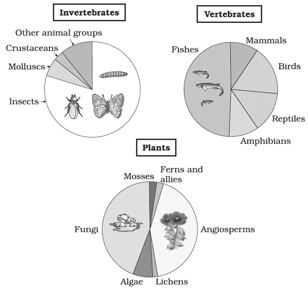
Representing global biodiversity: proportionate number of species of major taxa of plants, invertebrates and vertebrates
What is living?
The dog I can hear barking is alive, and so is the tree outside my window. However, snow falling from the clouds is not alive. The computer you’re using to read this article is not alive, and neither is a chair or table.
What is living?
Anything which shows characteristics of life is called living. The fundamental characteristics of life are- growth, reproduction, metabolism, cellular organization and consciousness. Living things are highly organized, meaning they contain specialized, coordinated parts.
Growth
Living things tend to grow in size. The increase in the number and mass of individuals are two unique characteristics of growth. Multicellular organisms exhibit growth by cell division. Growth varies in plants and animals. If we consider unicellular organisms, they tend to grow in size by cell division. Moreover, this can be easily detected by counting the cells during in vitro cultures under a microscope.
Reproduction
Reproduction is the process by which organisms replicate themselves.
Reproduction can primarily be of two types- sexual reproduction and asexual reproduction.
Reproduction generally refers to sexual reproduction. Asexual reproduction is used by the lower organisms like Bacteria that divide asexually via Binary fission, Virusestake control of host cells to produce more viruses; Hydras (invertebrates of the order Hydroidea) and Yeast are able to reproduce by Budding.
Sexual reproduction is a biological process that creates a new organism by combining the genetic material of two organisms in a process that starts with, meiosis a specialized type of Cell division
Metabolism
Metabolism is the sum of the chemical reaction that takes place within each cell of a living organism and that provides energy for vital processes and for synthesizing new organic material.
All living organisms are made up of chemicals. These chemicals, small and big belonging to various classes, size and function, etc are constantly being made and changed into other biomolecules.
There are thousands of metabolic reactions occurring simultaneously inside all living organisms, be they unicellular or multicellular.
Cellular organization
Cellular organization – All living organisms are made up of cells that perform all functions inside the body. The cellular organization is the components that make up the cell and how they are arranged inside it. Each component called an organelle, performs a specific function vital for the cell.
Consciousness
All living organisms have an inherent ability to sense their surroundings or environment and respond to their environmental stimuli which could be physical, chemical, or biological. Plants and animals both respond to external stimuli. Human beings are the only organisms who are aware of themselves, i.e., have self-consciousness.
What is living?
Chapter 1
The living world
What is living?
Biology is the study of biological organisms and processes. There is an incredible diversity of living organisms around the globe. Any system capable of executing processes including feeding, metabolizing, excreting, breathing, moving, growing, reproducing, and responding to external stimuli is characterized as living. Living refers to anything that exhibits the attributes of life. Growth, reproduction, metabolism, cellular organization, and consciousness are the basic features of life. On the other hand, self-consciousness is seen as the defining quality of living organisms. Let us take a detailed look at all the unique features of living organisms.
(A) Growth:
Growing is a process that occurs in all living creatures. The twin features of growth are an increase in bulk and an increase in the number of individuals. Cell division is a process that enables a multicellular organism to grow. Plants grow by the division of cells constantly throughout their lives. This growth is only visible in animals up to a particular age. Cell division, on the other hand, happens in some tissues to replace lost cells. Cell division is responsible for the growth of unicellular creatures. Growth and reproduction are mutually exclusive phenomena in the majority of higher animals and plants. It's important to note that growth is defined as a rise in bodily mass.
If we use body mass as a criterion for growth, non-living objects grow as well. Mountains, boulders, and sand mounds are all formed through erosion. Non-living objects, on the other hand, demonstrate this type of growth by accumulating material on the surface. Growth occurs from the inside out in living organisms. As a result, growth cannot be considered the only distinguishing characteristic of living beings. It must be described under what conditions it can be observed in all live species before we can grasp that it is a property of living systems. A dead creature can’t grow.
(B) Reproduction:
In multicellular organisms, reproduction refers to the generation of offspring with characteristics that are comparable to those of their parents. It refers to sexual reproduction invariably and implicitly. Asexual reproduction is also used by living species. Fungi, for example, can quickly proliferate and spread because they produce millions of asexual spores. We see budding in smaller species like yeast and hydra. True regeneration is observed in Planaria (flatworms), in which a fractured organism regenerates the missing section of its body and transforms into a new creature. Filamentous algae and moss protonema may all easily reproduce through fragmentation. In unicellular organisms such as bacteria, unicellular algae, and Amoeba, reproduction takes place through cell division (fission) which is synonymous with growth, or an increase in the number of cells.
Growth has already been characterized as a rise in cell number or mass. As a result, we find that the definitions of these two terms – growth and reproduction – are not always apparent in single-celled organisms. Furthermore, a large number of species do not reproduce (mules, sterile worker bees, infertile human couples, etc). As a result, reproduction alone cannot be considered a universal distinguishing trait of living entities. Of course, no non-living object can reproduce or replicate on its own.
(C) Metabolism:
Chemicals are found in all living things. Small and large compounds of diverse classes, sizes, functionalities, and other characteristics are constantly created and transformed into new biomolecules. Chemical or metabolic reactions are involved in these transformations. All living creatures, whether unicellular or multicellular, have millions of metabolic events going on at the same time. Metabolism is found in all plants, animals, fungi, and microorganisms. Metabolism is the sum of all chemical events that take place in our bodies. Metabolism does not exist in non-living objects.In cell-free systems, metabolic events can be demonstrated outside of the body. An isolated metabolic reaction(s) performed in a test tube outside the body of an organism is neither life nor non-living. While metabolism is a defining property of all living organisms, isolated metabolic reactions, referred to as in vitro, are not live objects but living reactions. As a result, the body's cellular organization is a fundamental defining property of all life forms.
(D) Self-consciousness:
The ability of all living organisms to detect their surroundings or environment and respond to these environmental stimuli, which can be physical, chemical, or biological, is perhaps the most evident and technically complex attribute. Our sense organs allow us to perceive our surroundings. External elements such as light, water, temperature, other species, contaminants, and so on affect plants. Environmental signals can be sensed and responded to by all species, from prokaryotes to the most complex eukaryotes. Seasonal breeders, both plants and animals, are affected by photoperiod. Chemicals that enter an organism's body are dealt with in the same way by all species. As a result, all organisms are 'conscious' of their surroundings. Humans are the only organisms that are aware of themselves or have self-consciousness.As a result, consciousness becomes the defining characteristic of living beings.
Underlying interactions are responsible for all life phenomena. Tissue properties are not found in the constituent cells but emerge as a result of interactions between them. Similarly, features of cellular organelles are not contained in the organelle's molecular elements but occur as a result of interactions among the organelle's molecular components. Emergent features emerge at a higher level of organization as a result of these interactions. This phenomenon occurs at all levels of the organizational complexity hierarchy.
As a result, living creatures can be described as self-replicating, evolving, and self-regulating interacting systems that can respond to external inputs. The narrative of life on Earth is told via biology. Biology is the study of how living species have evolved on our planet. All living species, present, past, and future, are related to one another to differing degrees via the sharing of similar genetic material.
Need for classification.
- Books Name
- ACME SMART COACHING Biology Book
- Publication
- ACME SMART PUBLICATION
- Course
- CBSE Class 11
- Subject
- Biology
Need for classification
- There are millions of plants and animals varying greatly in their form, structure and complexity.
- It is impossible to study all of them individually. To make the study of organisms possible and easier, scientists have divided organisms into different ranks or categories on the basis of similarities and differences.
- Classification is just like systematically arranged library where we can easily find out the required book, in the same way, if the organisms are arranged according to a system, it makes their study easy.
Some important needs are
- It is essential to understand the inter-relationship among different groups of organisms.
- It serves as a base for the development of other biological sciences like biogeography.
- Various applied biology fields also depend upon exact identification and classification.
Need for classification.
Diversity in the living world
The biological variety and variability of life on Earth are referred to as biodiversity. Looking around, one will see a wide range of living organisms, including potted plants, insects, birds, pets, and other animals and plants. Other species are invisible to the naked sight but are all around us. A species is represented by each different type of plant, animal, or organism you see. Between 1.7 and 1.8 million species have been identified and described. This refers to the diversity of species on the planet or the quantity and varieties of creatures that exist.
There are millions of plants and animals throughout the world that are known by their regional names. These regional names might differ from one location to the next, even within a country. When referring to organisms, such names can cause confusion. As confuse necessary to standardize the naming of living entities so that they are known by the same name all over the world. This is referred to as nomenclature.
This naming or nomenclature is only achievable if the organism is correctly characterized and we know to which organism the term refers. This is calledidentification.
Scientists have developed processes to assign a scientific name to each known organism in ordering the process. Scientific names for plants are determined by agreed-upon principles and criteria outlined in the International Code for Botanical Nomenclature (ICBN). Taxonomists have developed the International Code of Zoological Nomenclature (ICZN) for animals. Each creature has just one name thanks to the scientific names. Any organism's description should allow anyone (from all over the world) to come up with the same name. They also check to see if the name has been used for any other organism.
To give known creatures scientific names, biologists use broadly accepted rules. Each name is made up of two parts: a generic name and a specific epithet. Binomial nomenclature is a system for naming organismsconsisting of two parts. Carolus Linnaeus devised a naming system that is used by biologists all around the world. He is also known as the Father of Taxomy. This two-name approach was proven to be convenient.Mangiferaindica is the scientific name for the mango where, Mangifera denotes the genus, while indica denotes a specific species or epithet. The following are the universal rules of nomenclature:
1. Biological names are usually written in italics and written in Latin. Regardless of their origin, they are Latinized or derived from Latin.
2. In a biological name, the first component identifies the genus, whereas the second component denotes the specific epithet.
3. In handwriting, both words in a biological name are underlined or typed in italics to reflect their Latin origin.
4. The genus name begins with a capital letter, but the particular epithet begins with a tiny letter.
The author's name occurs after the specific epithet, i.e., the after the zoological name, and is shortened, e.g., Mangiferaindica Linn. It denotes that Linnaeus was the first to describe this species.
Classification is the procedure of grouping things into useful categories based on plainly observable characteristics. Plants and animals, as well as dogs, cats, and insects, are easily identifiable. These phrases link certain characteristics to the organisms in that group. When we talk about mammals,' we're talking about animals with external ears and body hair. As a result, terminology like "Dogs," "Cats," "Mammals," "Wheat," "Rice," "Plants," and "Animals," among others, are useful categories for studying creatures. Taxa is the scientific word for these classifications. A taxon can denote a variety of categories at various levels. Plants are classified as a taxon. 'Wheat' is a taxon as well. 'Animals,' 'Mammals,' and 'Dogs,' for example, are all taxa. As a result, the terms 'animals,"mammals,' and 'dogs' all refer to taxa at various levels.
As a result, all living species can be divided into different taxa based on their properties. Taxonomy is the term for this classifying procedure. The external and internal structure of organisms, as well as cell structure, development process, and ecological information, are essential and form the basis of modern taxonomic investigations. As a result, the fundamental stages of taxonomy are characterization, identification, categorization, and nomenclature. Humans, fora long, have been learning more about different types of organisms, particularly concerning their own Hence, the early classifications were based on the diverse utilities of organisms.
Humans have long been fascinated not only by diverse types of species and their diversity but also by the links that exist between them. The term "systematics" was coined to describe this field of study. The name "systematics" comes from the Latin word "systema," which means "an orderly arrangement of organisms." The title of Linnaeus' publication was SystemaNaturae. Identification, naming, and classification was later added to the scope of systematics. Systematics is the study of organisms' evolutionary relationships.
Biodiversity.
- Books Name
- ACME SMART COACHING Biology Book
- Publication
- ACME SMART PUBLICATION
- Course
- CBSE Class 11
- Subject
- Biology
Biodiversity
- From a current estimation, approximately 1.7 million species have been scientifically named and classified. These include nearly 1.2 million animals and over 0.5 million species of plants.
- Amongst animals, insects form the largest group, i.e., over 10,25,000 species. It is estimated that majority of species diversity is confined to tropical rain forests and huge diversity exist in under water reef formations in tropical oceans.
- Existing living species are the outcome of about 3.5 billion years of evolutionary process on this earth. Nearly 15,000 new organisms are discovered every year.
- A clearer understanding of this huge variety of organisms can be studied by dividing these into smaller groups or sub-groups (categories) and each group or sub-group comprising of organisms with more or less similar characters.
- Method of placing organisms into groups or sub-groups depending upon extent of similarities and differences is called classification.
- The division of organisms into different groups follows certain rules, that is why the term called taxonomy is used for classification of organisms following certain rules or principles.
Biodiversity.
Taxonomic Categories
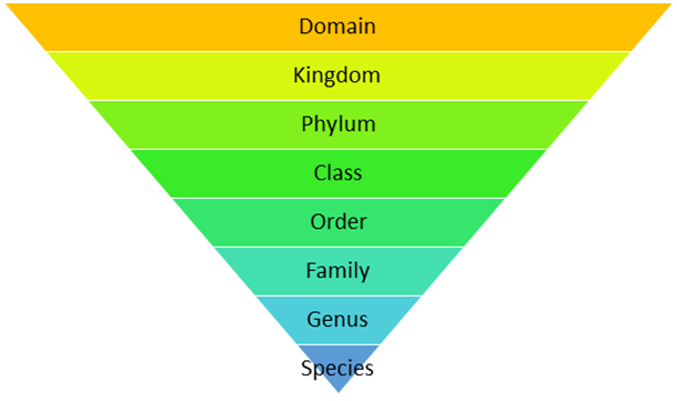
Classification is not a one-step procedure, but rather a series of stages, each representing a rank or category. The category is called a taxonomic category because it is a part of the larger taxonomic arrangement, and all categories combined make up the taxonomic hierarchy. Each category, also known as a classification unit, indicates a rank and is frequently referred to as a taxon (plural taxa). A taxonomic category is a category that is part of a taxonomic arrangement, and all categories combined make up the taxonomic hierarchy. Thus, organisms are divided into kingdoms, phyla or divisions, classes, orders, families, genera, and species.
Insects are a group of organisms with three pairs of jointed legs and other characteristics in common. It indicates that insects are recognizable concrete objects that can be categorized and so have a rank or category assigned to them. The categories are represented by groups of people. Rank is also indicated by category. Each rank, or taxon, is a classification unit. These taxonomic groups/categories are biological entities in their own right, not just morphological aggregations.
The establishment of common categories such as kingdom, phylum or division (for plants), class, order, family, genus, and species have resulted from taxonomic investigations of all known organisms. Species is the lowest level of classification for all organisms, including those in the plant and animal kingdoms. The knowledge of characteristics of an individual or group of organisms is required to categorize an organism into distinct categories. This aids in the identification of similarities and differences between individuals of the same species as well as individuals of different species.
Let us take a detailed look at different taxonomic categories:
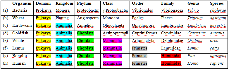
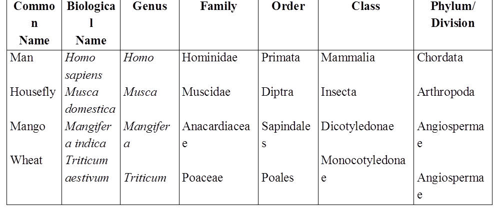
(A)Species: A species is a group of individual beings that share fundamental characteristics. Based on morphological features, one species is separated from other closely related species. Mangiferaindica, Solanum tuberosum (potato), and Pantheraleo are just a few examples (lion). The first three words, Mangifera, Solanum, and Panthera, are genera and denote a higher level of taxon or category, while the first three words, indica, tuberosum, and leo, are specific epithets. Each genus may have one or more specific epithets that refer to various creatures that share morphological characteristics. Panthera, for example, has the specific epithet tigris, and Solanum comprises species such as nigrum and melongena. Humans are members of the species sapiens, which is part of the genus Homo.As a result, the scientific designation for a human being is Homo sapiens. Species is regarded as the least inclusive and highly specific category of the taxonomic hierarchy.
(B) Genus: A genus is a collection of related species that share more characteristics than those from other genera. Genera can be defined as groups of closely related species. Potato and brinjal, for example, are two separate species that both belong to the Solanum genus. Panthera species include the lion (Pantheraleo), leopard (Pantherapardus), and tiger (Pantheratigris), all of which share several characteristics. This genus is distinct from the Felis genus, which includes cats.
(C) Family:In comparison to genus and species, a family consists of a set of related genera with fewer similarities. In plants, vegetative and reproductive characteristics are used to classify them into families. Solanum, Petunia, and Datura are three separate genera of plants that belong to the Solanaceae family. The genus Panthera, which includes the lion, tiger, and leopard, is placed in the family Felidae with the genus Felis (cats). Similarly, if you compare and contrast the characteristics of a cat and a dog, you would notice some parallels and variances. Felidae and Canidae are the two different families that they belong to.
(D) Order:Species, genus, and family are all based on a set of comparable characteristics. The aggregates of characters are used to identify the order and other higher taxonomic groups. The group of families that have a few similar characteristics is called order, which is a higher category. When compared to the different genera in a family, there are fewer comparable characters. Plant groups such as Convolvulaceae and Solanaceae are classified as Polymoniales based on their floral characteristics. Carnivora is an animal order that includes families like Felidae and Canidae.
(E) Class: A class is a taxonomic rank (a taxon) that contains species with similar characteristics; it is further divided into one or more orders. A class is a major taxonomic rank below the phylum (or division) and above the order in the biological categorization of life. This category includes orders that are related. Order Primata, which includes monkeys, gorillas, and gibbons, is placed in class Mammalia alongside order Carnivora, which includes tigers, cats, and dogs. Other orders also exist in the Mammalia class.
(F) Phylum/Division:A phylum is a classification or taxonomic rank that falls between kingdom and class. The Phylum is the next higher classification, which includes animals such as fish, amphibians, reptiles, birds, and mammals. All of these are classified as members of the phylum Chordata because they share characteristics such as the presence of a notochord and a dorsal hollow neural system. Classes with a few comparable features in plants are given to a higher category called Division.
In biological classification, the division is a taxonomic rank that is employed differently in zoology and botany. The division is a level similar to a phylum in botany and mycology. The term division is used in zoology to refer to an optional rank that is subordinate to the infraclass and superordinate to the cohort.
(G) Kingdom:In the classification system of animals, all animals belonging to distinct phyla are placed in the top category, Kingdom Animalia. The Kingdom Plantae, on the other hand, is unique in that it includes all plants of diverse divisions. These two groups are today referred to as the animal and plant kingdoms.
(H) Domain:A domain is the largest of all groupings in the classification of life in biology. At this level, the Archaea domain, Bacteria domain, and Eukarya domain are the three agreed-upon categories. The domain is considered to be the most inclusive category of the hierarchy.
The amount of shared features decreases as we progress from species to kingdom. The lower the taxon, the more features the members of the taxon share. The more difficult it is to determine a taxon's relationship to other taxa at the same level, the higher the category. As a result, the classification problem gets more complicated. Therefore, the taxonomists have also created sub-categories in the hierarchy to make the placement of distinct taxa more sound and scientific.
taxonomy and systematics; concept of species and taxonomical hierarchy.
- Books Name
- ACME SMART COACHING Biology Book
- Publication
- ACME SMART PUBLICATION
- Course
- CBSE Class 11
- Subject
- Biology
Taxonomy and Systematics
Taxonomy: The branch of science dealing with the study of principles and procedures of classification is called taxonomy. The term taxonomy was coined by A.P. de Candolle. Linnaeus is considered as Father of Taxonomy. Santapau is considered as Father of Indian Taxonomy. The fundamental elements of taxonomy are as follows :
- Characterisation and identification : It is the determination of the similarities of an organism with an already known organism, based upon specific characters.
- Nomenclature: It is the determination of correct name of an organism according to established universal rules.
- Classification: It is grouping of organisms into convenient categories on the basis of easily observable characters.
-
The classical taxonomy is based on observable morphological characters, however the modern taxonomy deals with several characters for the classification of organisms like
(a) External and internal structure alongwith the structure of cell.
(b) Development process.
(c) Ecological information of organisms
Systematics : The word "systematics" is derived from Latin word systema which means systematic arrangement of organisms. It was first used by Carolus Linnaeus. According to him, "systematics is the discipline of biology which deals with the kind and diversity of all organisms and the existing relationships amongst them."
Generally, the terms such as classification, systematics and taxonomy are used interchangeably but some taxonomists like Simpson (1961) relate them with a separate field. He defined systematics as
"The study of diversity of organisms and all their comparative and evolutionary relationships based on comparative anatomy, comparative ecology, comparative physiology and comparative biochemistry."
The main uses of systematics are as given below :
(i) It helps in providing knowledge of great diversity of animals and plants. It provides information regarding evolution which took place among plants and animals by knowing the distinction, relationship, habitat and habits. It thus, gives a vivid picture of entire organic diversity.
(ii) It helps in the identification of fossils which gives useful information about the phylogeny of organisms.
(iii) Newly discovered organisms can be identified through systematics.
TAXONOMIC CATEGORIES
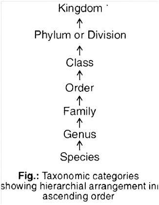
- Classification is not a single step process. It involves hierarchy of steps in which each step represents a rank or category.
- The category is a part of overall taxonomic arrangement. All categories together make taxonomic hierarchy.
- Each category is also termed as a unit of classification. It represents a rank and is commonly called as taxon.
- The taxon must be recognisable and order should belong to a category.
- Taxonomic hierarchy is a series of different ranks placed in ascending or descending order.
- It was Linnaeus who for the first time introduced five categories in the taxonomic hierarchy, viz., class, order, genus, species and variety.
- Later on three more categories, viz., kingdom, division or phylum and family were added.
- Variety was Species discarded to make a hierarchy of seven obligate categories.
- Taxonomic categories kingdom -division (in plants) or phylum (in animals) -class -order -family -genus -species. (descending order)
- Higher the category, higher is the number of organisms in it. Higher the category, fewer will be the number of common characters and greater is the difficulty of determining the relationship to other taxa at the same level. Hence, the problem of classification becomes more complex.
- Taxonomic categories and hierarchy can be illustrated by an example. Insects represent a group of organisms sharing common features like three pairs of jointed legs.
- It means insects are recognisable concrete objects which can be classified, and thus were given a rank or category.
- Similarly, mammals represent animals with external ears, body hairs, mammary glands etc. Dog, mammals, animals are all taxa but of different categories.
- Taxon dog, mammals and animals represent categories like species, class and kingdom respectively.
Species:
- Term given by John Ray. It is lowest category of classification. It is a group of closely related individuals with similar morphological, anatomical, biochemical and cytological characters.
- It is a group of naturally interbreeding population with the ability to produce fertile offsprings.
- Individuals of a species share common gene pool.
- It is reproductively isolated, thus genetically closed system.
e.g, Pisum sativum – Pea
Mangifera indica – Mango
Solanum tuberosum – Potato
Panthera leo – Lion
Homo sapiens – Human being
In these, sativum, indica, tuberosum, leo, sapiens represent the specific epithet, while Pisum, Mangifera, Solanum, Panthera and Homo represent genus.
Genus:
- Genus is a group of related species. Species has more characters in common in comparison to species of other genera.
- e.g., Potato, (Solanum tuberosum), makoi (S. nigrum) and brinjal (S. melongena) are three different species belonging to same genus Solanum.
- Lion, leopard and tiger with several common features belong to the same genus Panthera.
Family:
It is a group of related genera with still less number of similarities as compared to genus and species. Families are characterised on the basis of both vegetative and reproductive features of plants.
- For example, four related genera Solanum, Petunia, Datura and Atropa belong to the family Solanaceae. Genus Panthera and Felis (cat) are put together in family Felidae.
Order:
- It is a group of related families which exhibit a few similar characters. The similar characters are less in number as compared to different genera included in a family.
- For example, plant families like Convolvulaceae and Solanaceae are included in order Polymoniales mainly based on floral characters.
- In animals, order carnivora includes families like Felidae (cat) and Canidae (dog).
Class:
- It is a group of related orders.
- For example , plants order like Sapindales (mango) and Polymoniales are included in Class -Dicotyledonae, Order -Volvocales (Volvox) and Conjugales (Spirogyra) are included in Class -Chlorophyceae (green algae).In animals, order Primata (man, monkey) and Carnivora (cat, dog) are included in class -Mammalia.
Phylum/Division :
- It is a group of related classes.
- The phylum Chordata of animals contains not only the class mammalia but also aves (birds), reptilia (reptiles) amphibia (amphibians) and osteichthyes (fishes).
- In case of plants, classes with few similar characters like dicots and monocots constitute division -Angiospermae.
- The category phylum is used in animalia while division is used in plantae.
Kingdom:
- It is highest category in hierarchy with related phylums or divisions.
Kingdom animalia includes all animals belonging to different phyla. Kingdom Plantae includes all plants of various divisions.
Concept of Species :
Biological concept of species (was given by Ernst Mayr) : Species is the fundamental unit of classification. A species is a group of organisms (i) which are closely related (structurally and functionally) sharing a common gene pool (ii) which can interbreed freely in nature and produce fertile offspring in a natural environment. This concept of species is based upon reproductive isolation and called biological concept.
Some important interspecific hybrids (exception of biological concept of species)
(i) Sterile Hybrid (under natural conditions)
Mule = Between male donkey and female horse (Mare)
Hinny = Between male horse (Stal'lion) and female donkey
(ii) Fertile Hybrid (under captive conditions)
Tigon = Between male tiger and female lion
Liger = Between male lion and female tiger
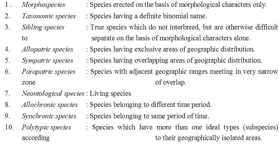
Types of Species :
TAXONOMICAL AIDS
- The laboratory and field studies are required for identification of various species and their placement in taxonomical hierarchy.
- The information thus gathered about the species, needs to be stored for future use.
- The taxonomical aids developed by biologists have established certain procedures and techniques to store and preserve the information as well as the specimens
Binomial nomenclature.
- Books Name
- ACME SMART COACHING Biology Book
- Publication
- ACME SMART PUBLICATION
- Course
- CBSE Class 11
- Subject
- Biology
NOMENCLATURE
There is a need to standardize the naming of living organisms, such that a particular organism is known by same name all over the world. A variety of nomenclatural methods are described below :
1.Vernacular name: Names in local or common language are called vernacular names. So, many vernacular names exist for an organism in different languages. These may vary from place to place.
2.Scientific names: These names were based on definite rules and criteria. These are of following types :
(i) Polynomial nomenclature
(ii) Trinomial nomenclature [both given in concept builder]
(iii) Binomial system of nomenclature:
a.Swedish naturalist Carolus Linnaeus established binomial nomenclature though, it was first proposed by Casper Bauhin in his book PINAX.
b. In binomial nomenclature, the first word is a generic name and second word is a specific epithet like Mangifera indica Linn.
c. After end of biological name, the name of author is written in abbreviated form who gave the name of the organism.
d.Scientific names are in Latin, because Latin was the language of scholars at the time of Linnaeus, and no change is possible in the language because this language has no synonyms.
e.Linnaeus gave some principles of the binomial nomenclature in Philosophia Botanica.
f. The nomenclature was used first in Species Plantarum (1753), where names and description of 5900 species of plants were given.
g.He published Systema Naturae (1758), where 4326 species of animals were described.
A. International Code of Nomenclature
Scientific names have been standardised through some international agencies, viz., International Code of Botanical Nomenclature (ICBN, 1961) and International Code of Zoological Nomenclature (ICZN, 1964), International Code for Nomenclature of Bacteria (ICNB), International Code of Nomenclature for Cultivated Plants (ICNCP) and currently being developed is International Committee for the Taxonomy of Viruses (ICTV).
B. Rules for Binomial Nomenclature
ICBN and ICZN formulated certain rules and regulations for giving scientific names to all organisms. These rules are as follows :
- The valid name of an organism has two components, i.e., a generic name and a specific epithet. The generic name should begin with a capital letter and species name should begin with a small letter.
- Both the words in a biological name when handwritten are separately underlined or printed in italics to indicate their Latin origin.
- The name of the author should be written after the scientific name in Roman type with capital letter without any comma in between and is written in an abbreviated form, e.g., Homo sapiens Linn. is the complete scientific name for modern man. This shows that Linnaeus was the first scientist who named man as Homo sapiens.
- Scientific names should not contain less than three and more than twelve letters.
- Principle of priority: It is the most important of all the rules of ICBN. If first name given to the organism is valid (in terms of rules), that will be considered at the first preference. Any other valid name given after that will be considered as synonym. No names are recognised prior to those used by Linnaeus in 1758 in the 10th edition of Systema Naturae for animals and 1753 for plants.
- All the three words (generic name, species epithet and author citation) collectively form Binomial epithet.
- If a species name has two or more words in its name, a hyphen is put between these. Such names are compound specific names (e.g., Hibiscus rosa-sinensis).
zoological parks.
- Books Name
- ACME SMART COACHING Biology Book
- Publication
- ACME SMART PUBLICATION
- Course
- CBSE Class 11
- Subject
- Biology
Zoological Parks
- Zoos or zoological gardens (parks) are protected areas or enclosed space where live wild animals are kept, under human care. This enables us to learn their food habits and behaviour.
- Objectives are public exhibition to understand wild life, recreation, education, ex situ conservation and breeding of rare fauna.
- Largest zoo of the world is situated in Kruger (S. Africa).
- National Zoological Park (Delhi) is one of the finest zoo of Asia.
Herbaria.
- Books Name
- ACME SMART COACHING Biology Book
- Publication
- ACME SMART PUBLICATION
- Course
- CBSE Class 11
- Subject
- Biology
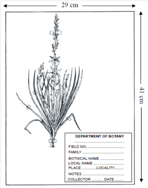
Herbarium (Dry Garden)
- It is defined as "a store house of collected plant specimens that are dried, pressed and preserved on sheets."
- These sheets are arranged in the sequence of an accepted classification system.
- These specimens, along with their description on herbarium sheets, become a store house or repository for future use. The herbarium sheet contains a label on the right-hand side at lower corner.
- Label provides information about date and place of collection, English, local and botanical names, family, collector's name etc.
- Herbaria also serve as quick referral systems in taxonomical studies.
Herbarium Sheet

Functions of a Herbarium
The two primary functions of herbarium are accurate identification and alpha taxonomic research (based on gross morphology).
The secondary functions include closer interaction between the student of general systematics and the herbarium.
Other important functions of a herbarium are –
1.To preserve plant wealth including type material and palaeobotanical collections.
2.To carry out exchange and loan of preserved plant material for research, exhibitions etc.
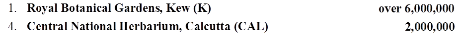
A list of important herbaria of the world is given below along with their standard abbreviations and the approximate number of specimens they hold
TYPE SPECIMEN (HERBARIUM SHEET)
Type specimen (Herbarium Sheet) of newly discovered plant should be place in herbarium (Dry garden). Standered size of herbarium sheet is 11.5 × 16.5 inches.
Holotype - Herbarium sheet on which the first description of plant is based.
Isotype - Duplicate of holotype - In presence of holotype a second herbarium sheet prepared from the original plant is called isotype.
Paratype - Additional herbarium sheet used in the first description of plant is called paratype. It is prepared from some other plant of same species having some variations.
Lectotype - In case of holotype is lost, second herbarium sheet prepared from the original plant is called lectotype.
Neotype - In case of holotype and original plant is lost, then herbarium sheet prepared from some other plant of same species is called neotype.
Syntype - In case of holotype and original plant is lost then many herbarium sheet prepared from many plants of same species is called syntype.
Note : Nomenclature is invalid in absence of Herbarium sheet.
Botanical gardens.
- Books Name
- ACME SMART COACHING Biology Book
- Publication
- ACME SMART PUBLICATION
- Course
- CBSE Class 11
- Subject
- Biology
Botanical Gardens
- From the time of Theophrastus, gardens have contributed to the science of botany. But, there was an impetus to the botanical explorations only in the Post-Linnean period.
- In ancient Indian culture, cultivation of food and medicinal plants is known since 4000 to 2000 B.C. The 'Hanging Gardens of Babylon' are amongst the wonders of the ancient world.
- During the Middle Ages, from A.D. 600-1600, there was a lapse in learning and introduction of plants.
- In the seventeenth century, there was a revival in the interest and by eighteenth century, most of the famous Botanical Gardens known today had already been established.
The functions of a botanical garden are:
- Provide records of local flora for monographic work.
- Provide facilities for collections and identification of living plant material for biosystematic studies / references.
- Supply seeds and material for botanical investigations.
- Botanical gardens have an aesthetic appeal and attract a large number of visitors for observing general plant diversity.
- Provides means of ex-situ conservation strategies.
- There are about 525 botanical gardens in various countries, but only about 125 have documented collections of authenticated taxa.
6.The International Association of Botanical Gardens was established in 1962. This association has published the International Directory of Botanical Gardens (1983).
BOTANICAL GARDENS OF INDIA
- The Indian Botanical Garden, Kolkata, India: It was founded in 1787, by Lt.Col. Robert Kyd. It covers an area of 273 acres and contains collections of world's tropical plants.
- It is one of the greatest botanical gardens of the world and one of the first to be established in tropics.
- William Roxburgh, 'Father of Indian Botany' was its director from 1793 to 1813.
- It has the largest herbarium of east and is famous for the Great Banyan tree, Ficus benghalensis, which is two centuries old, the palm houses, nurseries and the Amazon lily, Victoria amazonica (Nymphaeaceae), the plant with the largest leaves.
It is now under control of BSI (Botanical Survey of India).
Other botanical gardens of India are -
Lloyd Botanical Garden – Darjeeling
National Botanical Garden – Lucknow
Lalbag Gardens – Bangalore
Saharanpur Botanical Garden – Saharanpur
Diversity in the Living World
- Books Name
- ACME SMART COACHING Biology Book
- Publication
- ACME SMART PUBLICATION
- Course
- CBSE Class 11
- Subject
- Biology
Diversity in the Living World
From a current estimation, approximately 1.7 million species have been scientifically named and classified. These include nearly 1.2 million animals and over 0.5 million species of plants.
Amongst animals, insects form the largest group, i.e., over 10,25,000 species. It is estimated that majority of species diversity is confined to tropical rain forests and huge diversity exist in under water reef formations in tropical oceans.
Existing living species are the outcome of about 3.5 billion years of evolutionary process on this earth. Nearly 15,000 new organisms are discovered every year.
A clearer understanding of this huge variety of organisms can be studied by dividing these into smaller groups or sub-groups (categories) and each group or sub-group comprising of organisms with more or less similar characters.
Method of placing organisms into groups or sub-groups depending upon extent of similarities and differences is called classification.
The division of organisms into different groups follows certain rules, that is why the term called taxonomy is used for classification of organisms following certain rules or principles.
Diversity in the Living World
Diversity in the Living World
Diversity in the world can be experienced everywhere on the earth. Each different kind of plant, animal, or organism that you see represents a species. The number of species that are known and described ranges from 1.71.7 to 1.81.8 million, which is referred to as biodiversity. Every individual is unique with respect to structure, body functions, genetic make-up, etc
Living organisms inhabiting different habitats have different structural organs or functions developed according to the conditions of the habitat. The warm and humid regions of the earth have diverse organisms and thus are known as the region of mega biodiversity.
Terminology utilized as a part of classification includes Nomenclature, Classification, Taxonomy and Systematics.
The process of naming an organism is known as nomenclature. A principle and criterion were given for naming the organisms, by scientists known as International Code for Botanical nomenclature (ICBN). For animals, International Code of Zoological Nomenclature. This helps scientists from all over the world to interact with each other using the same name.
Each name consists of two parts the first part is the generic name and the second part is the name of the species. This is known as Binomial Nomenclature. This was given by a scientist known as Carolus Linnaeus.
Examples: Mangifera Indica (Mango); Homo sapiens (Man)
In this case, Mangifera and Homo are the generic names; indica and sapiens are specific epithets.
Rules of Nomenclature:
- Each biological name has two words-The principal word speaks about the genus and the second speaks to the specific epithet.
- The expressions of the name ought to be independently underlined when manually written and ought to be in italics when printed.
- The generic name ought to begin with a capital letter and the specific epithet ought to begin with a little letter.
- The names ought to be either Latin or Latinized.
Classification: It is the process of assigning creatures to particular groups or classes taking into account some defined characters. These classes are called Taxa (sing. taxon)
Taxonomy: It is the investigation of identification, nomenclature and arrangement of life forms taking into account outer and inside the structure with cell structure, advancement process and biological data.
systematics: It is the investigation of life forms with reference to identification, nomenclature, arrangement and evolutionary correspondence.
Diversity in the Living World
Diversity in the living world
The biological variety and variability of life on Earth are referred to as biodiversity. Looking around, one will see a wide range of living organisms, including potted plants, insects, birds, pets, and other animals and plants. Other species are invisible to the naked sight but are all around us. A species is represented by each different type of plant, animal, or organism you see. Between 1.7 and 1.8 million species have been identified and described. This refers to the diversity of species on the planet or the quantity and varieties of creatures that exist.
There are millions of plants and animals throughout the world that are known by their regional names. These regional names might differ from one location to the next, even within a country. When referring to organisms, such names can cause confusion. As confuse necessary to standardize the naming of living entities so that they are known by the same name all over the world. This is referred to as nomenclature.
This naming or nomenclature is only achievable if the organism is correctly characterized and we know to which organism the term refers. This is calledidentification.
Scientists have developed processes to assign a scientific name to each known organism in ordering the process. Scientific names for plants are determined by agreed-upon principles and criteria outlined in the International Code for Botanical Nomenclature (ICBN). Taxonomists have developed the International Code of Zoological Nomenclature (ICZN) for animals. Each creature has just one name thanks to the scientific names. Any organism's description should allow anyone (from all over the world) to come up with the same name. They also check to see if the name has been used for any other organism.
To give known creatures scientific names, biologists use broadly accepted rules. Each name is made up of two parts: a generic name and a specific epithet. Binomial nomenclature is a system for naming organismsconsisting of two parts. Carolus Linnaeus devised a naming system that is used by biologists all around the world. He is also known as the Father of Taxomy. This two-name approach was proven to be convenient.Mangiferaindica is the scientific name for the mango where, Mangifera denotes the genus, while indica denotes a specific species or epithet. The following are the universal rules of nomenclature:
1. Biological names are usually written in italics and written in Latin. Regardless of their origin, they are Latinized or derived from Latin.
2. In a biological name, the first component identifies the genus, whereas the second component denotes the specific epithet.
3. In handwriting, both words in a biological name are underlined or typed in italics to reflect their Latin origin.
4. The genus name begins with a capital letter, but the particular epithet begins with a tiny letter.
The author's name occurs after the specific epithet, i.e., the after the zoological name, and is shortened, e.g., Mangiferaindica Linn. It denotes that Linnaeus was the first to describe this species.
Classification is the procedure of grouping things into useful categories based on plainly observable characteristics. Plants and animals, as well as dogs, cats, and insects, are easily identifiable. These phrases link certain characteristics to the organisms in that group. When we talk about mammals,' we're talking about animals with external ears and body hair. As a result, terminology like "Dogs," "Cats," "Mammals," "Wheat," "Rice," "Plants," and "Animals," among others, are useful categories for studying creatures. Taxa is the scientific word for these classifications. A taxon can denote a variety of categories at various levels. Plants are classified as a taxon. 'Wheat' is a taxon as well. 'Animals,' 'Mammals,' and 'Dogs,' for example, are all taxa. As a result, the terms 'animals,"mammals,' and 'dogs' all refer to taxa at various levels.
As a result, all living species can be divided into different taxa based on their properties. Taxonomy is the term for this classifying procedure. The external and internal structure of organisms, as well as cell structure, development process, and ecological information, are essential and form the basis of modern taxonomic investigations. As a result, the fundamental stages of taxonomy are characterization, identification, categorization, and nomenclature. Humans, fora long, have been learning more about different types of organisms, particularly concerning their own Hence, the early classifications were based on the diverse utilities of organisms.
Humans have long been fascinated not only by diverse types of species and their diversity but also by the links that exist between them. The term "systematics" was coined to describe this field of study. The name "systematics" comes from the Latin word "systema," which means "an orderly arrangement of organisms." The title of Linnaeus' publication was SystemaNaturae. Identification, naming, and classification was later added to the scope of systematics. Systematics is the study of organisms' evolutionary relationships.
Taxonomic Categories
- Books Name
- ACME SMART COACHING Biology Book
- Publication
- ACME SMART PUBLICATION
- Course
- CBSE Class 11
- Subject
- Biology
TAXONOMIC CATEGORIES
Classification is not a single step process. It involves hierarchy of steps in which each step represents a rank or category.
The category is a part of overall taxonomic arrangement. All categories together make taxonomic hierarchy.
Each category is also termed as a unit of classification. It represents a rank and is commonly called as taxon.
The taxon must be recognisable and order should belong to a category.
Taxonomic hierarchy is a series of different ranks placed in ascending or descending order.
It was Linnaeus who for the first time introduced five categories in the taxonomic hierarchy, viz., class, order, genus, species and variety.
Later on three more categories, viz., kingdom, division or phylum and family were added.
Variety was Species discarded to make a hierarchy of seven obligate categories.
Taxonomic categories kingdom -division (in plants) or phylum (in animals) -class -order -family -genus -species. (descending order)
Higher the category, higher is the number of organisms in it. Higher the category, fewer will be the number of common characters and greater is the difficulty of determining the relationship to other taxa at the same level. Hence, the problem of classification becomes more complex.
Taxonomic categories and hierarchy can be illustrated by an example. Insects represent a group of organisms sharing common features like three pairs of jointed legs.
It means insects are recognisable concrete objects which can be classified, and thus were given a rank or category.
Similarly, mammals represent animals with external ears, body hairs, mammary glands etc. Dog, mammals, animals are all taxa but of different categories.
Taxon dog, mammals and animals represent categories like species, class and kingdom respectively.
Species:
Term given by John Ray. It is lowest category of classification. It is a group of closely related individuals with similar morphological, anatomical, biochemical and cytological characters.
It is a group of naturally interbreeding population with the ability to produce fertile offsprings.
Individuals of a species share common gene pool.
It is reproductively isolated, thus genetically closed system.
e.g, Pisum sativum – Pea
Mangifera indica – Mango
Solanum tuberosum – Potato
Panthera leo – Lion
Homo sapiens – Human being
In these, sativum, indica, tuberosum, leo, sapiens represent the specific epithet, while Pisum, Mangifera, Solanum, Panthera and Homo represent genus.
Genus:
Genus is a group of related species. Species has more characters in common in comparison to species of other genera.
e.g., Potato, (Solanum tuberosum), makoi (S. nigrum) and brinjal (S. melongena) are three different species belonging to same genus Solanum.
Lion, leopard and tiger with several common features belong to the same genus Panthera.
Family:
It is a group of related genera with still less number of similarities as compared to genus and species. Families are characterised on the basis of both vegetative and reproductive features of plants.
For example, four related genera Solanum, Petunia, Datura and Atropa belong to the family Solanaceae. Genus Panthera and Felis (cat) are put together in family Felidae.
Order:
It is a group of related families which exhibit a few similar characters. The similar characters are less in number as compared to different genera included in a family.
For example, plant families like Convolvulaceae and Solanaceae are included in order Polymoniales mainly based on floral characters.
In animals, order carnivora includes families like Felidae (cat) and Canidae (dog).
Class:
It is a group of related orders.
For example , plants order like Sapindales (mango) and Polymoniales are included in Class -Dicotyledonae, Order -Volvocales (Volvox) and Conjugales (Spirogyra) are included in Class -Chlorophyceae (green algae).In animals, order Primata (man, monkey) and Carnivora (cat, dog) are included in class -Mammalia.
Phylum/Division :
It is a group of related classes.
The phylum Chordata of animals contains not only the class mammalia but also aves (birds), reptilia (reptiles) amphibia (amphibians) and osteichthyes (fishes).
In case of plants, classes with few similar characters like dicots and monocots constitute division -Angiospermae.
The category phylum is used in animalia while division is used in plantae.
Kingdom:
It is highest category in hierarchy with related phylums or divisions.
Kingdom animalia includes all animals belonging to different phyla. Kingdom Plantae includes all plants of various divisions.
Concept of Species :
Biological concept of species (was given by Ernst Mayr) : Species is the fundamental unit of classification. A species is a group of organisms (i) which are closely related (structurally and functionally) sharing a common gene pool (ii) which can interbreed freely in nature and produce fertile offspring in a natural environment. This concept of species is based upon reproductive isolation and called biological concept.
Some important interspecific hybrids (exception of biological concept of species)
(i) Sterile Hybrid (under natural conditions)
Mule = Between male donkey and female horse (Mare)
Hinny = Between male horse (Stal'lion) and female donkey
(ii) Fertile Hybrid (under captive conditions)
Tigon = Between male tiger and female lion
Liger = Between male lion and female tiger
Types of Species :
Concept Builder
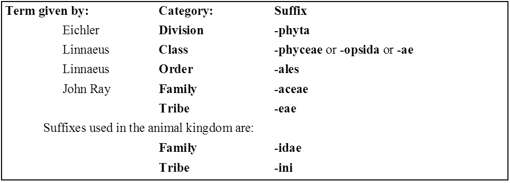
In accordance with International Code of Botanical Nomenclature, the names of different categories must end in the standard endings (suffixes) given below :
Tournefort gave the term genus and John Ray gave the term species
(b) Names of some families are changed according to ICBN rules for suffixes.
Old Name New Name
Palmae Arecaceae
Graminae Poaceae
Leguminosae Fabaceae
Compositae Asteraceae
Cruciferae Brassicaceae
Umbelliferae Apiaceae
Labiatae Lamiaceae
Guttiferae Clusiaceae
(c) Tribe is an intermediate category between sub-family and genus.
(d) In asexually reproducing organisms, physical resemblances and differences are used for delimiting species.
(e) Basic function of biological classification :
- (a) Recognition and description of species.
- (b) Grouping the species on the basis of similarities and relationship.
- (c) To establish evolutionary relationship amongst different organisms.
(f) The genera which have more than one specific epithets, are known as polytypic.
Lion – Panthera leo
Leopard – Panthera pardus
Tiger – Panthera tigris
(g) There are 7 obligate categories and about 21 intermediate categories. Prefixes: sub-and super-used for intermediate categories.
(h) Species name is given on the basis of some characters or habit, colour and distribution, e.g., niger (black), alba (white), tuberosum (tuber).
(i) Taxon is a group of real organisms which is assigned for any category. It is the unit of classification for any rank. Term taxon was introduced by ICBN in 1956 and defined by Mayr (1964).
(j) Category is an abstract term and represents only rank or level in a hierarchy and does not represent the living organisms. Example: Reptile is taxon but reptilia is category.
(k) Typological concept of Species
It was proposed by “Aristotle” and “Plato”. According to this concept, “There is a definite type or pattern of characters are present in the each species and all the members of species shows maximum resemblance with this pattern”. The species in which one fixed pattern of characters is present are called as monotypic species. e.g., Bacteria, Blue green algae if more than one type of pattern of characters are present. These are called “Polytypic species” or “Macrospecies”. e.g., Brassica oleracea Cauliflower, Cabbage, Knol-Knol.
Type of Polytypic Species :
Biotype : Members of same species inhabiting similar environment and having some genetic variations are known as biotypes. Variations found in these members are permanent. These members can not interbreed among themselves. e.g. Cauliflower, Cabbage, Knol-Khol are three biotypes of one species
Ecotypes : Members of same species inhabiting different environment and having some genetic variations are known as ecotypes. Variations are permanent. These members can interbreed among themselves but due to geographical barrier they can not interbreed.
e.g., Crow (Corvus splendense) found in different regions are ecotype of one species
Corvus splendense splendense - Indian crow
Corvus splendense insolense - Myanmar crow
Corvus splendense protegatus - Srilankan crow
Ecospecies : It contains one or more ecotype which although interfertile (capable of interbreeding), but do not produce viable offsprings due to some natural interruption (mountain, ocean etc).
Ecads or Ecophenes : Members of same species having some non genetic variation due to environment. These variations are temporary.
e.g.,

Taxonomic Categories
Taxonomic Categories
Classification is not a one-step procedure, but rather a series of stages, each representing a rank or category. The category is called a taxonomic category because it is a part of the larger taxonomic arrangement, and all categories combined make up the taxonomic hierarchy. Each category, also known as a classification unit, indicates a rank and is frequently referred to as a taxon (plural taxa). A taxonomic category is a category that is part of a taxonomic arrangement, and all categories combined make up the taxonomic hierarchy. Thus, organisms are divided into kingdoms, phyla or divisions, classes, orders, families, genera, and species.
Insects are a group of organisms with three pairs of jointed legs and other characteristics in common. It indicates that insects are recognizable concrete objects that can be categorized and so have a rank or category assigned to them. The categories are represented by groups of people. Rank is also indicated by category. Each rank, or taxon, is a classification unit. These taxonomic groups/categories are biological entities in their own right, not just morphological aggregations.
The establishment of common categories such as kingdom, phylum or division (for plants), class, order, family, genus, and species have resulted from taxonomic investigations of all known organisms. Species is the lowest level of classification for all organisms, including those in the plant and animal kingdoms. The knowledge of characteristics of an individual or group of organisms is required to categorize an organism into distinct categories. This aids in the identification of similarities and differences between individuals of the same species as well as individuals of different species.
Let us take a detailed look at different taxonomic categories:
(A)Species: A species is a group of individual beings that share fundamental characteristics. Based on morphological features, one species is separated from other closely related species. Mangiferaindica, Solanum tuberosum (potato), and Pantheraleo are just a few examples (lion). The first three words, Mangifera, Solanum, and Panthera, are genera and denote a higher level of taxon or category, while the first three words, indica, tuberosum, and leo, are specific epithets. Each genus may have one or more specific epithets that refer to various creatures that share morphological characteristics. Panthera, for example, has the specific epithet tigris, and Solanum comprises species such as nigrum and melongena. Humans are members of the species sapiens, which is part of the genus Homo.As a result, the scientific designation for a human being is Homo sapiens. Species is regarded as the least inclusive and highly specific category of the taxonomic hierarchy.
(B) Genus: A genus is a collection of related species that share more characteristics than those from other genera. Genera can be defined as groups of closely related species. Potato and brinjal, for example, are two separate species that both belong to the Solanum genus. Panthera species include the lion (Pantheraleo), leopard (Pantherapardus), and tiger (Pantheratigris), all of which share several characteristics. This genus is distinct from the Felis genus, which includes cats.
(C) Family:In comparison to genus and species, a family consists of a set of related genera with fewer similarities. In plants, vegetative and reproductive characteristics are used to classify them into families. Solanum, Petunia, and Datura are three separate genera of plants that belong to the Solanaceae family. The genus Panthera, which includes the lion, tiger, and leopard, is placed in the family Felidae with the genus Felis (cats). Similarly, if you compare and contrast the characteristics of a cat and a dog, you would notice some parallels and variances. Felidae and Canidae are the two different families that they belong to.
(D) Order:Species, genus, and family are all based on a set of comparable characteristics. The aggregates of characters are used to identify the order and other higher taxonomic groups. The group of families that have a few similar characteristics is called order, which is a higher category. When compared to the different genera in a family, there are fewer comparable characters. Plant groups such as Convolvulaceae and Solanaceae are classified as Polymoniales based on their floral characteristics. Carnivora is an animal order that includes families like Felidae and Canidae.
(E) Class: A class is a taxonomic rank (a taxon) that contains species with similar characteristics; it is further divided into one or more orders. A class is a major taxonomic rank below the phylum (or division) and above the order in the biological categorization of life. This category includes orders that are related. Order Primata, which includes monkeys, gorillas, and gibbons, is placed in class Mammalia alongside order Carnivora, which includes tigers, cats, and dogs. Other orders also exist in the Mammalia class.
(F) Phylum/Division:A phylum is a classification or taxonomic rank that falls between kingdom and class. The Phylum is the next higher classification, which includes animals such as fish, amphibians, reptiles, birds, and mammals. All of these are classified as members of the phylum Chordata because they share characteristics such as the presence of a notochord and a dorsal hollow neural system. Classes with a few comparable features in plants are given to a higher category called Division.
In biological classification, the division is a taxonomic rank that is employed differently in zoology and botany. The division is a level similar to a phylum in botany and mycology. The term division is used in zoology to refer to an optional rank that is subordinate to the infraclass and superordinate to the cohort.
(G) Kingdom:In the classification system of animals, all animals belonging to distinct phyla are placed in the top category, Kingdom Animalia. The Kingdom Plantae, on the other hand, is unique in that it includes all plants of diverse divisions. These two groups are today referred to as the animal and plant kingdoms.
(H) Domain:A domain is the largest of all groupings in the classification of life in biology. At this level, the Archaea domain, Bacteria domain, and Eukarya domain are the three agreed-upon categories. The domain is considered to be the most inclusive category of the hierarchy.
The amount of shared features decreases as we progress from species to kingdom. The lower the taxon, the more features the members of the taxon share. The more difficult it is to determine a taxon's relationship to other taxa at the same level, the higher the category. As a result, the classification problem gets more complicated. Therefore, the taxonomists have also created sub-categories in the hierarchy to make the placement of distinct taxa more sound and scientific.

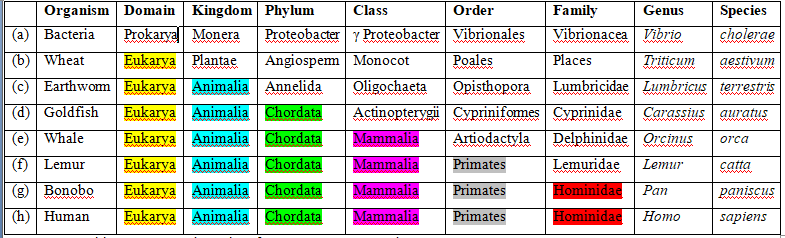
Taxonomic Categories
Taxonomy
Taxonomy Definition
Taxonomy is the branch of biology that classifies all living things. It was developed by the Swedish botanist Carolus Linnaeus, who lived during the 18th Century, and his system of classification is still used today. Linnaeus invented binomial nomenclature, the system of giving each type of organism a genus and species name. The Father of taxonomy was Carolus Linnaeus. He also developed a classification system called the taxonomic hierarchy, which today has eight ranks from general to specific: domain, kingdom, phylum, class, order, family, genus, and species.
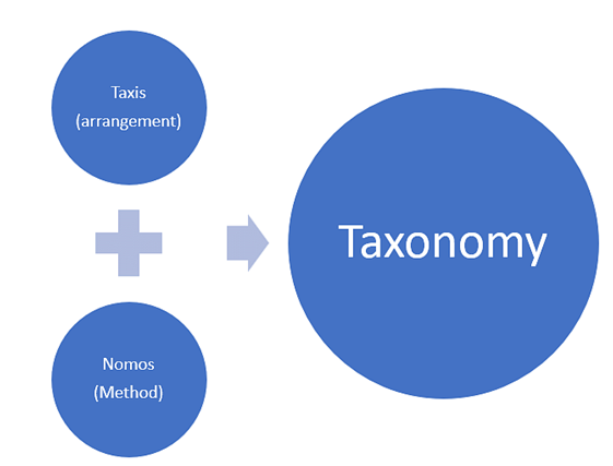
The Taxonomic Hierarchy
A taxon (plural: taxa) is a group of organisms that are classified as a unit. This can be specific or general. The hierarchy has two categories i.e., obligate and intermediate. Obligate is the one that is followed strictly and ranges from kingdom to species. Intermediate is not observed strictly and they. Also, they are added to the obligate lists like subdivision, superfamily, superclass, suborder, subspecies etc.
The following is a brief description of the taxonomic ranks of the taxonomic hierarchy.
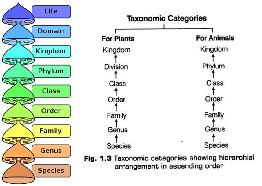
Domain
A domain is the highest (most general) rank of organisms. Linnaeus did invent some of the taxonomic ranks, but he did not invent the domain rank, which is relatively new. The term domain wasn’t used until 1990, over 250 years after Linnaeus developed his classification system in 1735.
The three-domain system of Carl Woese, introduced in 1990, with top-level groupings of Archaea, Bacteria, and Eukaryota domains.
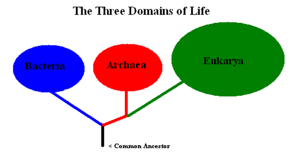
Kingdom
Before domains were introduced, the kingdom was the highest taxonomic rank. In the past, the different kingdoms were Animalia, Plantae, Fungi, Protista, Archaea, and Bacteria (Archaea and Bacteria were sometimes grouped into one kingdom, Monera)
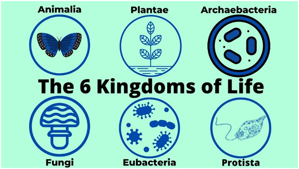
Phylum
Phylum (plural: phyla) is the next rank after kingdom; it is more specific than kingdom but less specific than class. There are 35 phyla in the kingdom Animalia, including Chordata (all organisms with a dorsal nerve cord), Porifera (sponges), and Arthropoda (arthropods).
Class
Class was the most general rank proposed by Linnaeus; phyla were not introduced until the 19th Century. There are 108 different classes in the kingdom Animalia, including Mammalia (mammals), Aves (birds), and Reptilia (reptiles), among many others.
Order
Order is more specific than class. Some of Linnaeus’ orders are still used today, such as Lepidoptera (the order of butterflies and moths). There are between 19-26 orders of Mammalia, depending on how organisms are classified—sources differ.
Genus
Genus (plural: genera) is even more specific than family. It is the first part of an organism’s scientific name using binomial nomenclature; the second part is the species name. An organism’s scientific name is always italicized, and the genus name is capitalized while the species name is not. Genus and species are the only taxonomic ranks that are italicized. The scientific name for humans is Homo sapiens. Homo is the genus name, while sapiens is the species name.
Species
Species are the most specific major taxonomic rank; species are sometimes divided into subspecies, but not all species have multiple forms that are different enough to be called subspecies. There are an estimated 8.7 million different species of organisms on Earth, While each genus name is unique, the same species names can be used for different organisms. For example, Ursus americanus is the American black bear, while Bufo americanus is the American toad.
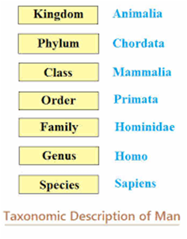
Taxonomical Aids
- Books Name
- ACME SMART COACHING Biology Book
- Publication
- ACME SMART PUBLICATION
- Course
- CBSE Class 11
- Subject
- Biology
TAXONOMICAL AIDS
The laboratory and field studies are required for identification of various species and their placement in taxonomical hierarchy.
The information thus gathered about the species, needs to be stored for future use.
The taxonomical aids developed by biologists have established certain procedures and techniques to store and preserve the information as well as the specimens.
1. Botanical Gardens
From the time of Theophrastus, gardens have contributed to the science of botany. But, there was an impetus to the botanical explorations only in the Post-Linnean period.
In ancient Indian culture, cultivation of food and medicinal plants is known since 4000 to 2000 B.C. The 'Hanging Gardens of Babylon' are amongst the wonders of the ancient world.
During the Middle Ages, from A.D. 600-1600, there was a lapse in learning and introduction of plants.
In the seventeenth century, there was a revival in the interest and by eighteenth century, most of the famous Botanical Gardens known today had already been established.
The functions of a botanical garden are:
1. Provide records of local flora for monographic work.
2. Provide facilities for collections and identification of living plant material for biosystematic studies / references.
3. Supply seeds and material for botanical investigations.
4. Botanical gardens have an aesthetic appeal and attract a large number of visitors for observing general plant diversity.
5. Provides means of ex-situ conservation strategies.
There are about 525 botanical gardens in various countries, but only about 125 have documented collections of authenticated taxa.
6. The International Association of Botanical Gardens was established in 1962. This association has published the International Directory of Botanical Gardens (1983).
Concept Builder
Some of the important Botanical Gardens are listed below :
1. Royal Botanical Garden, Kew, England : It was founded in 1759 by William Alton, but was officially opened in 1841 and was called the Botanical Capital of the World. Sir J.D. Hooker was one of its directors. It is famous for its Alpine house, Rose Garden, Green house, Temperature house, Bamboo garden and Lily pond. It is associated with Jodrell laboratory of experimental taxonomy. It is spread in 200 acres. It is famous for collection of Acacias, Acer, Rhus, Citrus, Rosa, Prunus, Magnolia. It has Chelsa Physic Garden for horticulturists. Kew
2. Orto Botanico (Padua Gardens), Italy: It is said to be the first botanical garden. It is famous for collections of grasses, Alliums, Irids, Paeonias, succulents and hydrophytes.
3. Pisa, Italy: Almost as old as Padua Gardens, it was the first to introduce palaeontological practises; for the study of plants fossils. It is known for trees like Magnolia grandiflora, Liriodendron tulipifera etc. Caesalpino and John Ray have been associated with this garden.
4. Villa Taranto, Italy: It is the most beautiful garden of Italy and important from the horticultural study point of view.
5. Main Botanical Garden, Moscow: Largest Botanical Garden, spread over an area of 900 acres.
BOTANICAL GARDENS OF INDIA
The Indian Botanical Garden, Kolkata, India: It was founded in 1787, by Lt.Col. Robert Kyd. It covers an area of 273 acres and contains collections of world's tropical plants.
It is one of the greatest botanical gardens of the world and one of the first to be established in tropics.
William Roxburgh, 'Father of Indian Botany' was its director from 1793 to 1813.
It has the largest herbarium of east and is famous for the Great Banyan tree, Ficus benghalensis, which is two centuries old, the palm houses, nurseries and the Amazon lily, Victoria amazonica (Nymphaeaceae), the plant with the largest leaves.
It is now under control of BSI (Botanical Survey of India).
Other botanical gardens of India are -
Lloyd Botanical Garden – Darjeeling
National Botanical Garden – Lucknow
Lalbag Gardens – Bangalore
Saharanpur Botanical Gardens – Saharanpur
Herbarium (Dry Garden)
It is defined as "a store house of collected plant specimens that are dried, pressed and preserved on sheets."
These sheets are arranged in the sequence of an accepted classification system.
These specimens, along with their description on herbarium sheets, become a store house or repository for future use. The herbarium sheet contains a label on the right-hand side at lower corner.
Label provides information about date and place of collection, English, local and botanical names, family, collector's name etc.
Herbaria also serve as quick referral systems in taxonomical studies.
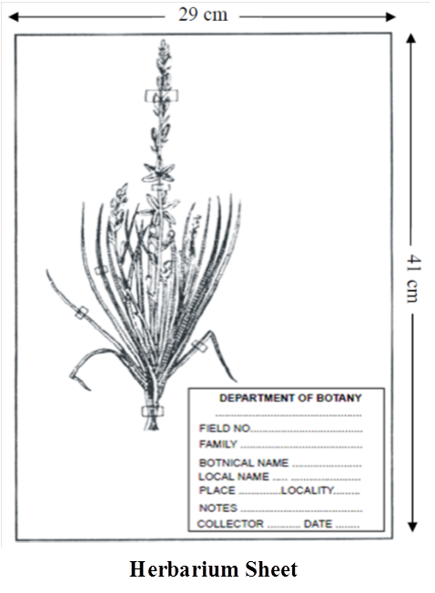
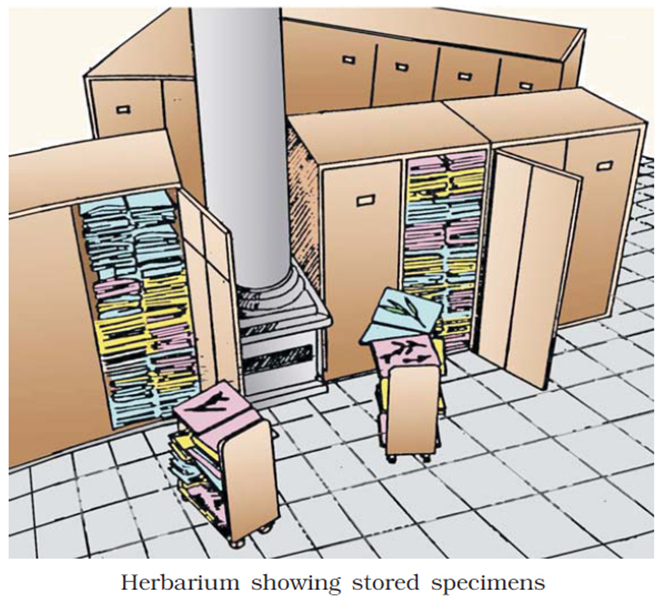
Concept Builder
Herbarium Technique
The herbarium technique involves the following steps:
- (a) Collection
- (b) Drying
- (c) Poisoning
- (d) Mounting
- (e) Stitching
- (f) Labelling
- (g) Deposition
(a) Collection: Collection of plant material is done with an aesthetic sense and scientific mind.
The material should be perfect and complete for determination, i.e., must have fully grown
leaves, complete inflorescence etc.
Woody elements are well represented by flowering twigs with 30-40 cm in lengths, while
herbaceous plants are collected alongwith underground parts.
Diseased plants, infected twigs should be avoided.
The collections are kept inside metallic vasculum or polythene bags.
(b) Drying: The plant collections are pressed in ordinary newspaper folders, avoiding overlapping. The folders, in turn, are pressed in a field press. The moistened folders are changed frequently to avoid blackening and decay of plant material.
(c) Poisoning: The specimens are poisoned to keep away the microbes. When the specimens are partially dehydrated, they are poisoned by using chemicals like 0.1% of corrosive, sublimate (HgCl2).
(d-f) Mounting, Stitching and Labelling : Dried specimens are glued and stitched on herbarium sheets made up of thick card sheets cut to the required size. The international size of the herbarium sheet is 41× 29 cm (16 ×11 inches) . The field data is entered on label on the right hand side lower corner of the herbarium sheet. Size of label is commonly 7 × 12cm. The small paper envelopes called fragment packets are often attached to the herbarium sheet to hold seeds, extra flowers or loose plant parts.
(g) Deposition: Arrangement of specimen, according to accepted classification, is called deposition. In India, herbaria are arranged according to Bentham and Hooker system of classification.
The specimens so preserved are sprayed with repellents or disinfectants such as DDT powder, copper sulphate solution at intervals of 4 to 6 months to keep off small insect pests such as silver fish.
Functions of a Herbarium
The two primary functions of herbarium are accurate identification and alpha taxonomic research (based on gross morphology).
The secondary functions include closer interaction between the student of general systematics and the herbarium.
Other important functions of a herbarium are
1. To preserve plant wealth including type material and palaeobotanical collections.
2. To carry out exchange and loan of preserved plant material for research, exhibitions etc.
A list of important herbaria of the world is given below along with their standard abbreviations and the approximate number of specimens they hold
1.Royal Botanical Gardens, Kew (K) over 6,000,000
4. Central National Herbarium, Calcutta (CAL) 2,000,000
TYPE SPECIMEN (HERBARIUM SHEET)
Type specimen (Herbarium Sheet) of newly discovered plant should be place in herbarium (Dry garden). Standered size of herbarium sheet is 11.5 × 16.5 inches.
Holotype - Herbarium sheet on which the first description of plant is based.
Isotype - Duplicate of holotype - In presence of holotype a second herbarium sheet prepared from the original plant is called isotype.
Paratype - Additional herbarium sheet used in the first description of plant is called paratype. It is prepared from some other plant of same species having some variations.
Lectotype - In case of holotype is lost, second herbarium sheet prepared from the original plant is called lectotype.
Neotype - In case of holotype and original plant is lost, then herbarium sheet prepared from some other plant of same species is called neotype.
Syntype - In case of holotype and original plant is lost then many herbarium sheet prepared from many plants of same species is called syntype.
Note : Nomenclature is invalid in absence of Herbarium sheet.
3. Keys [Given by John Ray]
The scheme for identification of plants and animals based upon similarities and dissimilarities is known as a key. It is based on the set of contrasting characters known as couplet, each character of couplet is called as lead. Separate taxonomic keys are required for each taxonomic category. Keys are generally analytical in nature and are of two types (1) Indented key (2) Bracketed key –
1. Indented Key: It has a sequence of choice between two or more statements of characters of species. These require great taxonomic skills to prepare, so are generally less followed.
2 Bracketed Key: In the Bracketed key the pairs of contrasting statements are used for identification. The bracketed number on the right side indicates the next choice of paired contrasting statements. These are most popular keys.
4. Zoological Parks
Zoos or zoological gardens (parks) are protected areas or enclosed space where live wild animals are kept, under human care. This enables us to learn their food habits and behaviour.
Objectives are public exhibition to understand wild life, recreation, education, ex situ conservation and breeding of rare fauna.
Largest zoo of the world is situated in Kruger (S. Africa).
National Zoological Park (Delhi) is one of the finest zoo of Asia.

5. Museums
These have collections of preserved plants and animals for study and reference.
Specimens are preserved in jars or containers in preservative solution.
Plant and animal specimens may also be preserved as dry specimens.
Insects are preserved in insect boxes after collecting, killing and pinning.
Larger animals are usually stuffed and preserved.
These often have collections of skeletons of animals too.
Museums are prepared to preserve algae, fungi, mosses, ferns and organs of gymnosperms since they cannot be kept in herbaria. These differs from parks because no living object is displayed in museums.
Some important Museums:
(i) Natural History Museum, London (England)
(ii) United States National Museum, Washington
(iii) National Museum of Natural History (NMNH), Delhi
(iv) Prince of Wales Museum, Mumbai etc.
Comperative Study of Botanical Garden, Herbarium and Museum :
6.Flora, Manuals, Monographs and Catalogues
Flora: Contains the actual account of habitat and distribution of plants of a given area. These provide the index to the plant species found in a particular area.
Some important flora are given below:
(i) Flora of British India by J.D. Hooker.
(ii) Flora of Delhi by J.K. Maheshwari.
(iii) Flora Indica by William Roxburgh.
(iv) Flora Simlensis by H. Collet
Manuals: The complete listing and description of the plants growing in a particular area.
e.g., Manual of Cultivated Plants by L.H. Bailey
Monographs: Contain information on anyone taxon.
e.g., The Genus Pinus by N.T. Mirov
Catalogues: This includes the alphabetical arrangements of species describing their features.
Concept Builder
1. Ex-situ / Off-site conservation strategies for organisms includes Botanical gardens and Zoological parks.
2. Succulent plants or those plants which are unsuitable for pressing and drying techniques, are fixed in suitable liquid preservative like 2.5% solution of formaldehyde, glacial acetic acid and ethyl alcohol [FAA].
3. Herbarium technique was first introduced by Caesalpino et. al.
4. Botanical gardens of world have 15% or 35,000 plants species of the world flora.
Tapiary -Beautifying areas
Bonsai trees -Dwarf trees are grown,in pots
5. According to WZCS (World Zoo Conservation Strategies), zoological parks are meant to conserve the species that are particularly endangered in their natural environment. India has about 200 zoological parks.
SUMMARY
1. The living world is rich in variety. Millions of plants and animals have been identified and described but a large number still remains unknown.
2. The very range of organisms in terms of size, colour, habitat, physiological and morphological features make us seek the defining characteristics of living organisms.
3. In order to facilitate the study of kinds and diversity of organisms, biologists have evolved certain rules and principles for identification, nomenclature and classification of organisms.
4. The branch of science dealing with these aspects is referred to as taxonomy.
5. The taxonomic studies of various species of plants and animals are useful in agriculture, forestry, industry and in general for knowing our bio-resources and their diversity.
6. The basics of taxonomy like identification, naming and classification of organisms are universally evolved under international codes.
7. Based on the resemblances and distinct differences, each organism is identified and assigned a correct scientific/biological name comprising two words as per the binomial system of nomenclature.
8. An organism represents/occupies a place or position in the system of classification. There are . many categories/ranks and are generally referred to as taxonomic categories or taxa. All the categories constitute a taxonomic hierarchy.
9. The basis of modern taxonomic studies is : External and internal structure, along with the structure of cell, development process and ecological information of organisms
10. Taxonomists have developed a variety of taxonomic aids to facilitate identification, naming and classification of organisms. These studies are carried out from the actual specimens which are collected from the field and preserved as referrals in the form of herbaria, museums and in botanical gardens and zoological parks. It requires special techniques for collection and preservation of specimens in herbaria and museums.
11. Live specimens, on the other hand, of plants and animals, are found in botanical gardens or in zoological parks.
12. Taxonomists also prepare and disseminate information through manuals and monographs for further taxonomic studies.
13. Taxonomic keys are tools that help in identification based on characteristics.
Taxonomical Aids
Taxonomic Aids
Taxonomic studies of various plant, animal, and other organism species are useful in agriculture, forestry, and industry, and they aid in our understanding of biodiversity. These studies necessitate accurate organism taxonomy and identification. Identifying organisms necessitates extensive laboratory and field research. Taxonomic aids are collections of samples or preserved species that aid in the extended investigation for taxonomic hierarchy identification. The primary source of taxonomic studies is the collection of genuine specimens of plant and animal species. These are also necessary for systematics training and are crucial to studies. It's used to classify organisms, and the data acquired is saved alongside the specimens. In some situations, the specimen is saved for further research. Some popular taxonomic aids are discussed below
(I) Herbarium: A herbarium is a reference collection of properly selected and dried plants that are connected to standard-sized paper sheets and filed systematically so that they can be quickly found for study. It's a repository for plant specimens that have been collected, dried, pressed, and preserved on sheets. These sheets are organized according to a widely accepted classification scheme. These specimens, coupled with their descriptions on herbarium sheets, form a repository or storehouse for future use. A label on the herbarium sheets provides information such as the date and location of collection, English, local, and botanical names, family, and collector's name, among other things. In taxonomic investigations, herbaria also function as rapid referral systems.
The Museum of Natural History in Paris, The Royal Botanic Gardens in Kew, England, The New York Botanical Garden, and The Komarov Botanical Institute in St. Petersburg, Russia, have some of the world's largest herbaria, each having 7 to 9.5 million specimens.

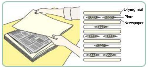
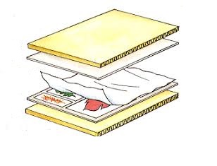
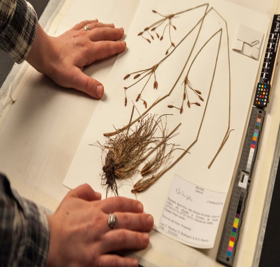
Figure 3: Different stages of herbarium preparation.
(II) Botanical Gardens:
For reference, these specialty gardens have collections of living plants. Plant species are planted in these gardens for identification, and each plant is labeled with its botanical/scientific name as well as its family. Botanical gardens commit their resources to plant research and conservation, as well as public awareness of the world's plant diversity. Royal Botanical Garden, Kew (England), Indian Botanical Garden, Howrah (India), and National Botanical Research Institute, Lucknow are all well-known botanical gardens.
(III) Museum:
A museum is a building where historical, artistic, or scientific objects are displayed, maintained, or researched. Modern museums are designed to collect, preserve, interpret, and show objects of aesthetic, cultural, or scientific significance for public study and education. Biological museums are most commonly found at educational institutions like schools and colleges. For study and reference, museums have collections of preserved plant and animal specimens. Preservative solutions are used to preserve specimens in containers or jars. Dry specimens of plants and animals can also be preserved. After capturing, killing, and pinning insects, they are kept in insect boxes. Stuffed and preserved larger creatures, such as birds and mammals, are common. Animal skeletons are frequently shown in museums.With 19 museums and galleries, the National Zoological Park, and many examination stations, the Smithsonian is the world's largest exploration and museum complex. The company's headquarters are in Washington, D.C.
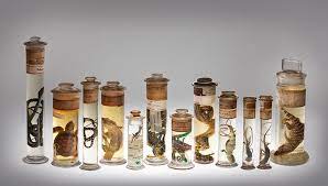
(IV) Zoological Parks:
Zoological parks are protected areas where animals are kept under human supervision and cared for. In zoological parks, many endangered species that are on the verge of extinction are rescued. These are facilities where wild animals are housed in safe conditions under human supervision so that we can learn about their eating habits, behavior, etc. Animals at zoos are kept in settings that are as close to their natural habitats as possible. These parks also contribute to the preservation of the natural balance by increasing the percentage of rainfall. They play a critical role in the conservation of animals. There are a total of 164 zoological parks in India. The Arignar Anna Zoological Park in Chennai is India's largest zoological park covering about 1300 acres with rare species of flora and fauna.
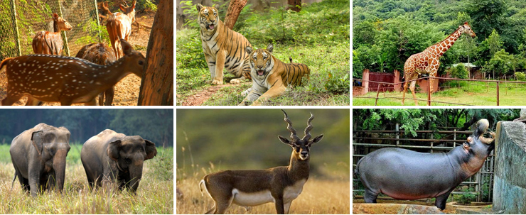
(V) Key:
Keys are used foridentifying and classifying various plants and animals based on their similarities and variances in characteristics. As a result, only one is accepted while the other is rejected. A lead is a name given to each assertion in the key. For identification purposes, separate taxonomic keys are necessary for each taxonomic group such as family, genus, and species. The majority of keys are analytical these are used to distinguish between classes, orders, families, genera, and species. It is split into pairs of characters that are either present or absent.
Other methods of recording descriptions include flora, manuals, monographs, and catalogs. They also assist with proper identification. Flora includes a detailed description of a certain area's habitat and plant distribution. These provide an index to the plant species that can be found in a certain area. A Catalogue is a comprehensive list, pamphlet, or register that contains a collection of traits and their variants found in diverse taxa.Manuals are important for identifying the names of species that can be found in a given area. Monographs are documents that focus on a single taxon.
Taxonomical Aids
Taxonomic Aids: Taxonomic studies of animals, plants, microorganisms and other species are highly useful in the field of forestry, industry, agriculture and the study of biodiversity. Hence, the organism should be identified, and classified and information about the organism should be stored for future studies and references. This storehouse of information and specimens that helps in taxonomic studies are called Taxonomic Aids.
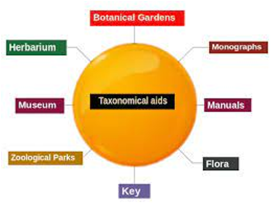
Herbarium: A herbarium is the storehouse of plant specimens.
It is a collection of dried, pressed, plant specimens that are arranged systematically according to a universally accepted system of classification. The Bentham and Hooker system is followed in most of Herbaria. The sheets on which the specimens are mounted are called Herbarium sheets
The total number of plant specimens are about 6.56.5 million.
Another Botanical Herbarium called Central National Herbarium is at Indian Botanical Garden, Howrah, Kolkata.
Some of the other examples are as follows:
a. National Botanical Garden, Lucknow
b. Lalbagh Botanical Garden, Bangalore, Karnataka
c. Government Botanical Garden, Ooty
d. Tropical Botanical Garden and Research Institute, Trivandrum, Kerala.
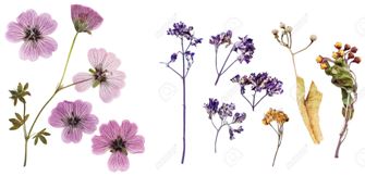
Botanical Garden: Botanical gardens are the place where varieties of plants collected from different places of the world are grown together in a protected environment. It may consist, of many rare, elite, exotic species. It shows rich biodiversity and plays an important role in germplasm conservation. In the Botanical Garden, all plants carry identification plates, having a common name, botanical name and the name of the family that belongs
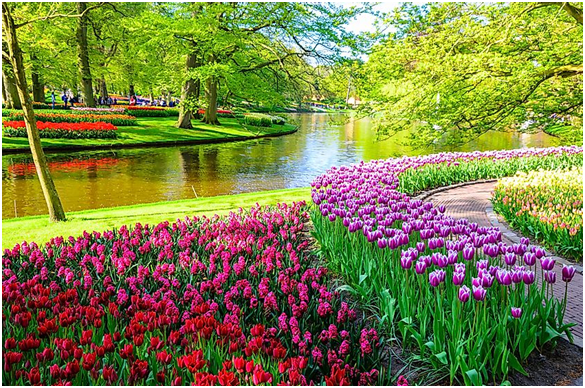
Museum: It is the collection of preserved plants and animal specimens for study and reference. Specimens are preserved in jars using preservative solutions of alcohol and formalin. Only those plants that cannot be kept in the herbarium are preserved. Some of the specimens include algae, fungi, mosses, ferns, succulents, fruits, etc. Even fishes, reptiles, amphibians, etc., are stored. Large animals are preserved in skeleton forms. Insects are dried out and mounted over pins inside the insect boxes. Others are preserved as dry specimens in the Biological Museum.
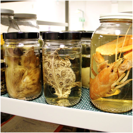
Zoological Park: Zoological Park is an enclosed area where wild animals are kept in an open, natural, but protected environment under human care. Zoological parks are the best places to study the behavior and food habitats of wild animals. Zoological parks help in conserving endangered animals and their multiplication through captive breeding. In India, there are around 200200 Zoological parks.
Zoological Parks are also important spots for children, students and the public to see wild animals
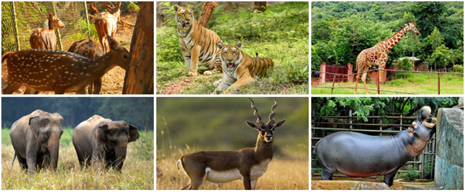
Keys: Key is another taxonomical aid used for the identification of plants and animals based on similarities and dissimilarities.Keys are of two types, i.e., dichotomous keys (bracketed key and indented key) and poly clave keys. Dichotomous Keys is the one in which the choices allow only two alternative couplets. There are two types of dichotomous keys – Indented Keys, also called yoked and Bracketed Keys. Poly clave key is relatively a new alternative to dichotomous keys and becoming increasingly popular, especially because of the ease of computerizing them and is also termed multiple access or synoptic key. This key is based on the identification of species by the process of elimination.
Kingdom Systems of Classification
- Books Name
- ACME SMART COACHING Biology Book
- Publication
- ACME SMART PUBLICATION
- Course
- CBSE Class 11
- Subject
- Biology
KINGDOM SYSTEMS OF CLASSIFICATION
(1) Two Kingdom Classification: It was given by Linnaeus. Traditionally all the organisms of the world were divided into two kingdoms -the animal kingdom (Animalia) and the plant kingdom (Plantae). The major criterion of classification was the presence or absence of cell wall. Other criterias were locomotion, mode of nutrition, response to external stimuli etc
Concept Builder
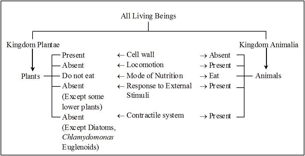
Shortcomings of two-kingdom system of classification:
This system did not distinguish between the eukaryotes and prokaryotes, unicelled and multicelled organisms, photosynthetic and non-photosynthetic organisms.
There are few organisms like Chlamydomonas, Euglena and the slime moulds which have been claimed by both zoologists and botanists (organisms which share characteristics of both animals and plants).
Since there are certain organisms that do not fall naturally into either plant or animal kingdom, it was proposed that a new kingdom is to be established to accommodate such organisms.
(2) Three Kingdom Classification:
Haeckel, a German zoologist (1866), suggested that a third kingdom Protista, be created to include all unicellular microorganisms.
This includes a wide variety of unicellular, mostly aquatic eukaryotes like -Fungi, Protozoa, Algae, Bacteria and Slime moulds.
Thus, he proposed three kingdoms, namely -Plantae, Protista and Animalia.
(3) Four Kingdom Classification:
Copeland (1956) gave four kingdom of classification and included Monera as fourth kingdom.
Copeland originally called it as kingdom 'Mychota'.
It was called 'Monera' by Daugherty and Allen.
Kingdom Monera includes all the prokaryotic organisms i.e., eubacteria (including cyanobacteria, formerly known as blue-green algae) and archaebacteria.
The actinomycetes (filamentous bacteria) are also included in this kingdom.
(4) Five Kingdom Classification:
According to five-kingdom concept proposed by R.H. Whittaker.
Whittaker (1969), the organisms are divided into five kingdoms namely Monera, Protista, Fungi, Plantae, Animalia, on the basis of the following criteria
(a) Complexity of cell structure: prokaryotic vs eukaryotic organisation of cells.
(b) Complexity of body organisation: unicellularity vs multicellularity; simple multicellular forms to complex multicellular forms.
(c) Mode of nutrition: Autotrophic vs heterotrophic (parasitic or saprobic or ingestive organisms). It was the major criteria of this classification system.
(d) Reproduction.
(e) Phylogenetic or evolutionary interrelations
Table 1 : Comparative account of different characteristics of the Five Kingdoms
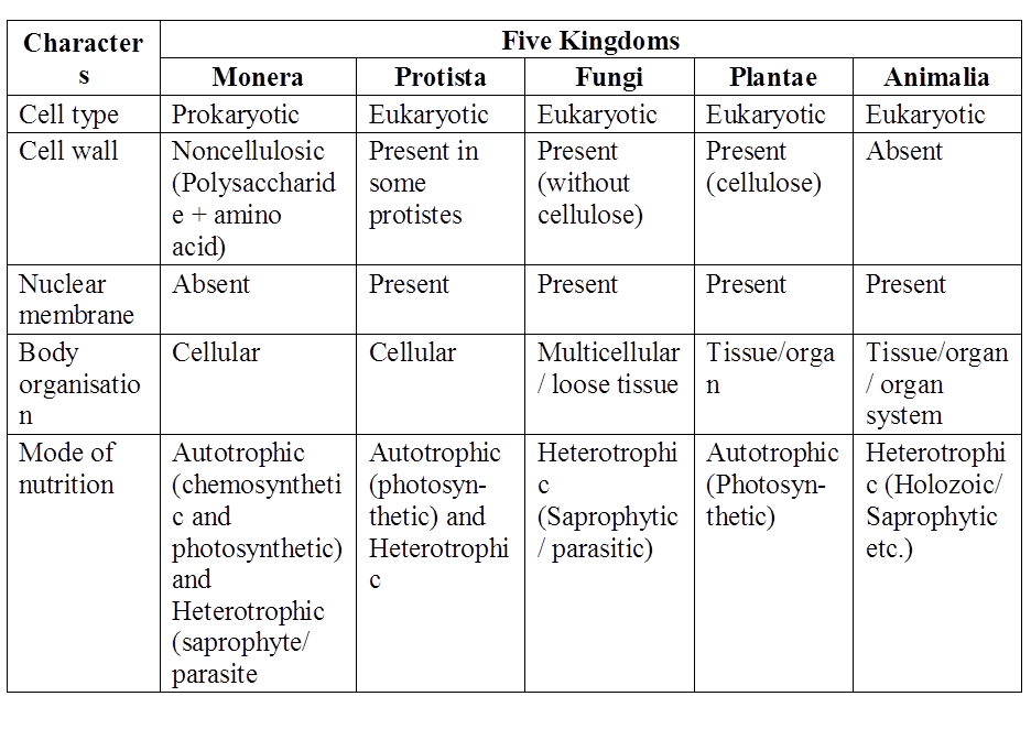
(5) Six Kingdom Classification :
Carl Woese proposed six kingdom classification.
These six kingdoms are Archaebacteria, Eubacteria, Protista, Fungi, Plantae and Animalia.
He separated the archaebacteria from eubacteria on the basis of some major differences such as the absence of peptidoglycan in the cell walls of the former and the occurrence of branched chain lipids (a monolayer instead of a phospholipid bilayer) in the membrane.
Based on the sequence of 16S ribosomal RNA genes, Woese found that the six kingdoms naturally cluster into three main categories.
He called these categories as domains of life.
These domains are Bacteria, Archae and Eukarya and are believed to have originated from common ancestor called progenote.
Three Domains of life
Based on the sequence of 16 S ribosomal RNA genes, woese found that the six kingdoms naturally cluster into three domains.
These domains are Archae, Bacteria and Eukarya, and are believed to be originated from common ancestor called Progenate. Domain is a category higher than kingdom.
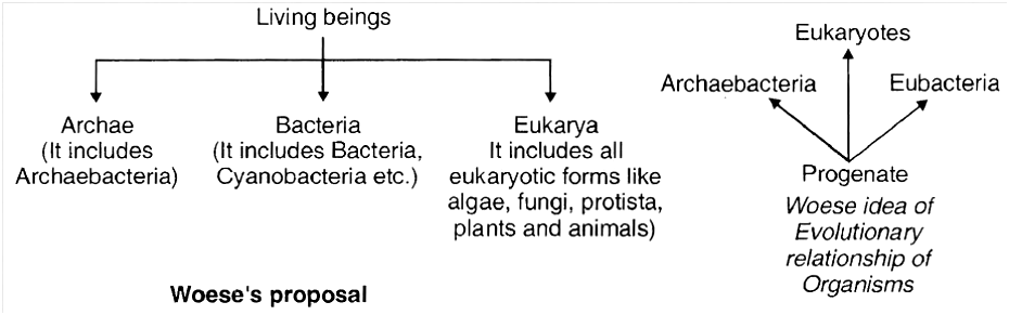
Kingdom Systems of Classification
Chapter 2
Biological Classification
Biological classification is the practice of categorizing living and extinct organisms into groups based on shared traits. Classifying organisms is necessary because it allows us to better understand biodiversity. It aids in the identification of living entities as well as the comprehension of their diversity. Classification aids in our understanding of different types of plants and animals and their characteristics, similarities, and differences. It allows us to see how complex organisms develop from simpler organisms. Different categories are used to understand and research the characteristics, similarities, and differences among various living species. Classification is the tool that allows us to manage a wide range of living organisms. Understanding the inter-relationships between different groups of organisms is critical. Other biological sciences are built on the foundation of classification.
Kingdom Systems of Classification:
Aristotle was the first to attempt to classify things on a more scientific basis. He classified plants into trees, shrubs, and herbs based on simple physical characteristics. He also separated the animals into two groups: those with red blood and those without. A Two Kingdom system of classification was devised during Linnaeus's time, with Plantae and Animalia kingdoms encompassing all plants and animals, respectively. Eukaryotes and prokaryotes, unicellular and multicellular organisms, photosynthetic (green algae), and non-photosynthetic (fungi) species were all lumped together in this system. Plant and animal classifications were simple to implement and understand, but a huge number of creatures did not fit into either group. As a result, the long-used two-kingdom classification was determined to be inadequate. Aside from gross appearance, there was a need to include other characteristics such as cell structure, wall nature, manner of nourishment, habitat, reproduction strategies, evolutionary linkages, and so on. As a result, classification systems for living organisms have experienced several alterations over time. Though the plant and animal kingdoms have remained consistent throughout all systems, the understanding of which groups/organisms should be included inside these kingdoms has evolved; the number and character of additional kingdoms have also been interpreted differently over time by different scientists.
A Five Kingdom Classification was proposed by R.H. Whittaker in 1969. Monera, Protista, Fungi, Plantae, and Animalia were the kingdoms he defined. Cell structure, body arrangement, manner of nourishment, reproduction, and phylogenetic links are among his key classification criteria. A three-domain approach has also been proposed, which divides Kingdom Monera into two domains and leaves the remaining eukaryotic kingdoms in the third domain, resulting in a six-kingdom categorization.
Let's take a look at this five-kingdom classification to see what concerns and considerations affected the system. Plants included bacteria, blue-green algae, fungi, mosses, ferns, gymnosperms, and angiosperms in previous classification systems. The fact that all of the species in this kingdom had a cell wall in their cells served as a unifying characteristic. This grouped people who had a lot of things in common but also had a lot of things in common. It brought together prokaryotic bacteria and blue-green algae (cyanobacteria), as well as other eukaryotic species. It also combined unicellular and multicellular species together, such as Chlamydomonas and Spirogyra being categorized together under algae.The classification did not distinguish between the heterotrophic fungus and the autotrophic green plants, albeit they did have a distinct difference in the composition of their cell walls, with the fungi having chitin and the green plants having a cellulose cell wall. When these qualities were taken into account, the fungi were given their own kingdom: Kingdom Fungi. Kingdom Monera was created to unite all prokaryotic species together, while Kingdom Protista was created to group all unicellular eukaryotic organisms together. Chlamydomonasand Chlorella (both of which have cell walls and were previously put in Algae within Plants) have been combined with Paramoecium and Amoeba in the kingdom Protista (which were earlier placed in the animal kingdom which lack cell walls). It has brought together organisms that were previously classified into various kingdoms.
Kingdom: Monera
- Books Name
- ACME SMART COACHING Biology Book
- Publication
- ACME SMART PUBLICATION
- Course
- CBSE Class 11
- Subject
- Biology
KINGDOM: MONERA
The Kingdom Monera includes all prokaryotes.
Monerans are the most primitive forms of life, originating from more ancient living stock termed progenote.
The kingdom Monera includes eubacteria and archaebacteria.
Eubacteria includes Cyanobacteria, Actinomycetes, Mycoplasma, Rickettsiae, Chlamydiae and Spirochaetes etc.
Classification of Monera
(1) In 4 kingdom system, a new kingdom was created to accommodate all prokaryotic organisms i.e, eubacteria and archaebacteria. Copeland called it kingdom Mychota. It was called 'Monera' by Daugherty and Allen.
(2) Actually, archaebacteria differ from eubacteria in many respects and resemble eukaryotes in some ways.
(3) (i) Carl Woese separated the archaebacteria from eubacteria on the basis of some major differences such as the absence of peptidoglycan in the cell walls of the former and the occurrence of branched chain lipids (a monolayer instead of a phospholipid bilayer) in the membrane.
(ii) Therefore, 6 kingdoms given by Carl Woese are
Kingdom-1 -Archaebacteria Kingdom-2 -Eubacteria
Kingdom-3 -Protista Kingdom-4 -Fungi
Kingdom-5 -Plantae Kingdom-6 -Animalia
Salient Features of Monera
1. These are unicellular, colonial, multicellular prokaryotic organisms without nuclear membrane, nucleolus, chromatin and histone proteins.
2. Cell wall is made up of peptidoglycan (exceptions are Archaebacteria and Mycoplasma).
3. Membrane bound organelles are absent.
4. Cyclosis is absent and ribosomes are of 70 S type .
5. Respiratory enzymes are found associated with plasma membrane.
6. Nucleoid or genophore or incipient nucleus or prochromosome is composed of naked DNA, RNA and nonhistone proteins.
7. Reproduction by asexual method.
8. Cell division is amitotic type and lacks spindle formation.
Let us discuss various monerans in detail :
1. EUBACTERIA
Bacteria are cosmopolitan and occur in every habitat wherever living or dead organic matter is present.
Anton Von Leeuwenhoek discovered bacteria in rain water which had been allowed to stand for many days and tartar scrapped from teeth.
In 1695, he published his work "The Secrets of Nature".
A.V. Leeuwenhoek termed these microorganisms as dierkens which was later translated as animalcules by the Royal society.
Term microbe for animalcules was coined by Se' dillot, but the term microorganism was proposed by Pasteur.
Concept Builder
Ehrenberg first of all coined the word 'bacteria'
Louis Pasteur is considered father of modern microbiology. He introduced the term aerobic and anaerobic for the life in the presence or absence of oxygen respectively.
Robert Koch, a German doctor, demonstrated that the anthrax disease of sheep was caused by bacteria. Koch had followed four experimental steps (Koch's postulates) which help to establish a relationship between a microorganism and a disease.
Smallest bacterium : Dialister pneumosintes
Largest filamentous bacterium : Beggiatoa mirabilis
Shapes of Bacteria
Bacteria occur in four basic forms or shapes.
These are spherical (Cocci), rod shaped (Bacilli), Vibrio and Spiral.
Though most bacterial species have cells that are of a fairly constant and characteristic shape, some species are pleomorphic (i.e., these can exhibit a variety of shapes), e.g., Rhizobium leguminosarum.
(a) Coccus : Spherical or nearly spherical, aflagellate, sub-divided into six groups on the basis of cell arrangement:
Monococcus -Only single cell represents the bacterium, e.g., Micrococcus luteus, M. roseus.
Diplococcus -Cocci divide in one plane and remain attached in pairs, e.g., Meningococcus, Gonococcus, Diplococcus pneumoniae.
Streptococcus -Cocci remain attached to form chains of different lengths, e.g., Streptococcus lactis.
Tetracoccus -Cocci divide in two planes at right angles to one another and form groups of four, e.g., Tetracoccus, Neisseria.
Staphylococcus -Cocci divide in several planes resulting in formation of irregular bunches of cells, sometimes resembling a cluster of grapes, e.g., Staphylococcus aureus.
Sarcinae -Cocci divide in 3 planes at right angles to one another and resemble cubical packets of 8 or more cells forming three dimensional geometrical figures, e.g., Sarcina lutea.
(b) Bacillus : Rod-like forms, either singly or may be arranged differently. They are generally flagellate. It is the most common of all the shapes. They are of following types :
(i) Monobacillus -The bacteria occur singly, e.g., Bacillus anthracis, Lactobacillus.
(ii) Diplobacillus -Bacteria are arranged in pairs. e.g., Bacillus subtilis
(iii) Streptobacillus -Bacteria form a chain of rods, e.g., Streptobacillus.
(iv) Palisade-like -If the cells are lined side by side like match sticks and at angles to one another.
e.g., Corynebacterium diphtheriae.
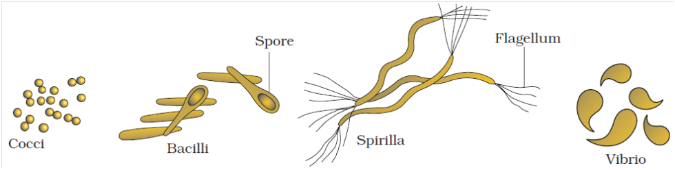
(c) Spiral bacteria : Coiled forms of bacteria exhibiting twists with one or more turns are called spirilla, e.g., Spirillum volutans.
(d) Vibrio : Bacteria with less than one complete twist or turn are called vibrio. These resemble a comma (,) in appearance, e.g., Vibrio cholerae.
(e) Stalked bacteria : The body of bacterium possesses a stalk, e.g., Caulobacter.
(f) Budding bacteria : The body is swollen at places, e.g., Rhodomicrobium
Bacterial Cell Structure
Bacterial cell structure is very simple although they are very complex in behaviour. They show the most extensive metabolic diversity. Electron microscope can only reveal the detailed structure of bacterial cell. It consists of following structures:
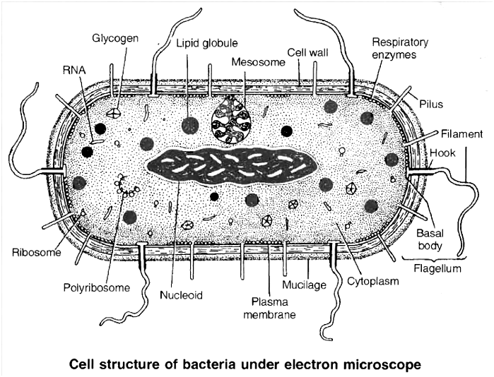
1. Glycocalyx:
(i) It is outermost part of cell envelope (Glycocalyx, cell wall, plasma membrane)
(ii) Represented by either slime layer or capsule
(a) Slime layer is composed of dextran, dextrin and lavan sugars and protect the cell against desiccation and loss of nutrients.
(b) Capsule is made up of polysaccharides and D-glutamic acid. It provides gummy or sticky character and virulent property to the cell.
2. Cell wall :
It is present outside the cell membrane and is a rigid structure.
Due to its rigidity, it protects the internal structures of the cell and provides shape to the cell.
However, its main function is to prevent the cell from expanding and bursting because most bacteria live in hypotonic environments, and are likely to take in much water and eventually burst.
The cell walls of almost all the eubacteria (true bacteria) are made up of peptidoglycan, also called murein or mucopeptide.
It is found only in prokaryotes.
As the name suggests, the peptidoglycan consists of two components-a peptide portion which is composed of amino acids connected by peptide linkages, and a glycan or sugar portion.
The glycan portion, which forms the backbone of peptidoglycan, is composed of alternating units of amino sugars N-acetyl-glucosamine (NAG) and N-acetylmuramic acid (NAM) joined together by b-1, 4 linkages.
The peptidoglycan chains are laterally linked by short chains of four amino acids which are attached to N-acetylmuramic acid residues.
The four amino acids of this tetrapeptide are D-alanine, L-alanine, D-glutamic acid and L-Iysine (in Gram +ve bacteria) or diaminopimelic acid (in Gram -ve bacteria).
The tetrapeptide chains are also interlinked by a peptide bridge between the carboxyl group of an amino acid in one tetrapeptide chain and amino group of an amino acid in another tetrapeptide chain.
The cross linkages can occur between tetrapeptides in different chains, as well as between adjacent tetrapeptide chains. As a result, peptidoglycan forms a rigid, multilayered sheet.
Another component, teichoic acid, an acidic polymer consisting of a carbohydrate (e.g., glucose), phosphate and an alcohol is found in cell walls of Gram +ve bacteria.
Teichoic acid has several functions such as binding metals, acting as receptor sites for some viruses and maintaining cells at low pH to prevent degradation of cell walls by self-produced enzymes.
The walls of Gram positive bacteria contain very little amount of lipids.
The cell walls of Gram negative bacteria are much more complex.
The peptidoglycan layer is very thin making up only 10% or less of the cell wall.
However, the most interesting feature is the presence of an outer membrane that covers a thin underlying layer of peptidoglycan.
The outer membrane is a bilayered structure consisting chiefly of phospholipids, proteins and lipopolysaccharides (LPS).
The outer membrane serves as a barrier to prevent the escape of important enzymes from the space (periplasmic space) between the cytoplasmic membrane and the outer membrane.
It also prevents the entry of various chemicals that could damage the cell.
It acts as main surface antigen in cell wall.
However, permeability of outer membrane to nutrients is provided by proteins called porins which form channels in the membrane through which substances of hydrophilic nature and low molecular weight can diffuse.
Christian Gram (1884) developed a staining method for bacteria, using Gram stain (crystal violet).
On the basis of stainability with Gram Stain, bacteria are classified into two groups; Gram positive and Gram negative.
Flow chart representing Gram staining technique
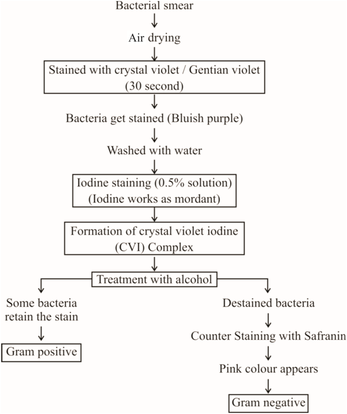
3. Surface Appendages : These include flagella and fimbriae (or pili).
(a) Flagella are long, fine, wavy, filamentous appendages that protrude through the cell wall, responsible for the motility of bacteria. These are much thinner than the flagella or cilia of eukaryotes.
Structure of Flagella: The entire flagellar apparatus is made up of three distinct regions: basal body, hook and filament.
Basal body: It is most complex portion of flagellum and has four rings (L, P, S and M), only two rings S and M are present in gram +ve bacteria. L and P rings in cell wall constitute distal set, while S and M rings are present in plasma membrane, forming proximal set.
Hook: Made up of different protein units.
Filament: Bacterial flagella are made up of identical spherical subunits of a protein called flagellin. Longitudinal chains of flagellin molecules run longitudinally around each other to form a wavy helical or rope-like structure. Therefore, a cross section of the flagellum reveals a number of flagellin molecules around a central space.
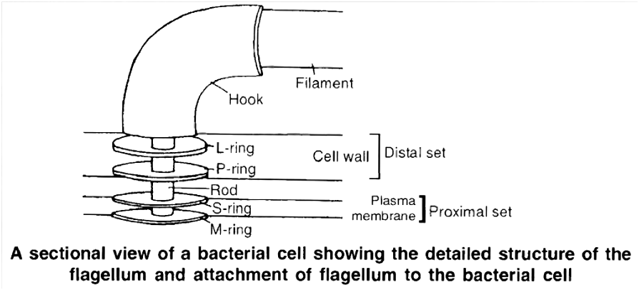
Concept Builder
Depending upon the presence or absence, number and position, following types of flagellar arrangements are observed among bacteria :
Atrichous: Flagella absent, e.g., Pasteurella, Lactobacillus.
Monotrichous: Only one flagellum attached at one pole of the organism, e.g.,Thiobacillus, Vibrio.
Amphitrichous: One flagellum at both ends, e.g., Nitrosomonas.
Cephalotrichous: Two or more flagella attached at one end, e.g., Pseudomonas fluorescence.
Lophotrichous: Two or more flagella attached at both ends, e.g., Spirillum volutans.
Peritrichous: Flagella distributed all over the surface of the cell, e.g., Escherichia coli, Clostridium tetani.
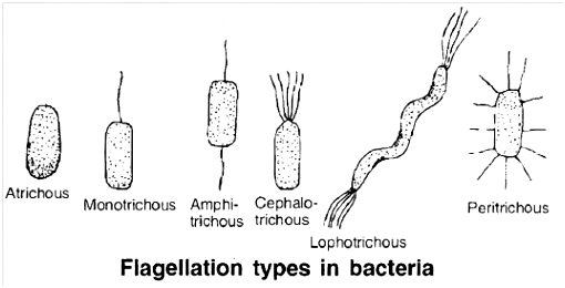
(b) Pili and fimbriae are hollow, non helical, filamentous appendages projecting from the walls of Gram-negative bacteria. These are thinner and shorter and more in number than the flagella. These are made up of specific proteins called pilin.
There are different types of pili which serve different functions. One type, known as type I pili, (somatic pili) play a major role in infection by facilitating the attachment of bacterial cell to the host cell. Another type, termed sex pili, serve as portals of genetic material from donor to recipient cell during conjugation.
Differences between Gram positive and Gram negative bacteria
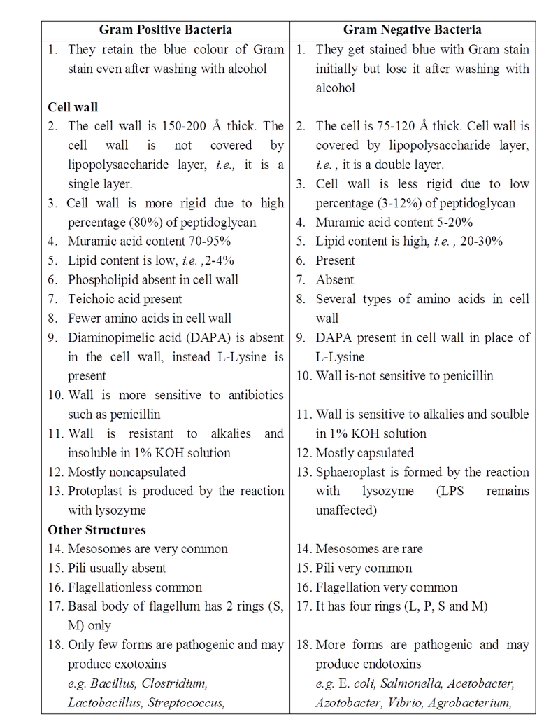
4. Protoplast: Cell wall encloses the protoplast, the living matter. It includes (i) Cell membrane (ii) Cytoplasm, (iii) Nucleoid and may have plasmid and episome.
(i) Cell membrane:
It lies inner to the cell wall, actually representing the outermost layer of the protoplast.
It is living and semipermeable, controlling the movements of various dissolved substances in and out of the cells.
Functionally, the cell membrane of bacteria resembles mitochondria of eukaryotic cells as respiratory ETS enzymes and succinate dehydrogenase (Kreb's Cycle) are associated with the membrane.
The cell membrane gets invaginated and folded to form a structure called mesosome (chondroid) in some bacteria, particularly the Gram positive bacteria.
These may be central or peripheral in position and they are supposed to play a role in replication of DNA during cell division, as these are often attached to the nuclear body.
Besides, these increase the surface area of absorption and help in septa formation during binary fission.
(ii) Cytoplasm:
It is homogenous colloidal mass of carbohydrates, fats, proteins, lipids, nucleic acids, minerals and water.
It does not show streaming movements.
It lacks sap vacuoles and gas vacuoles (may be present in some bacteria which live in aquatic condition).
Typical membrane bound organelles of eukaryotic cells like endoplasmic reticulum, mitochondria, golgi complex and plastids are absent.
The cytoplasm appears granular due to the presence of ribosomes.
However, these are 70S type in bacteria as compared to 80S type in eukaryotes.
Ribosomes lie scattered freely in the cytoplasm, but sometimes may form a small chain of 4-6 ribosomes attached to mRNA constituting polyribosome or polysome.
Various non living inclusions like glycogen particles, fat bodies, volutin granules (polymetaphosphate -source of energy) and lipid molecules acting as food reserve lie dispersed in the cytoplasm.
The cytoplasm is usually colourless, lacking pigments.
However, in photosynthetic bacteria, the cytoplasm contains pigments like bacteriochlorophyll and bacterioviridin.
The pigments either lie dispersed in the cytoplasm or present in membrane bound spherical vesicles called chromatophores.
These pigments are capable of entrapping solar energy for photosynthesis
(iii) Nucleoid (Prochromosome, Genophore, Incipient nucleus) –
Bacterial cell lacks a well organized nucleus.
It consists of a long double stranded DNA molecule repeatedly folded with the help of RNA to form a circular ring.
DNA has no free ends and not associated with histone proteins (polyamines present). Circular DNA ring, without histones is often termed bacterial chromosome.
Plasmid (Minichromosome) :
Term plasmid was given by Lederberg and Hays.
These are small, extrachromosomal, non-essential, circular, double stranded, free naked DNA molecules.
The genes present on them have no vital role in survival and growth of bacteria.
These perform autonomous replication.
If plasmids temporarily integrate with bacterial chromosome, then they are called episomes.
Concept Builder
Types of plasmids :
(a) F-Plasmid: It forms sex pilus and is responsible for process of conjugation or fertility factor transfer.
(b) R-Plasmid: These plasmids have resistance gene (Resistance Transfer Factor, RTF) for antibiotics like penicillin, tetracycline.
(c) Col-Plasmid: Genes of this plasmid are responsible for production of colicins (bacteriocin) for killing other bacteria.
(d) Ti Plasmid: From Agrobacterium tumefaciens, used in genetic engineering
(e) Degradative plasmid of Pseudomonas putida (superbug) helps to decompose hydrocarbons of petroleum in oil spills
5. Bacterial Life Processes
Discussion of bacterial life processes revolves around the study of the prominent metabolic activities like respiration and nutrition.
(A) Respiration: On the basis of mode of respiration, the bacteria are divided into two main groups: i.e., aerobes and anaerobes. Each group is further of two types i.e. strict or obligate and facultative.
(a) Obligate or strict aerobes : These bacteria can live only in presence of oxygen they possess the enzyme system for aerobic respiration only. In the absence of oxygen, they cannot respire and thus, die, e.g., Bacillus subtilis.
(b) Facultative anaerobes : They normally respire aerobically. However, they are capable of switching over to anaerobic mode to get energy for their survival, if sufficient oxygen to sustain aerobic respiration is not available in the environment, e.g., Pseudomonas.
(c) Obligate or strict anaerobes: These bacteria respire anaerobically only. The growth of such bacteria will certainly be slower as anaerobic respiration liberates much less amount of energy as compared to aerobic respiration. They lack enzymes necessary for carrying out aerobic respiration e.g., Clostridium botulinum.
(d) Facultative aerobes: They normally respire anaerobically, but are capable of respiring aerobically as well, if oxygen is available. Most of the photosynthetic bacteria are facultative aerobes e.g., photosynthetic bacteria Chlorobium.
Concept Builder
(i) Aerotolerant anaerobes: Bacteria that continue to perform anaerobic respiration even in the presence of oxygen, e.g., Lactic acid bacteria.
(ii) Anaerotolerant aerobes: Aerobic bacteria continue to perform aerobic respiration even in absence of free oxygen by using oxygen of oxidised salts, e.g., Denitrifying bacteria
(B) Nutrition :
Nutrition in bacteria is of two types i.e. autotrophic and heterotrophic.
Bacteria having autotrophic mode of nutrition may be photoautotrophs and chemoautotrophs, carrying out photosynthesis and chemosynthesis, respectively.
(I) Autotrophic Nutrition
(i) Photoautotrophic bacteria:
These bacteria are capable of entrapping solar energy and utilizing it for the synthesis of complex food materials due to the presence of pigments like bacteriochlorophyll (bacteriopurpurin) and bacterioviridin.
Purple sulpher bacteria (e.g., Thiospirillum) and green sulpher bacteria (Chlorobium limicola) are the most familiar examples containing pigment bacteriochlorophyll, bacteriopurpurin and bacterioviridin respectively.
Concept Builder
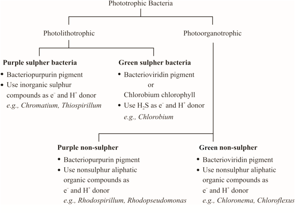
Bacterial photosynthesis, however, differs from photosynthesis of higher plants in not liberating oxygen.
This type of photosynthesis, characteristic of bacteria, is termed as anoxygenic.
Normal photosynthesis, occurring in higher plants, is termed as oxygenic.
In bacterial photosynthesis water is not the source of electron that acts as reducing power to convert CO2 into glucose.
The bacteria obtain reducing power from various compounds such as hydrogen sulphide, thiosulphate or even some organic compounds.
No oxygen is evolved as it does not involve splitting of water.
Hydrogen released by various compounds mentioned above is picked up by NAD+ which gets reduced to NADH2 acting as reducing power.
NADH2 alongwith ATP, produced generally by entrapping solar energy are used to reduce CO2 to glucose.
Simple equation for anoxygenic photosynthesis may be written as follows :
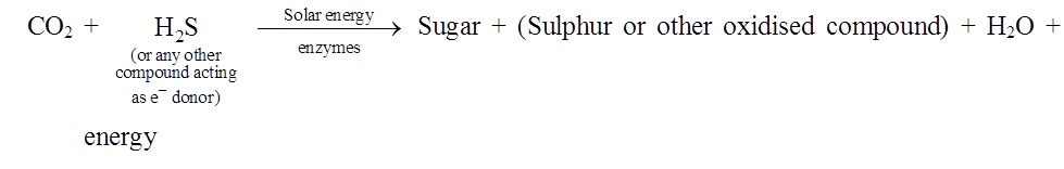
Most of the photosynthetic bacteria are anaerobes (facultative aerobe
(ii) Chemoautotrophic Bacteria:
Bacteria belonging to this category obtain energy for the synthesis of food by oxidising certain inorganic substances like ammonia, nitrates, nitrites, ferrous ions etc.
Thus, they do not utilise light as energy source.
The chemical energy thus obtained, is trapped in ATP molecules.
This energy is then used in carbon assimilation with the help of hydrogen from some source other than water, e.g., hydrogen bacteria, nitrifying bacteria, sulphur bacteria, etc.
They play a great role in recycling nutrients like nitrogen, phosphorous, iron, sulphur.
(a) Hydrogen bacteria. These bacteria oxidise hydrogen in the presence of oxygen, e.g., Hydrogenomonas.
(b) Nitrifying bacteria obtain energy by oxidizing ammonia into nitrate, oxidation of ammonia occurs in two steps. Each step is carried out by a specialised group of bacteria.
In the first step, ammonia is oxidised into nitrites by the species of the genus Nitrosomonas and Nitrococcus.
In the second step, the nitrite is converted into nitrate. This is brought about by species of the genus Nitrobacter and Nitrocystis which use this energy for chemosynthesis.
(c) Sulpher bacteria. These bacteria obtain energy either by the oxidation of elemental sulphur or oxidation of H2S.
Oxidation of elemental sulpher. Sulpher bacteria (Thiobacillus thioxidans) oxidise elemental sulpher to sulphuric acid and utilise energy produced in this process. These bacteria can survive even in extreme acidic environment.
Oxidation of H2S to S. Some bacteria like Beggiatoa use the energy from oxidation of H2S and store the sulphur so produced in the form of granules.
(d) Iron bacteria. These bacteria (e.g., Ferrobacillus, Leptothrix) inhabit water which contain iron compounds. These bacteria convert ferrous ions to ferric form. The ferric ion is deposited as insoluble ferric hydroxide. The energy so released, is utilised in the assimilation of CO2.
(e) Methane bacteria. Methanomonas, is one example which oxidises methane to carbon dioxide.
(II) Heterotrophic bacteria :
These bacteria are most abundant in nature and are incapable of synthesizing their own food from simple raw materials.
They obtain nourishment either from dead and decaying organic matter or directly from a living host.
All heterotrophic bacteria are segregated into three main categories, i.e., saprophytic, symbiotic and parasitic forms.
(i) Saprophytic bacteria :
They are free living bacteria, obtaining nourishment from organic remains such as dead animals, animal excreta, fallen leaves, decaying vegetables, fruits, bread and other products of animal and plant origin.
These bacteria secrete digestive enzymes into the substrate and the complex insoluble Substances are converted into simple soluble compounds like water, hydrogen sulphide, ammonia, CO2 etc.
Some of the simpler substances are absorbed and assimilated by the bacteria, whereas the others are added to the soil and atmosphere to complete the nature's material cycle.
Anaerobic breakdown of carbohydrates and proteins is termed fermentation and putrefaction respectively.
Aerobic breakdown of organic compounds is called decay.
(ii) Symbiotic bacteria :
They are mainly Gram negative type.
A familiar example of symbiotic bacteria is Rhizobium leguminosarum, associated with roots of leguminous plants.
They are capable of fixing atmospheric nitrogen as ammonia, inside the nodule only and not in free state.
However, some bacteria like Azotobacter, Beijerinckia, Klebsiella are free living, aerobic and capable of nitrogen fixation in free state, enriching the soil.
Clostridium pasteurianum is anaerobic N2 fixing bacteria.
(iii) Parasitic bacteria
These bacteria draw nourishment and obtain special organic compounds required for growth from living organisms, either plants or animals, called hosts.
The disease causing bacteria are termed pathogenic and the ones not causing any disease are termed as non-pathogenic.
6.. Reproduction
Bacteria reproduce mainly by asexual method and also show sexual recombination (True sexual reproduction is absent).
A. Asexual Reproduction :
Bacteria produce several types of asexual spores like, sporangiospores, oidia, conidia and endospores. However, the most common mode of asexual reproduction is binary fission.
Under favourable conditions of nutrient availability, moisture and temperature, daughter cells may repeat binary fission many times and may forms a large population.
Fortunately, such a rapid rate is seldom achieved.
The process gradually slows down and ultimately stops because of:
(i) Shortage of space.
(ii) Lack of nutrient availability.
(iii) Accumulation of waste products (making environmental conditions unfavourable for growth).
(iv) Development of bacteriophages, destroying bacteria.
(a) Binary Fission :
It is the most common method under favourable conditions of temperature, moisture and availability of nutrients.
Mature bacterial cell divides into two daughter cells.
In this process the cell division is amitotic type i.e., not involving the spindle formation
(b) Endospores :
Cells of certain bacteria, e.g., Bacillus, Clostridium etc. form thick-walled, highly resistant bodies within the cell, called endospores.
One bacterial cell normally produces only a single endospore.
The endospores may be spherical or oval in shape and are terminal or central in position.
Anticoagulant nature of endospore is due to the presence of Ca-dipicolinic acid in cortex layer of wall.
Concept Builder
Structure of Endospore :
The endospore consists of a central core made up of nuclear material and spore cytoplasm.
The cytoplasm includes DNA, RNA and proteins, lipid, Ca, Mn.
The central core is surrounded by a delicate membrane called core wall.
Around the wall is another layer, which is much thicker and is of relatively low density.
This layer, called cortex, mainly consists of peptidoglycan and calcium dipicolinic acid (CaDPA).
It is an anticoagulant which provides resistance to heat.
This complex may also be present in spore cytoplasm.
Endospore is highly resistant to desiccation, chemicals and radiations.
The cortex, in tum, is enclosed in spore coat which may be smooth, grooved, or raised into ridges. The whole structure may be wrapped in an exosporium.
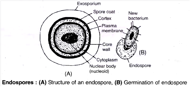
The endospores are formed probably upon induction by the exhaustion of nutrients (unfavourable conditions).
During endospore formation, a part of the protoplast containing nuclear body undergoes dehydration, stores food material and gets separated from rest of the protoplast to form endospore.
It gets surrounded by different layers.
These can withstand temperature as high as 1000e or as low as -100°C, so these can remain unharmed during pasteurisation.
The remarkable resistance shown by endospores is due to :
(i) Thick and impermeable spore coat
(ii) Low water content
(iii) Low metabolic activity
(iv) Ca-DPA Complex
Under favourable conditions, endospore absorbs water, becomes metabolically active, ruptures thick spore coat and the bacterium surrounded by thin cell wall emerges out.
Endospores are actually the means of perennation and not reproduction as only one endospore is formed per cell and subsequently each endospore grows into single bacterium cell after the commencement of favourable conditions.
B. Sexual Recombination (Genetic Recombination) :
The bacteria exhibit a primitive form of sexual reproduction which differs from eukaryotic sexual reproduction because there is no gamete formation and fusion.
However, the essential feature of sexual reproduction, i.e., exchange of genetic material does take place and is called genetic recombination.
Three methods are known by which genetic recombination is achieved by bacteria.
In the order of their discovery, these are transformation, conjugation and transduction.
(a) Transformation :
Griffith (1928) worked on the effect of Diplococcus or Streptococcus pneumoniae bacteria on mice and discovered the process of transformation.
In transformation, the donor and recipient do not come in contact.
The donor cell releases a piece of DNA which is actively taken up by the recipient cell from the solution.
This ability to pick up DNA from the solution is called competence.
Two strains of D. pneumoniae are :
- Capsulated or S-III (Virulent strain) and Non-capsulated or R-II (non-virulent strain).
- Four steps were performed in experiment:
![]()
![]()
![]()
![]()
(But R-II is not virulent and S-III lost its virulent capacity upon heating).
Griffith concluded that 'something' passed from heat killed S-III to R-II bacteria, so that non virulent strain changed or transformed into virulent bacterial strain.
Avery, Macleod and McCarty (1948) repeated this experiment using various enzymes and proved that the transformation principle is DNA of heat killed S-III strain.
They proved that DNA is a genetic material.
(b) Conjugation:
Lederberg and Tatum (1946) demonstrated in E. coli that during conjugation, one cell containing F-plasmid acts as donor (F+ or male) cell and the other lacking F-plasmid as recipient (F– or female) cell.
The plasmid contains fertility factor or F gene which produces protrusions termed sex pili.
These help the donor F+ cell in attaching to the recipient cell.
The plasmid replicates and a replica is transferred to recipient cell, changing it into F+.
Often the plasmid integrates with bacterial chromosome, converting it into Hfr (High frequency of recombination) cell or super male and a part or whole of bacterial chromosome is transferred to recipient cell through-conjugation tube.
Such association of episome with the endogenote increases the efficiency of genetic transfer.
The number of genes transferred depends upon the time for which the two cells remain joined together.
When F-conjugates with super male, the frequency of recombination increases by 1000 times, that is why it is called as Hfr (super male).
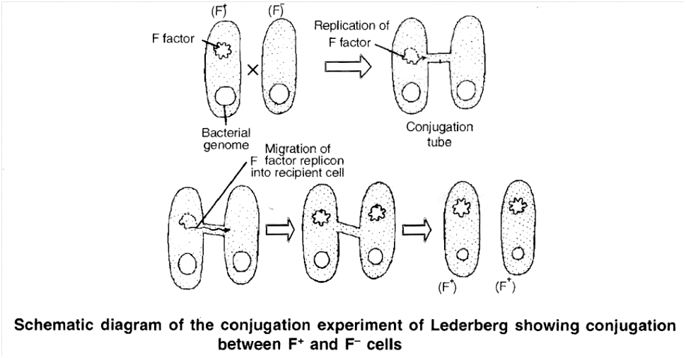
(c) Transduction :
During transduction, a small double stranded piece of DNA is transferred from donor to recipient by a bacteriophage.
This mode of genetic recombination in bacteria was first demonstrated by Zinder and Lederberg (1952) while working with Salmonella typhimurlum.
Some viruses have the ability to integrate their DNA with bacterial DNA, which is replicated at the same time as the host DNA and is passed from one bacterial generation to the next.
Such bacteria carrying phage (viral) DNA with their own DNA are called lysogenic bacteria.
Occasionally, the phage DNA becomes active and codes for the production of new virus particles.
A number of phage particles are synthesised followed by the destruction of the host cell and release of phage particles.
Upon release, the phage particles attack sensitive bacterial cells, multiply and release more phage particles.
However, sometimes faulty deatchment of phage DNA from bacterial DNA results in the incorporation of a small amount of bacterial DNA into the phage DNA.
Subsequent infection of another bacterium with this aberrant phage called transducing phage, introduces the piece of foreign bacterial DNA into the recipient's chromosomes, producing a genetic change.
Types of transduction:
The ability of the bacteriophage to carry the genetic material from any region of bacterial DNA is called generalised transduction, e.g., T4-phage.
On the other hand, there are bacteriophages such as lambda phage (l) of E coli which can carry only a specific region of the bacterial DNA to a recipient.
This is called specialised transduction (or restricted transduction).
Sometimes, the DNA brought by the phage does not integrate with the genome of the recipient bacterium and is lost after one or two generations.
Such a transduction is called abortive transduction
7. Economic Importance of Bacteria
Bacteria play significant role in day to day activities of human beings.
A. Beneficial activities
(a) Role in agriculture
(i) Decay and decomposition of organic matter :
They bring about decay and decomposition of dead remains of plants and animals.
These are the most important for mineral cycling to occur.
(ii) Sewage disposal :
The bacteria decompose the organic matter present in the sewage, converting into simpler inorganic substances.
The inorganic substances thus formed, being soluble, pass out through filter alongwith water which is highly useful for irrigation purposes, e.g., Clostridium, E. coli.
(iii) Nitrogen cycle :
The proteins present in dead remains of living organisms are converted into amino acids by different types of saprophytic bacteria.
Amino acids are converted into ammonia by ammonifying bacteria (Bacillus vulgaris, B. ramosus).
Nitrifying bacteria convert ammonium salts first into nitrites (Nitrosomonas, Nitrococcus) which are subsequently converted into nitrates (Nitrobacter).
In presence of denitrifying bacteria (Pseudomonas denitrificans), nitrates and nitrites of soil are converted to gaseous nitrogen.
(iv) Nitrogen fixation :
It is the biological process in which atmospheric nitrogen is converted into nitrogenous compounds by nitrogen fixing bacteria.
These bacteria are of 2 types :
(a) Free living
(b) Symbiotic
(a) Free living bacteria are Azotobacter and Beijerinckia (aerobic) & Clostridium (anaerobic).
(b) Common symbiotic bacteria are Rhizobium leguminosarum and Xanthomonas. These convert nitrogen into ammonia which is directly converted into amino acids by plants.
(v) Manure preparation :
Saprotrophic bacteria help in preparation of farmyard manure by converting farm refuse, dung and other organic wastes into humus.
(b) Role in Industry :
Man has utilized the metabolic activities of bacteria in preparation of a number of industrial products as listed below :
(i) Butter milk and sour cream
(ii) Yoghurt
(iii) Cheese
(iv) Vinegar
(v) Retting of fibres : Retting is a controlled microbial decomposition for separation of fibres. The tissues are immersed in water tanks where anaerobic butyric acid bacteria dissolve the pectin of middle lamella of cells, thus, separating the fibres. Clostridium perfringens and Pseudomonas fluorescence are useful in this process.
(vi) Curing of leaves: To improve the flavour and taste in tea, using Micrococcus candidans and in tobacco leaves by Bacillus megatherium.
(vii) Single cell proteins (SCP) : Like -Methylophilus methylotropus and Rhodopseudomonas capsulata.
(c) Role in Medicine :
Bacteria have been used extensively in preparation of antibiotics, vaccines, serums and vitamins.
(i) Antibiotics :
The term antibiotic was given by Waksman, who discovered streptomycin.
These are the organic substances produced by microorganisms which inhibit the growth of other organisms (mostly pathogens) but do not affect the growth of organisms secreting these.
The first commercial antibiotic penicillin was discovered by Flemming (1959) from a fungus called Penicillium.
Some antibiotics of eubacterial origin:
(a) Bacitracin ........................... Bacillus licheniformis
(b) Polymixin ........................... Bacillus polymyxa
(c) Gramicidin ........................... B. brevis
(d) Subtilin ........................... B. subtilis
(ii) Vaccine production
Vaccines and serums against typhoid, cholera, TB, pertussis, tetanus and diphtheria are made with the help of bacteria or their toxins e.g., DPT (against diphtheria, pertussis and tetanus), TI(against tetanus), BCG (Bacille of Calmette-Guerin against TB), DT (against diphtheria and tetanus).
(iii) Vitamins :
Escherichia coli present in human intestine produces large quantities of B-complex vitamins and vitamin K. Bacteria are utilized in industrial production of a number of vitamins like riboflavin from Clostridium butylicum, Cobalamine (B12) from Bacillus megatherium and Pseudomonas denitrificans.
(iv) Pollution control :
Pseudomonas putida degrades petroleum wastes.
Flavobacterium can decompose 2, 4-D. DDT can be decomposed by Acetobacter aerogens.
Ganga's water contains Bdellovibrio bacteriovorus that maintains purity of its water.
(v) Poly--hydroxybutyrate is used to produce biodegradable plastic.
B. Harmful activities
(i) Spoilage of food : Saprophytic bacteria cause decay of vegetables, fruit, meat, bread and other foods, making these unfit for human consumption. Some bacteria even produce strong toxins in the infected food stuffs which cause food poisoning when consumed.
(ii) Deterioration of Domestic Articles : Some saprophytic bacteria like Cellulomonas, Spirochaete cytophaga cause deterioration of domestic articles of daily use such as leather, woolen, canvas articles etc.
(iii) Denitrification of Soils : Denitrifying bacteria like Thiobacillus denitrificans, Micrococcus denitrificans and Pseudomonas convert nitrates and nitrites present in the soil into gaseous nitrogen, thus depleting the soil nitrogen, thereby decreasing soil fertility.
(iv) Desulphurification : Desulphovibrio desulphuricans.
(v) Diseases : Common disease of humans, animals and plants are listed below respectively –
Table: 2
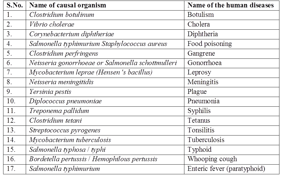
Table : 3
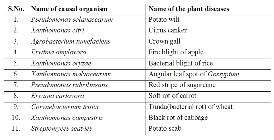
II. CYANOBACTERIA
Cyanobacteria are Gram negative photosynthetic prokaryotes, being the most primitive organisms to have oxygenic photosynthesis.
They added oxygen to the atmosphere, which is indispensible for the existence of aerobic forms of living organisms.
They are also known as BGA (Blue green algae) and are classified variously under cyanophyceae or myxophyceae.
Occurrence
They are mainly fresh water forms, though few are marine.
Red sea is named so because of abundant occurrence of a cyanobacterium Trichodesmium erythraeum, which imparts red colouration to water.
They occur in symbiotic association with almost every group of eukaryotes i.e. green algae, fungi, bryophytes like mosses and Anthoceros, ferns, gymnosperms, angiosperms, sponge, shrimps, mammals etc.
Anabaena azollae is associated with Azolla, an aquatic fern.
Anabaena cycadeae is associated with coralloid roots of Cycas.
In many lichens (symbiotic association of algae and fungi), the algal partner may be a cyanobacterium.
When they live endozoically in protozoans they are called cyanelle.
Structural Organization
These may be unicelled or multicelled. The latter may be filamentous or colonial.
Filamentous form consists of one or more cellular strands, called trichomes, surrounded by mucilagenous sheath.
Cyanobacteria are characterised by the absence of flagellum throughout life cycle.
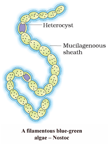
Cell Structure
- The cell structure in cyanobacteria is typically prokaryotic.
- The cell lacks a well defined nucleus and the chromatin material is centrally located, resembling the bacterial chromosome.
- The cell wall is 4 layered and is invariably covered by mucilagenous sheath, composed largely of mucopeptides.
- Protoplasm in cyanobacterial cell can be distinctly divided into two parts the centroplasm and chromoplasm.
- The central colourless centroplasm contains the chromatin material.
- The peripheral protoplasm is coloured or pigmented because of the presence of thylakoids, called as chromoplasm.
- The protoplast lacks membrane-bound organelles like endoplasmic reticulum, golgi bodies, mitochondria, lysosomes, plastids and contains 70S ribosomes.
- Similar to the mesosome of bacteria, a group of coiled membrane called lamellasome is found which connects nucleoid to the cell membrane.
- It helps in respiration and replication of DNA.
- The cell membrane lack sterols. The sap vacuoles are absent.
- Instead, the cell may contain gas filled vacuoles which help to regulate the buoyancy of the organism in water.
- The characteristic feature of cyanobacterium cell is the presence of a system of photosynthetic lamellae called thylakoids.
- The characteristic photosynthetic pigments present in the thylakoids are chlorophyll a and phycobilins i.e., phycocyanin (blue coloured), phycoerythrin (red coloured) and allophycocyanin (light blue coloured).
The cyanobacterial cell contains reserve food material in the following forms
(i) Cyanophycean granules (Protein)
(ii) ![]()
(iii) Cyano-or myxophycean starch or -granule (Similar to glycogen but negative to iodine test)
(iv) Volutin body (Reserve phosphate)
(v) Polyhedral body (Rubisco rich)
Concept Builder
Gaidukov's phenomenon or complementary chromatic adaptation -Cyanobacteria or blue green algae can adaptively change their body colour according to different wavelengths of available light, e.g., Trichodesmium erythraeum. It is also known as "red sea" causing alga.
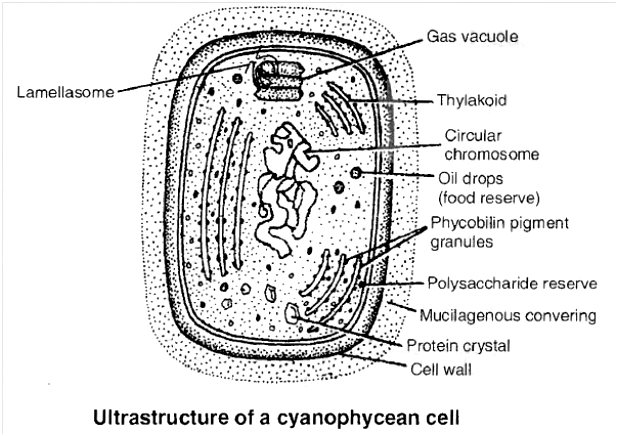
Metabolism
They are the most self-dependent organisms, because most of these are capable of converting atmospheric nitrogen into ammonium compounds besides utilizing atmospheric CO2 for synthesis of organic food during photosynthesis.
Biological nitrogen fixation is an anaerobic process as nitrogenase enzyme required for the process acts efficiently in the absence of oxygen.
Nitrogen fixation under anaerobic conditions occurs mainly in large, specialized cells called heterocysts as in Nostoc.
Heterocyst has terminal pores which at maturity develops a polar granule.
Thickened cell wall of these cells is impermeable to oxygen so this creates anaerobic environment in the cell even under aerobic conditions.
Besides this, heterocysts also lack PS II activities and CO2 fixation is done only by vegetative cells.
Oxygen is not evolved due to absence of PSII.
However, PSI remains active in heterocyst which generates ATP required to fix nitrogen.
Besides N2 fixation heterocyst promotes fragmentation.
This property of nitrogen fixation, most of the BGA enrich the soil by releasing nitrogenous compounds in the surroundings.
Reproduction
Cyanobacteria reproduce asexually. Typical sexual reproduction is absent.
Asexual reproduction occurs by following methods :
(i) Binary fission: It occurs in unicellular forms. The daughter cells formed by amitotic division separate immediately after the division.
(ii) Fragmentation: It occurs in filamentous forms. The filament breaks up into short pieces or fragments which grow to form new filaments.
(iii) Heterocysts : Under special conditions, the heterocysts germinate to form new filaments.
(iv) Hormogonia: Due to the formation of biconcave, mucilage filled dead cells called necridia, in between living cells of trichome, the filament breaks into hormogonia.
(v) Akinetes: Vegetative cells are transformed into thick walled akinetes due to the deposition of food material followed by the thickening of wall. On the arrival of favourable conditions, they germinate to form new filaments.
Importance of Cyanobacteria
(i) They are the most ancient organisms having oxygenic photosynthesis and thus, played a significant role in the evolution of aerobic forms of life.
(ii) They convert atmospheric nitrogen into ammonium compounds and excess of these compounds is excreted out, enriching the soil. The death and decay of these also increase the soil fertility, particularly the nitrogen content of the soil. Tolypothrix and Aulosira fix N2 non-symbiotically in rice fields .
Cyanobacteria like Nostoc and Anabaena have been used for reclaiming usar soils. As they can live in damp or aquatic habitat, they enrich the root environment in any wetland condition as in rice fields.
(iii) Cyanobacteria are associated in symbiotic relationship with almost every group of plants. They benefit the partner by providing nitrogenous compounds because of their capability of nitrogen fixation.
(iv) Some cyanobacteria serve as food to several aquatic animals. Spirulina is edible, non-toxic, fast growing cyanobacterium. It is cultivated in tanks as source of protein rich animal food (SCP).
(v) Extract of Lyngbya is used for the manufacture of antibiotic.
(vi) Some cyanobacteria like Microcystis aeruginosa, Anabaena flos-aquae, Aphanizomenon flos-aquae are known to cause algal blooms in water bodies. These also secrete toxins into the surroundings, which are harmful to aquatic animals and even to human beings. Water from such sources is harmful and may even prove fatal for organisms drinking it. They also deplete the oxygen from the water reservoir and thereby, cause large scale death of the fishes and other aquatic animals.
III. MYCOPLASMA
E. Nocard and E.R. Roux (1898)-two French Scientists, discovered these organisms from pleural fluid of cattles suffering from pleuropneumonia.
These are pleomorphic and were called PPLO (Pleuropneumonia Like Organisms) or Jokers of plant kingdom.
This organism was later on given the name Asterococcus mycoides by Borrel et al. (1910).
Nowak (1929) placed Asterococcus mycoides under the genus Mycoplasma.
All such organisms are now called Mycoplasma, or MLO's (Mollicutes like organisms).
These are sometimes placed in a separate class called Mollicuta.
Mycoplasma infects animals (e.g., dog, sheep, mice and man) and plants (e.g., potato, corn, brinjal etc.).
- They are generally found in soil, sewage water, plants and animals.
Structure:
These are unicellular, simplest free living prokaryotes.
They do not have cell wall so they are highly pleomorphic and can assume various shapes like spherical, granular, filamentous, coccoid etc.
Cell membrane is the outermost limiting layer.
It is trilamellar unit membrane structure.
In culture, colonies of mycoplasma show a characteristic fried egg appearance with an opaque central area and translucent peripheral zone.
The cells are generally non motile, but a few are gliding type.
The protoplasmic matrix contains ribosomes (70S type), fatty acid substances and proteins.
Organized nucleus, endoplasmic reticulum, plastids, mitochondria, golgi bodies, Iysosomes, centrioles, flagella, etc. are absent.
Mycoplasma has both RNA and DNA.
RNA is single stranded, present in both ribosomes and cytoplasm and DNA is double stranded, long coiled thread extending almost throughout the cell.
Enzymes are present freely in the cytoplasm as well as associated with the plasma membrane.
Replicating disc assist in replication and separation of the genetic material.
Nature of Mycoplasma:
Mycoplasma can pass through bacteriological filters and lack cell wall.
This shows that they are not bacteria.
Since they can multiply in abiotic medium having sterols, so they are not considered as virus.
Due to many similarities with bacteria they are said to be "Bacteria with their coats off".
Mode of nutrition is heterotrophic.
Some are saprophytic, but mostly they are parasitic.
They are parasitic, because they are unable to synthesize required growth factors, e.g., M. gallisepticum (0.3 to 0.5 µm, smallest prokaryote).
They can survive without oxygen.
Sensitivity to Antibiotics:
Mycoplasma are Gram negative, insensitive to penicillin but sensitive to streptomycin, erythromycin, chloramphenicol (metabolic inhibitors) etc.,
They are insensitive to penicillin because they are wall less and penicillin interferes in the synthesis of peptidoglycan, a component of cell wall of bacteria.
Reproduction - Much is not known about reproduction of Mycoplasma but they mainly reproduce by means of elementary bodies.
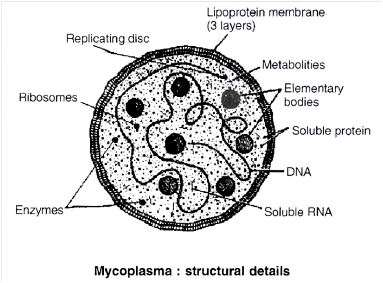
Concept Builder
Mycoplasmas cause various diseases in plants, animals and human beings.
Some are given below :
1. Plant diseases: The Mycoplasma diseases are generally transmitted through insects such as leaf hopper, mites and flies.
(i) Witches' broom (ii) Aster yellow
(iii) Brinjal little leaf (iv) Bunchy top of Papaya
(v) Sesame phyllody (vi) Sandal spike
2. Diseases in animals
(i) Pleuropneumonia in cattle
(ii) Inflammation of genitals
3. Diseases in human beings
(i) Infertility in man
(ii) Primary atypical pneumonia
IV. ARCHAEBACTERIA
They are believed to have evolved immediately after the origin of life on earth, as even now these are living under extremely adverse conditions such as extreme salty areas (halophiles), hot springs (thermoacidophiles) and marshy areas (methanogens).
Very few other organisms can survive under such environmental conditions. So these are termed as "living fossils".
These possess introns in DNA, their ribosomal proteins are highly acidic, these prokaryotes possess histone proteins different from that of eukaryotes.
These being the most primitive and ancient most bacteria.
Archaebacteria differ from other bacteria in having a different cell wall structure and this feature is responsible for their survival in extreme conditions.
The cell wall in archaebacteria contains proteins and non-cellulosic polysaccharides.
It lacks peptidoglycan, the characteristic cell wall material in bacteria and cyanobacteria.
It consists of glycoprotein, pseudomurein and non cellulosic polysaccharide.
Pseudomurein is like bacterial peptidoglycan, but contain N-acetyltalosaminuronic acid instead of NAM and lacks D-Amino acid.
The cell membrane contains branched chain lipids (phytanyl side chains) which decreases membrane fluidity.
This chemical composition of the cell membrane enables these organisms to withstand extremes of temperature and pH.
Archaebacteria are divided into three groups· methanogens, halophiles and thermoacidophiles
(a) Methanogens:
They are obligate anaerobes occurring in marshy habitats.
They are capable of converting CO2, methanol and formic acid (HCOOH) into methane and hence the name methanogens.
This property is exploited commercially in the production of fuel gas and methane in gobar gas plants (biogas fermenters).
Some of the methanogens live in rumen of herbivorous animals like buffalo, cow etc. (ruminants).
These microorganisms assist in fermentation of cellulose in such animals, e.g., Methanococcus, Methanobacterium, Methanosarcina, Methanospirillum.
(b) Halophiles:
They are aerobic chemoheterotrophic coccoid forms and are Gram negative. They occur in high salt concentration medium like sea, salt lake, brines, marshes, salted field etc.
In high light intensity a reddish pigment bacteriorhodopsin develops in their membrane to trap sun light to produce ATP, but they cannot use this ATP in food synthesis.
Sap vacuoles are absent in halophiles, hence, they cannot get plasmolysed in high salt concentration.
They maintain a high osmotic concentration of KCl in their cells.
These bacteria get lysed if NaCl level falls below 10%, e.g., Halococcus, Halobacterium.
These can grow well in a medium containing 25-30% of NaCl.
(c) Thermoacidophiles:
They are capable of tolerating high temperature as well as high acidity and hence, the name thermoacidophiles.
They often live in hot-water springs where the temperature is as high as 80°C and the pH as low as 2.
They oxidise sulphur to sulphuric acid under aerobic conditions and the energy obtained in this reaction is utilized for the synthesis of organic food.
The medium becomes highly acidic due to the production of sulphuric acid. Under anaerobic conditions sulphur is reduced to H2S, e.g., Thermoplasma, Thermoproteus, Thermococcus. Hence, these are chemosynthetic in nature.
Concept Builder
Thermoacidophiles are capable of withstanding extremely low pH and high temperature due to:
(a) The cell membrane containing branched chain lipids
(b) The presence of resistant enzymes which can operate under acidic conditions.
It seems probable that archaebacteria, living in extreme environmental conditions got separated from the main line of bacterial evolution quite early. They survived through long period of geological changes successfully, existing at present and are perhaps the oldest "living fossil".
V. ACTINOMYCETES (Ray Fungi)
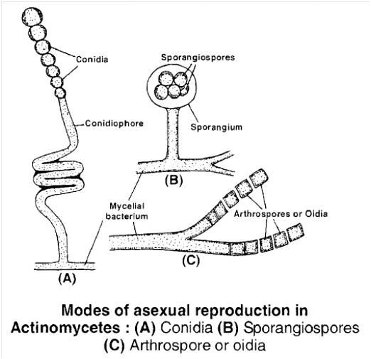
(1) These are branched, filamentous bacteria and are considered as intermediate form between bacteria and fungi, e.g., Nocardia, Mycobacterium, Corynebacterium, Frankia, Streptomyces.
(2) These filaments form radiating colonies in cultures so are also called mycelial bacteria.
(3) Cell wall contains peptidoglycan and is Gram positive in nature. It contains mycolic acid.
(4) They are facultative anaerobic, saprophytic, non-motile and are important decomposers of dead organic matter.
(5) They reproduce commonly by conidia, sporangiospore, oidia and fragmentation.
(6) They are acid fast in nature.
Concept Builder
Economic Importance of Actinomycetes : They are the source of more than 100 antibiotics. Most common antibiotics used in medicine are produced by filamentous bacteria and are listed below :
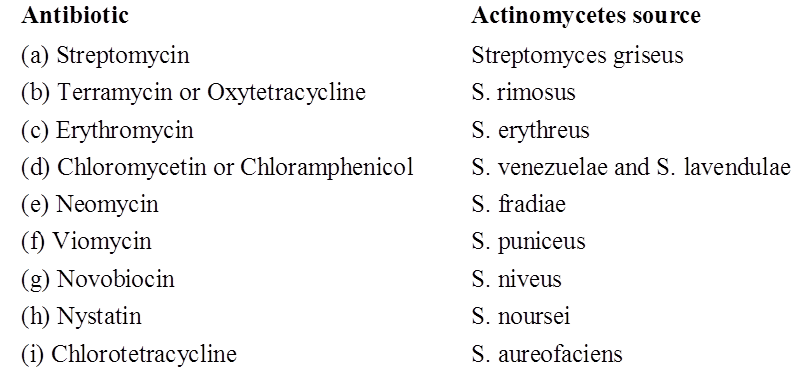
Frankia can fix N2 symbiotically by root nodule formation in non legumes like -Alnus, Myrica and Casuarina.
Earthy or musty smell of freshly ploughed soil or soil after first shower of rain, is due to oily secretion geosmin by some members like Nocardia, Micromonospora and Streptomyces
Concept Builder
1. In 1798, Edward Jenner used cowpox virus for immunising people against small pox.
2. Pasteurization simply kills bacteria but does not sterilize milk. It frees the latter of all vegetative cells of pathogens, but not the endospores.
3. Mycobacterium and Xanthomonas form nodules in leaves of Ardisia, Pavetta.
4. Bacterial cell membrane contains pentacyclic sterol like molecule termed as hopanoids as membrane stabiliser.
5. Kleinberger discovered tuberculosis and anthrax bacteria & developed "L-form" bacteria.
6. H. T. Ricketts described some new types of organisms (now considered a type of bacteria) in the blood of victims of Rocky Mountain spotted fever and similar organisms were called Rickettsiae, in honour of Ricketts. They occur as obligate intracellular parasites. The Q fever is caused by Coxiella burnetti. Rocky mountain fever is caused by Rickettsia rickettsii.
7. Chlamydiae: They are obligate intracellular parasite of vertebrates. They reproduce by elementary bodies. They do not have their own ATP generating system and obtain this energy from host. So that they are called energy parasite.
- Diseases
- Trachoma (eye disease) – C. trachomatis
- Conjuctivitis – C. trachomatis
- Psittacosis (influenza like) – C. psittaci
8. Joseph Lister developed the technique of aseptic cultures.
9. Nostoc develops symbiotic associations with Gunnera stem and Trifolium roots.
Kingdom: Monera
Kingdom Monera
Monera is a biological kingdom that is solely made up of prokaryotes. Their DNA is not enclosed within the nucleus as these single-celled organisms lack a true nucleus. The Monerans also do not possess any membrane-bound organelles.
The Kingdom Monera is made up entirely of bacteria. They are the most common microorganisms on the planet. Bacteria can be found practically anywhere. A handful of soil contains hundreds of bacteria. They also exist in harsh environments like hot springs, deserts, snow, and deep oceans, where only a few other species can survive. As parasites, many of them live in or on other organisms.
The spherical Coccus (pl.: cocci), the rod-shaped Bacillus (pl.: bacilli), the comma-shaped Vibrium (pl.: vibrio), and the spiral Spirillum are the four types of bacteria classified by their shape.
Although bacteria have a simple structure, their activity is extremely sophisticated. Bacteria, in comparison to many other creatures, have the greatest metabolic diversity.Some bacteria are autotrophic, meaning they produce their own nourishment from inorganic sources. They can be either photosynthetic or chemosynthetic autotrophs. The great majority of bacteria are heterotrophs, meaning they feed on dead organic matter or other species. Archaebacteria, Eubacteria, and Cyanobacteria are the three sub-kingdoms of the kingdom Monera.
A. Archaebacteria:
These bacteria are unique in that they survive in extremely salty environments (halophiles), hot springs (thermoacidophiles), and marshy environments (methanogens). Archaebacteria vary from other bacteria in that they have a unique cell wall construction that allows them to survive in harsh environments. Methanogens are bacteria that live in the guts of ruminant animals like cows and buffaloes and are responsible for the creation of methane (biogas) from their feces.
B. Eubacteria:
"True bacteria" is another name for Eubacteria. The existence of a hard cell wall and, if motile, a flagellum distinguishes them. A few bacteria have pili, which are small appendages on the cell surface that aid the bacterium in sexual reproduction. Pili also aids in the attachment of pathogens to their hosts. Depending on the type of the cell wall and the stain they take, they are classified as gram-positive or gram-negative.
C. Cyanobacteria:
Cyanobacteria are photosynthetic autotrophs with chlorophyll similar to green plants. They are unicellular, colonial, or filamentous algae that can be found in freshwater, marine, or terrestrial environments. Gelatinous sheath surrounds the colonies in general. Blooms are common in contaminated water bodies. Some of these species, such as Nostoc and Anabaena, can fix atmospheric nitrogen in specialized cells called heterocysts. Chemosynthetic autotrophic bacteria utilize the energy released by oxidizing inorganic compounds like nitrates, nitrites, and ammonia to produce ATP. They are vital in the recycling of nutrients such as nitrogen, phosphorus, iron, and sulfur.
In nature, heterotrophic bacteria are the most common. The majority of them are significant decomposers. Many of them have a major influence on human affairs.They aid in the creation of curd from milk, the manufacture of antibiotics, and the fixation of nitrogen in legume roots, among other things. Some of these are infections that harm humans, crops, farm animals, and pets. Bacterial infections such as cholera, typhoid, tetanus, and citrus canker are well-known. Bacteria mostly reproduce through fission. They may generate spores in unfavorable conditions. They also reproduce by adopting a rudimentary method of DNA transfer from one bacteria to the next, which is similar to sexual reproduction.
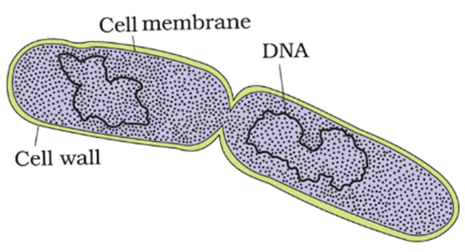
Mycoplasma are creatures that lack a cell wall entirely. They are the world's tiniest living cells, capable of surviving without oxygen. In animals and plants, many mycoplasmas are pathogenic.
Kingdom: Protista
- Books Name
- ACME SMART COACHING Biology Book
- Publication
- ACME SMART PUBLICATION
- Course
- CBSE Class 11
- Subject
- Biology
KINGDOM : PROTISTA
All unicellular eukaryotes, irrespective of their mode of nutrition, are included in the kingdom Protista in Whittaker's system. The term protista was coined by Ernst Haeckel. This kingdom forms a link between kingdom Monera on one hand and other three kingdoms i.e. , Plantae, Fungi and Animalia on the other hand. Protistans are ancestors of all multicellular eukaryotes (plants, fungi and animals).
Kingdom Protista includes
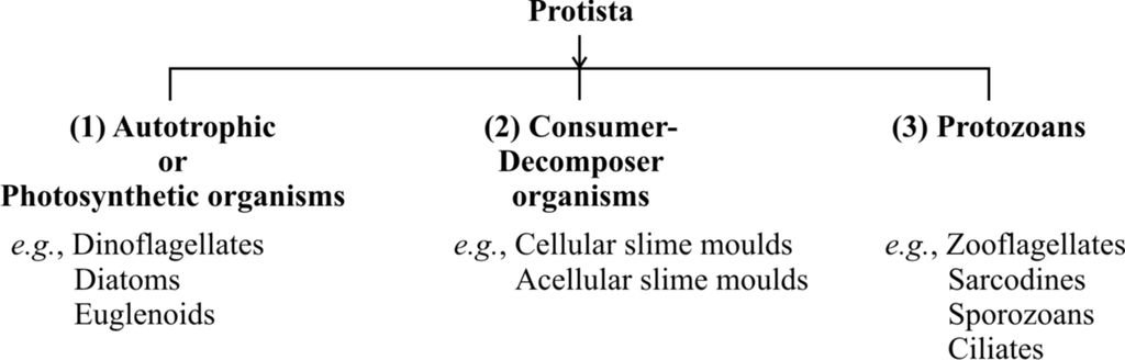
General Characteristics of Protista
1. Unicellular, eukaryotic organisms. Some are colonial without much cellular differentiation. Organisation at tissue level is absent.
2. Mostly aquatic organisms.
3. Cell structure is eukaryotic type having all kinds of membrane bound organelles and 80 S cytoplasmic ribosomes and cells may possess cellulosic cell wall.
4. Flagella and cilia have (9+2) pattern of microtubule organization consisting of tubulin protein.
5. Movement by pseudopodia, flagella or cilia where ciliary mode is fastest.
6. Mode of nutrition may be photosynthetic (holophytic), holozoic (ingestive), saprobic or parasitic (absorptive). Some have mixotrophic nutrition (photosynthetic and saprobic) as in Euglena.
7. Reproduction occurs by asexual and sexual means.
8. Life cycle is of 2 types-(i) Showing zygotic meiosis (ii) Showing gametic meiosis.
9. These are decomposers, photosynthetic or parasites. Parasitic protists may cause diseases like dysentery, malaria, sleeping sickness etc.
Photosynthetic protists and Slime moulds are described below:
(1) Photosynthetic Protists
These are popularly called protistan algae. Protistan algae constitute the major portion of the phytoplanktons.
A. Diatoms
Diatoms are golden brown photosynthetic protists and are called Chrysophytes (including both diatoms and desmids). They are both aquatic and terrestrial. Some are marine. They support much of marine life. Their important characters are
These are microscopic organisms possessing varying colours.
They are basically unicellular, but may form pseudofilament and colonies, lacking flagella except in the reproductive stage. They may be free floating (phytoplanktonic) due to presence of light weight lipids.
The cellulosic cell wall is impregnated with silica to form transparent siliceous shell, known as frustule. Depending upon the symmetry, diatoms may be pennate type, having bilateral symmetry (e.g., Navicula) and centric type, having radial symmetry (e.g., Melosira).
The cell wall is characteristic, made up of two halves; one half covering the other (epitheca over hypotheca) resembling a soap box.
The cell wall encloses the peripheral layer of cytoplasm (primordial utricle) surrounding a large central vacuole.
Nucleus lies in the central vacuole, suspended with the help of cytoplasmic strands.
Mode of nutrition is holophytic (photoautotrophic), photosynthetic pigments are chlorophyll a, chlorophyll c, b-carotene and special carotenoids containing fucoxanthin; xanthophylls like diatoxanthin, diadinoxanthin.
The reserve food is oil and a polysaccharide called leucosin (chrysolaminarin), volutin granules are also present.
They are responsible for almost 50% of the total organic matter synthesized in the biosphere.
Movement occurs by mucilage propulsion.
They mainly undergo asexual reproduction. The common mode of asexual reproduction is binary fission.
During binary fission, one half of the cell wall is retained by each of the daughter cells formed. The other half of the cell wall is secreted afresh.
Resting spores are called statospores (centric diatoms).
They reproduce sexually as well. Sexual reproduction varies from isogamy to oogamy. It involves gametic meiosis as diatoms are generally diploid (diplontic life cycle).
Concept Builder
1. Silica shells of dead diatoms are nearly indestructible and thus, get accumulated at the sea bed. Such huge rock-like deposits of hard shells of diatoms constitute diatomaceous earth, which is mined to obtain a whitish powder called diatomite or kieselguhr or diatomaceous earth. Diatomite is rough and gritty. Because of these features, it is used in filters in brewing industry, sugarcane refineries, in polishes for metals, tooth pastes, for making insulating bricks, in insulation of refrigerators, houses and for making the latter sound proof, in the manufacture of dynamite, water glass or sodium silicate and strong acids. This is added to paint to increase their night visibility.
2. They are very good indicators of water pollution. Common examples of diatoms are Triceratium, Melosira, Navicula, Cymbella.
B. Dinoflagellates
Dinoflagellates are golden brown photosynthetic protists, belonging to class Dinophyceae (Pyrrophyta). They are mainly marine, though few are fresh water forms.
They may appear red, yellow, green, brown or blue depending upon the main pigment present in cell.
General characters :
Unicellular, motile, biflagellate, golden brown photosynthetic protists (some are non-motile, amoeboid, palmelloid or filamentous).
They are mostly marine, some are found in fresh water.
The body is enclosed by a rigid coat called theca or lorica consisting of 2 to many articulated or sculptured plates of cellulose and pectin , hence are also called armoured dinoflagellates.
Theca has generally two grooves i.e., longitudinal called sulcus and transverse called cingulum or annulus or girdle.
Flagella are heterokont (different). One is longitudinal and other is transverse. The flagella pass out through the pores in the lorica and lie in the grooves. The transverse flagellum lies in the circular groove and the longitudinal flagellum in the longitudinal groove. The longitudinal flagellum is narrow, smooth directed posteriorly and the transverse flagellum is ribbon like
Both are oriented at right angle to each other producing spinning movements. Therefore, these protists are also called 'whirling whips'.
Most of the species have brown, green or yellow chromatophores with chlorophyll a, c, a-carotene, xanthophyll (e.g., Peridinin). Plastids are generally surrounded by 3-membrane envelope and contain 3-thylakoid lamellae. They are autotrophic or photosynthetic (Ceratium), a few are saprobic or parasitic.
Reserve food is carbohydrate and oils.
Nucleus is relatively larger in size, has condensed chromosomes even in interphase, chromosomes do not have histone. Nuclear envelope and nucleolus remain present even during cell division. This organisation is called Mesokaryon (Dodge, 1966).
A non-contractile vacuole called pusule is present near the flagellar base. It may have one or more vesicle and takes part in floatation and osmoregulation.
Some dinoflagellates possess trichocysts and cnidoblasts like those of coelentrates.
Reproduction is commonly asexual and occurs through cell division.
Isogamous and anisogamous sexual reproduction is reported from some dinoflagellates
e.g., Ceratium.
Life cycle involves zygotic meiosis (Ceratium, Gymnodinium). Gametic meiosis occurs in Noctiluca.
Concept Builder
1. Some marine dinoflagellates show bioluminescence, i.e., emit light, e.g., Noctiluca, Pyrodinium, Pyrocystis. Due to phosphorescence the sea glows at night.
2. Some dinoflagellates like Gonyaulax catenella produce a toxin called saxitoxin into the sea water which is highly poisonous to vertebrates, e.g., fishes and other aquatic animals. Marine shell fish consume dinoflagellates and accumulate the poison which is not harmful to the shell fish (mussel) but upon being consumed causes severe illness in man called paralytic shell fish poisoning (PSP) and even prove fatal.
3. Some dinoflagellates proliferate in large number and cause red tide of the sea, e.g., Gonyaulax, Gymnodinium.
C. Euglenoid (Euglena-like)
It is a group of chlorophyllous and non chlorophyllous flagellate protists. Largest genera being Euglena amongst them.
Euglenoids are unicellular, flagellate protists found in water or damp soil. Majority of them are fresh water organisms found in stagnant water.
Body is spindle shaped with blunt anterior end and pointed posterior end.
Cell wall is absent but a covering periplast or pellicle is present which is proteinaceous (elastic) in structure.
Locomotory organs are flagella.
The cell bears a single long tinsel type flagellum (stichonematic) arising at the anterior end. Actually, there are two flagella but one of these is reduced. The longer flagellum has two branches at the base each having its own basal granule. In the area of union of two flagella is present a photosensitive paraflagellar body.
Myonemes are oblique but parallely arranged strips in pellicle. Euglenoids perform creeping movement of contraction and expansion with the help of myonemes which is called metaboly or euglenoid movement.
The apical end of the cell bears an invagination with three distinct parts, i.e., mouth (cytostome), canal (gullet or cytopharynx) and reservoir. It helps in the ingestion of solid food particles.
Stigma or an eye spot is attached to the membrane of the reservoir at the level of paraflagellar body and along with it seems to be involved in perception of light stimulus. It contains photosensitive red-orange pigment called astaxanthin.
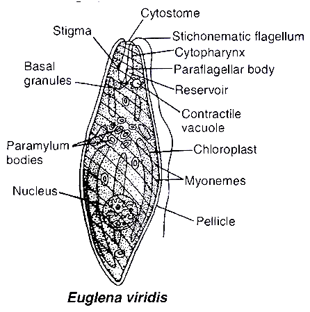
A contractile vacuole occurs in the anterior end of the cell just below the reservoir, meant for osmoregulation and excretion.
Single large nucleus lies near the centre of the protoplast.
Nutrition in Euglena viridis is photoautotrophic. However, it is capable of getting nourishment from dead and decaying organic matter in the substrate by secreting digestive enzymes (saprophytic nutrition) in the absence of light. This dual mode of nutrition is termed as mixotrophic. Holozoic nutrition is absent in Euglena. Some forms are holozoic (Paranema) or saprobic (Rhabdomonas).
Photosynthetic pigments are chlorophyll a, chlorophyll b, xanthophyll and b-carotene.
Reserve food material is paramylon, stored in cytoplasm in the form of paramylum granules. They are chemically b 1,3-glucans.
Under favourable conditions, they mainly reproduce by longitudinal binary fission. During unfavourable conditions, palmella stage and cysts are formed for perennation. Sexual reproduction is not known to occur in euglenoids, e.g., Euglena and Paranema.
Concept Builder
1. Euglena is producer-decomposer protist.
2. It is studied as plant as well as animal and is called as plant animal.
3. Plant characters of Euglena:
- Presence of chloroplast with photosynthetic pigments.
- Holophytic nutrition
4. Animal characters of Euglena:
- Absence of cell wall and presence of proteinaceous pellicle.
- Presence of stigma and paraflagellar body.
- Presence of contractile vacuole.
- Presence of longitudinal binary fission.
(2) Slime moulds or consumer-decomposer protists
They were included in class myxomycetes of fungi in two-kingdom classification. They were called mycetozoa by DeBary as they are closely related to animals. Mycologists include them in gymnomycota. Because of their nature they are called protistan fungi.
General characteristics of the slime moulds are :
They are usually free-living, creeping over debris like fallen leaves and rotting logs of wood.
They have naked protoplast, not covered by any cell wall in vegetative stage.
They lack chlorophyll and have saprobic or phagotrophic mode of nutrition.
During life cycle they are amoeboid and non-cellulosic, but spores have cellulosic wall so that their vegetative phase resembles with animals while reproductive phase resembles with plants.
Amoeboid plasmodial stage resembles protozoa and spore forming nature is like fungi.
Spores are extremely resistant and survive for many years, even under adverse conditions. The spores are dispersed by air currents.
Reproduction is both asexual and sexual. This group is represented by two separate types of organisms i.e. acellular and cellular.
A. Acellular or Plasmodial slime moulds
General characters:
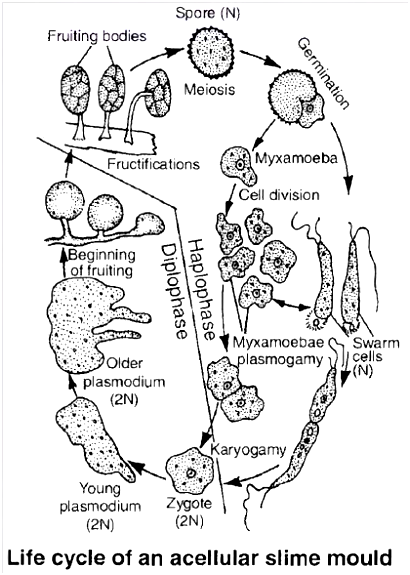
Slimy masses found on decaying leaves and lumber.
Somatic body is free living, multinucleate, naked, diploid mass called Plasmodium. Movement occurs by means of pseudopodia.
During unfavourable conditions, entire plasmodium forms many fructifications/fruting bodies (polycentric). The fruiting body is called sporocarp which contains a stalk having a sporangium at its tip. The wall of sporangium is called peridium.
Sporangium has an intricate network of cytoplasmic threads called capillitium.
DiploId protoplast forms haploid spores by meiosis.
Spore wall is double, outer wall is spiny and sculptured.
On germination, spores produce biflagellate swarm cells or non-motile myxamoebae which act as gametes.
Sexual reproduction is isogamous.
Diploid zygote directly forms the plasmodium which becomes multinucleate by repeated mitotic divisions of the diploid nucleus.
Chief mode of nutrition is saprotrophic.
Vegetative reproduction is by fission .
e.g., Physarum, Physarella, Fuligo, Dictydium, Lycogala
B. Cellular slime moulds or communal slime moulds
General characters :
Wall less, uninucleate myxamoebae present. Complete absence of flagellated cells during life cycle.
Formation of pseudoplasmodium stage as a result of chemotactic movement of myxamoebae due to release of cAMP and acrasin.
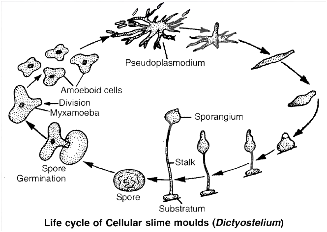
Sporangia are naked.
Spores have cellulosic wall.
Sexual reproduction is anisogamous.
Common cellular slime mould, Dictyostelium, is a colonial form in which hundreds of uninucleate, haploid amoeboid cells are aggregated without any fusion to form a colony.
The colony gives the appearance of single multinucleate mass of protoplasm and thus, called pseudoplasmodium.
Under exhausted food supply and stimulation by cAMP and chemical acrasin, many cells come close together by chemotactic movement during the formation of pseudoplasmodium.
Pseudoplasmodium exhibits primitive form of multicellularity and division of labour.
So these are also called as communal slime moulds.
On these basis cellular slime moulds are regarded as advanced protists and primitive fungi.
During unfavourable conditions, the myxamoebae may form a cyst called microcyst for perennation and dispersal.
Under dry conditions, the pseudoplasmodium produces stalked sporocarp, which may be branched or unbranched, each branch bearing single sporangium terminally (monocentric).
Sporangium is wall less. Within the sporangium, amoeboid cells become rounded to secrete a spore wall around.
On the approach of favourable conditions, spores are liberated.
Each spore germinates by rupturing cellulosic wall to form myxamoeba and the myxamoebae may live independently, multiply by repeated mitotic divisions or get aggregated to form pseudoplasmodium.
Sexual reproduction is anisogamous type. During sexual reproduction, a number of myxamoebae form a clump.
One of the myxamoeba becomes larger and engulfs the surrounding smaller myxamoebae.
The plasmogamy occurs and the fused protoplast secretes a thick wall around to form macrocyst.
In the macrocyst, karyogamy occurs and it thus, becomes zygote.
It is followed by meiosis and several mitotic divisions to form a large number of haploid myxamoebae, which are released by rupture of macrocyst wall.
- e.g., Dictyostelium, Polysphondylium.
Kingdom: Protista
Kingdom Protista
Kingdom Protista is a diverse group of organisms. It includes all eukaryotes that aren't animals, plants, or fungi, including single-celled and multicellular creatures. There are an estimated 60,000 to 200,000 protist species, with many more yet to be discovered. Apart from a relatively simple organization, the eukaryotes that make up this kingdom have little in common. Some protists are small and unicellular, like an amoeba, while others, like seaweed, are enormous and multicellular. Multicellular protists, on the other hand, lack highly specialized tissues and organs. Protists are distinguished from other eukaryotes, such as fungi, mammals, and plants, by their basic cellular arrangement. Protista is largely an aquatic phylum. This kingdom is linked to the kingdoms of plants, animals, and fungus.
The Protista cell body, like other eukaryotes, has a well-defined nucleus and other membrane-bound organelles. Some have flagella or cilia, while others don't. Cell fusion and zygote development are used by protists to reproduce both asexually and sexually. Many protists, such as algae, are photosynthetic and play an important role in ecosystems as primary producers. Other protists cause a variety of deadly human diseases, including malaria and sleeping sickness.Chrysophytes, Dinoflagellates, Euglenoids, Slime molds, andProtozoans come under Protista.
A. Chrysophytes:
Diatoms and golden algae (desmids) are members of this category. They can be found in both freshwater and marine habitats. They are tiny and float on water currents passively (plankton). They are mostly photosynthetic. The cell walls of diatoms produce two thin overlapping shells that fit together like a soapbox. Because the walls are embedded with silica, they are unbreakable. As a result, diatoms have left behind a vast amount of cell wall deposits in their habitat, which has been referred to as 'diatomaceous earth' over billions of years. Because this soil is rough, it is utilized for polishing and filtration of oils and syrups. In the oceans, diatoms are the primary 'producers.'
B. Dinoflagellates:
These organisms are mostly marine and photosynthetic.They appear yellow, green, brown, blue, or red dependingon the main pigments present in their cells. The cell wallhas stiff cellulose plates on the outer surface. Most ofthem have two flagella; one lies longitudinally and theother transversely in a furrow between the wall plates.Very often, red dinoflagellates (Example: Gonyaulax)undergo such rapid multiplication that they make thesea appear red (red tides). Toxins released in such largenumbers may even kill other marine animals such as fish.
C. Euglenoids:
The majority of them are freshwater organisms found in stagnant water. Instead of a cell wall, they have a protein-rich layer called pellicle which makes their body flexible.They have two flagella, a short and a long one. Thoughthey are photosynthetic in the presence of sunlight, whendeprived of sunlight they behave like heterotrophs bypredating on other smaller organisms. Interestingly, thepigments of Euglenoids are identical to those present in higher plants. Example: Euglena.
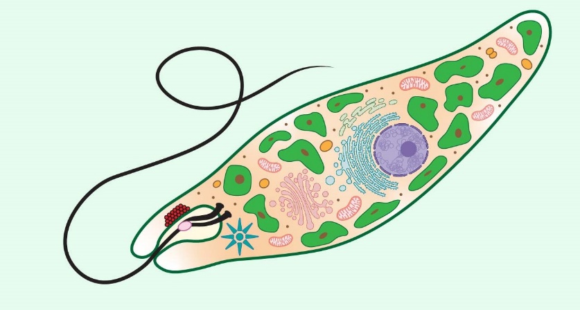
D. Slime Molds:
Slime molds are protists that feed on their surroundings. The organism eats organic matter as it glides along with rotting twigs and leaves. They create a plasmodium, which can grow and spread over severalfeet if the conditions are right. The plasmodium develops and generates fruiting bodies with spores at their tips in unfavorable conditions. True walls exist within the spores. They are highly hardy and may live for many years in harsh environments. Air currents disseminate the spores.
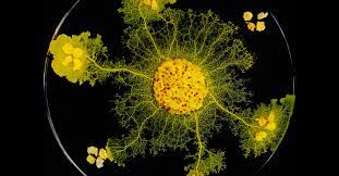
E.Protozoans:
They are all heterotrophic organisms that exist as predators or parasites. Animal relatives are thought to be their ancestors. Protozoans are divided into four categories.
1. Amoeboid protozoans dwell in freshwater, seawater, or moist soil. As in Amoeba, they move and seize their prey by putting out pseudopodia (false feet). Silica shells cover the surface of marine organisms. Some of them are parasites, such as Entamoeba.
2. Flagellated protozoans: This group includes both free-living and parasitic protozoans. They have flagella on their bodies. Sleeping sickness is caused by a flagellated parasitic protozoan. Trypanosoma is a good example.
3. Ciliated protozoans: Ciliated protozoans are aquatic, actively moving organisms with thousands of cilia. They have a cavity (gullet) on the cell surface that opens to the outside. The water-containing food is directed towards the gullet by the coordinated movement of rows of cilia. Paramoecium is a good example.
4. Sporozoans: This term refers to a group of organisms that have a spore-like stage in their life cycle. Plasmodium (malarial parasite), which causes malaria, is the most well-known. Malaria is a disease that has a devastating impact on the human population
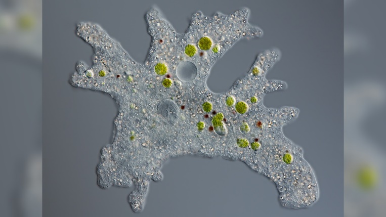
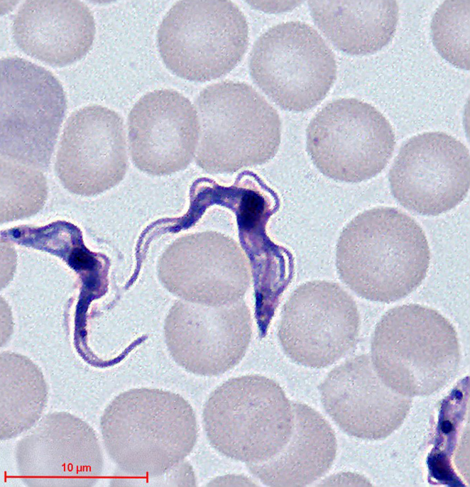

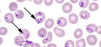
Figure 8 (a) Ameoba (b) Trypanosoma (c) Paramecium (d) Plasmodium
Kingdom: Fungi
- Books Name
- ACME SMART COACHING Biology Book
- Publication
- ACME SMART PUBLICATION
- Course
- CBSE Class 11
- Subject
- Biology
KINGDOM : FUNGI
This kingdom contains achlorophyllous, eukaryotic, heterotrophic, spore producing, thalloid organisms. The study of fungi is called mycology.
Pier Antonio Micheli is considered as father or founder of mycology.
Mycologist H.A. de Bary is the father of modern mycology.
The father of Indian mycology is E.J. Butler.
General Characters
They are cosmopolitan and occur in air, water, soil and on animals and plants. They are mostly terrestrial. They prefer to grow in warm and humid places.
They may grow on tree bark, dung, wood, burnt wood and keratinous material (e.g., hair, horns) and are called corticolous (bark), coprophilous (cow dung), epixylic (wood), xylophilous (burnt wood) and keratinophilous (keratin) respectively.
The body is haploid (n) and thalloid, i.e., not differentiated into root, stem and leaves. They are multicellular (except Yeast and Synchytrium).
The fungal body is made up of thread like elongated tubular structures, called hyphae. These cris-cross with one another to form a network known as mycelium.
The hyphae may be aseptate and multinucleate. Such a hypha is termed coenocytic. In most of the fungi, the mycelium is septate.
The septum, however, is not complete, but has a pore through which continuity of the cytoplasm of the adjoining cells is maintained.
The septum may have simple central pore as in ascomycetes, but in higher fungi (class basidiomycetes), the septum is dolipore septum, in which central pore possesses a barrel shaped inflation.
In septate mycelium, individual cell may contain single nucleus (monokaryotic -feature of primary mycelium) or an intermediate phase of two nuclei (dikaryotic -feature of secondary mycelium).
The cell wall of the hyphae is made up of chitin or fungal cellulose, which is a polysaccharide containing nitrogenous compound and it is basically made up of acetylglucosamine.
In some fungi, the cell wall is made up of cellulose (e.g., Phytophthora, Pythium and other oomycetes). Reserve food material is stored in the form of oil and glycogen.
Cells have unicisternal golgi bodies.
Mitosis in somatic cells is Karyochorisis type (mitosis with intranuclear spindle formation).
Nutrition is heterotrophic which includes saprophytes, parasites and symbionts.
In most of the fungi, there are two distinct phases in the life cycle, the vegetative or assimilative phase and the reproductive phase.
In vegetative phase, fungus is microscopic hidden in the substratum and is hardly visible to the naked eyes.
The fungus enters into reproductive phase after attaining maturity in the vegetative phase.
In unicellular yeasts, the same cell performs both assimilative and reproductive functions.
Such type of fungal bodies in which entire cell gets transformed into reproductive structures are known as holocarpic.
Fungal body is termed eucarpic in which a part of mycelium is used up in the development of reproductive structures.
Concept Builder
Modification of mycelium:
(a) Plectenchyma :
When hyphae of a mycelium grow together like plates and intertwine with one another forming a thick woven structure, it is called plectenchyma.
Plectenchyma may have :
(i) Prosenchyma : Loosely interwoven structure whose hyphal components lie more or less parallel to each other and are recognizable.
(ii) Pseudo parenchyma : Hyphae are compactly arranged and hyphal components have lost their identity and appear isodiametric and continuous in section resembling parenchyma of higher plants.

(b) Sclerotia (Singular Sclerotium) :
In some fungi like Claviceps the mycelium may pass into a dormant or resting stage by the formation of hard resting bodies resistant to unfavourable conditions.
Each sclerotium is composed of central prosenchymatous and peripheral pseudoparenchymatous arrangement which are again surrounded by a ring of pigmented hyphae.
(c) Rhizomorph:
- When the fungal hyphae aggregate together below surface they behave as an organized unit to form a root like strand with a thick hard cortex.
- It also develops a growing tip somewhat resembling that of a root tip, e.g., Agaricus.
(d) Appressorium: Terminal swollen structure of germ tube for attachment, and penetration.
(e) Haustoria: Terminal swollen structure for absorption of food, e.g., Albugo.
(f) Snares/hyphal traps: Helps in capturing nematodes in predaceous fungi, e.g., Arthrobotrys, Dactylaria
Reproduction in Fungi
- Fungi reproduce by all the three modes, i.e., vegetative, asexual and sexual.
1. Vegetative reproduction: It occurs by the following methods:
(a) Fragmentation : The mycelium breaks up into two or more fragments due to mechanical injury, decay or some other reasons. Each fragment grows into independent mycelium.
(b) Fission: Here, simple splitting of vegetative cells into two daughter cells takes place by simple constriction.
(c) Budding: Some fungi like yeast produce small outgrowths, i.e., buds from their vegetative body. Eventually, the buds are cut off from parent cell and mature to form new individuals.
2. Asexual reproduction:
It occurs through spores.
These are single celled specialized structures which separate from the organism, get dispersed and germinate to produce new mycelium after falling on suitable substrate.
The spores produced during asexual reproduction in fungi are formed by mitotic division and are thus termed, mitospores.
The various means of asexual reproduction are as follows :
(a) Zoospore :
Many fungi, especially aquatic members produce these types of spores.
Zoospore may be uniflagellate, e.g., Synchytrium or biflagellate, e.g., Saprolegnia, Pythium and are naked uninucleate structures formed in zoosporangia.
They germinate to give rise to new mycelium.
Biflagellate zoospores are of two kinds (e.g., Saprolegnia) pear shaped or pyriform with 2 flagella placed at anterior end (primary zoospore) and kidney shaped or bean shaped, bearing two laterally inserted flagella (secondary zoospore).
This phenomenon of having two types of zoospores is called diplanetism.
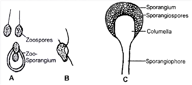
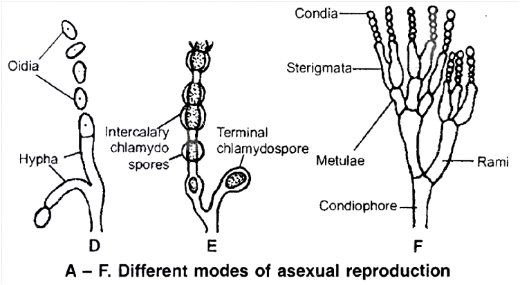
(b) Sporangiospore :
(c) Conidia:
Conidia are non motile, thin walled exogenous spores produced at the tips of erect hyphae called conidiophore.
They are arranged in chains upon the conidiophore, e.g., Aspergillus and Penicillium
(d) Chlamydospore:
In some fungi the hyphae under unfavourable conditions, forms thick walled resting resistant spores which later get separated from each other.
They may be terminal or intercalary.
They may remain viable for several years.
On return to favourable conditions they germinate to give rise to new individuals.
Thus, chlamydospores are structures for perennation also, e.g., Rhizopus.
(e) Oidia:
Non-motile thin walled spores developing under sugar rich conditions in medium.
Their budding condition is called torula stage.
3. Sexual reproduction:
Sexual reproduction is reduced in fungi and takes place by two fusing gametes.
It includes 3 stages :
(a) Plasmogamy:
There is union between two haploid protoplasts which results in bringing the fusing nuclei of different parents close together.
In some fungi, plasmogamy is immediately followed by karyogamy.
However, in Ascomycetes and Basidiomycetes, an intermediate dikaryotic (n + n) condition occurs. This phase is called dikaryophase.
(b) Karyogamy: The two haploid nuclei which come together in plasmogamy fuse and thus, a diploid zygote is produced.
(c) Meiosis: Reduction division takes place in the zygote thus, reducing the number of chromosomes to half.
Plasmogamy occurs by the following methods :
(a) Planogametic copulation / Gametic fusion:
This is the simplest form of sexual reproduction.
In this process, fusion of two gametes of opposite sex or strains takes place.
One or both of the fusing gametes are motile.
It results in the formation of a diploid zygote, e.g., Allomyces.
This process is usually of three types: Isogamy, Anisogamy, Oogamy.
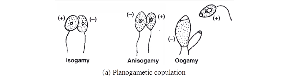
(b) Gametangial contact :
In this process two gametangia come in contact with one another.
A fertilization tube is developed to facilitate the migration of entire contents of male gametangium into the female gametangium.
Both the gametangia never fuse together losing their identity, e.g., Pythium, Albugo (Oomycetes).
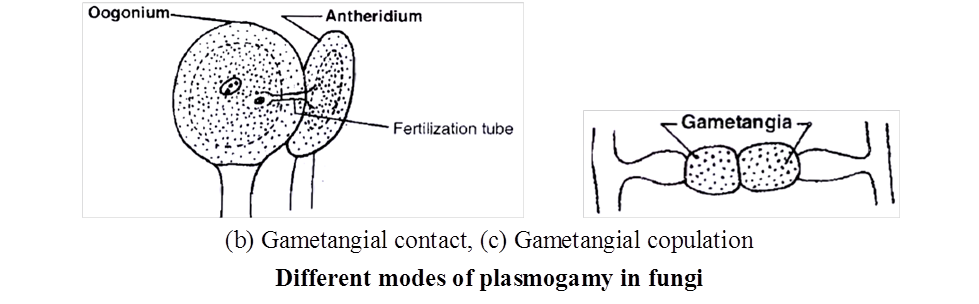
(c) Gametangial copulation :
In this process, direct fusion of entire contents of two gametangia is accomplished by dissolution of their common walls resulting in the formation of a single cell, in which protoplasts of two gametangia fuse, e.g., Mucor, Rhizopus (Zygomycetes).
(d) Spermatization :
Some fungi produce many minute, spore like, single-celled structures called spermatia (non motile male gametes) on spermatiophores (hyphae).
These structures are transferred through agencies like water, wind and insects to special female receptive, hyphae (Basidiomycetes).
The contents migrate into receptive structure.
Thus, dikaryotic condition is established, e.g., Puccinia.
(e) Somatogamy :
This takes place in most of the higher true fungi, where formation of gametes is absent.
In such fungi, direct fusion of somatic hyphal cells occur to establish dikaryophase, e.g., Agaricus
Classification of Fungi
A number of criteria are used for classifying fungi.
Morphology of mycelium, mode of spore formation and fruiting bodies form the basis for the classification of this kingdom.
A common systematic presentation is given below
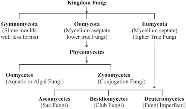
I. Oomycetes : The algal fungi
Hyphal wall contains cellulose and other glucans in many members.
The mycelium is coenocytic (multinucleate and aseptate).
Asexual reproduction involves the formation of spore containing sacs or sporangia. In aquatic forms, the sporangia produce zoospores.
Zoospores generally have two laterally inserted flagella with heterokont condition, in which one flagellum is smooth (whiplash) while the other is of tinsel type (having fine surface outgrowths called mastigonemes).
Sexual reproduction is by planogametic fusion or gametangial contact.
The product of sexual reproduction and site of meiosis is oospore.
Concept Builder
(i) Phytophthora infestans causes late blight of potato and occasionally of tomato as well. Great Irish famine of 1845 -1847 was due to late blight of potato.
(ii) Albugo candida (Cystopus candidus) causes white rust of crucifers and is characterised by the appearance of irregular white blisters on the leaves and stems.
(iii) Pythium debaryanum causes damping off disease in seedlings of tomato, chillies, castor, mustard.
(iv) Sclerospora graminicola causes downy mildew in cereals particularly, Pennisetum typhoides (vern. Bajra) green ear disease.
(v) Peronospora parasitica causes downy mildew in a number of plants, such as mustard, spinach, onion etc.
(vi) Saprolegnia causes salmon disease of gills in fishes.
II. Zygomycetes: The conjugation fungi
It is class of terrestrial fungi which are mostly saprotrophic and rarely parasitic.
Hyphal wall contains chitin or fungal cellulose.
The mycelium is coenocytic (multinucleate, aseptate) like the one found in Oomycetes.
Motile cells (zoospores or planogametes) are absent.
Mitospores are non motile. They are called sporangiospores as the spores are formed inside sporangia that are borne at the tips of special hyphae called sporangiophores.
Sexual reproduction occurs through gametangial copulation or conjugation. Because of it, zygomycetes are also called conjugation fungi.
The gametes are commonly multinucleate and are called coenogametes.
Sexual reproduction produces a resting diploid spore called zygospore. Because of the presence of zygospore, the group of fungi is called zygomycetes. Zygospore differs from oospore in that, for its formation a distinct food laden, non motile, large female gamete is not produced.
Zygospore is the site of meiosis and does not give rise to new mycelium directly. Instead it produces a new sporangium called germ sporangium (previously called zygosporangium). Germ sporangium forms meiospores called germ spores.
Sometimes, gametangia fail to fuse. Gametangia become surrounded by a thick wall resulting in formation of azygospore (parthenogenetically produced zygospore).
Concept Builder
(i) Rhizopus stolonifer (= R. nigricans) is popularly known as black bread mould. Mucor mucedo is called dung mould or pin mould. Rhizopus and Mucor are the common saprotrophic fungi that attack a variety of food stuffs. Soft rot or leek disease of strawberry, apple, jack fruit, sweet potato etc. is due to Rhizopus. Mucor pusillus causes infection of internal organs in human beings. Absidia corymbifera causes bronchomycosis.
(ii) Ramysin (antibiotic) is obtained from Mucor ramannianus.
Life Cycle of Rhizopus
Rhizopus is a saprophytic fungus, commonly found on dead organic matter rich in carbohydrates.
Mycelium is made up of white narrow thread like hyphae growing on the surface of substratum.
Two types of vegetative hypha (Rhizoidal and Stoloniferous) arise from definite points called hold fast / apparent nodes.
Third hypha is asexual, called Sporangiophore and fourth type is sexual hypha called zygophore.
The absorptive hyphae are called rhizoidal hyphae which penetrate into the substratum.
The hyphae appeared over the surface of substratum are in the form of stolons.
From the nodes of stolons arise branched rhizoidal hyphae on the under surface and a group of aerial structures called sporangiophore (asexual hypha) from the upper surface of 'apparent nodes'.
The apical portion of each sporangiophore ends into a swollen structure called sporangium which is filled with spores.
The hyphae are coenocytic, aseptate and branched. It has many nuclei, oil drops, glycogen bodies and vacuoles in the cytoplasm.
The reproduction in Rhizopus is vegetative, asexual and sexual.
- Vegetative reproduction: This takes place by fragmentation of hyphae.
- Asexual reproduction :
Asexual reproduction takes place by the formation of non-motile spores inside a sporangium.
The tip of aerial hypha swells in which cytoplasm migrates with nuclei.
The tip swells considerably and the nuclei divide repeatedly.
The contents of the young swollen tip differentiate into a central zone called columellaplasm mainly filled with vacuolated cytoplasm surrounded by a peripheral zone called sporangioplasm containing dense cytoplasm and many nuclei.
Vacuoles ultimately form a continuous vacuolated layer by fusing laterally one after the other and ultimately develop into a dome shaped septum known as columella (sterile part).
In the meantime, cleavage of sporangioplasm takes place resulting into innumerable, small 2-10 nucleate portions which round up, become invested with spore membranes, and develop into nonflagellate spores, the sporangiospores.
These are formed under most favourable conditions. Thus, the sporangium is large, globose and contains many spores.
Spores are dispersed by bursting of the thin wall of the sporangium due to pressure that is set up in the columella.
Other two asexual spores are oidia and chlamydospores (formed under unfavourable conditions).
The spores on germination produce a germ tube giving rise to new mycelium.
3. Sexual Reproduction :
Sexual reproduction in Rhizopus takes place by the formation of two multinucleate gametangia.
Both the gametangia are similar externally but are different physiologically i.e. they are of (+) and (-) type.
It is called heterothallism (This was discovered by Blakeslee in Rhizopus stolonifei ).
Another species called Rhizopus sexualis is a homothallic species.
When two mycelia of opposite strains, (+) behaving as male and (-) behaving as female respectively, come near one another, under influence of a chemical called trisporic acid, this stimulates the formation of special sub-aerial hypha called zygophores.
This hypha produces small outgrowths, called progametangia.
Their apical ends are swollen and filled with multinucleate protoplasm.
This apical portion of these progametangia, comes in contact with one another.
A septum is laid down, separating the terminal portion which is now termed gametangium. The remaining part of progametangia is called suspensor.
The multinucleate undifferentiated protoplast of each gametangium is termed as coenogamete.
As the gametangia mature, the separating wall dissolves from the middle to outward and intermingling of the contents of two gametangia takes place (Gametangial copulation).
Nuclear pairing and fusion of one (+) and another (-) nuclei gives rise to a large number of diploid nuclei.
The young zygospore lying within the parent gametangial wall enlarges considerably and secretes several layers of thick wall (5 layered, 2 layered exosporium and 3 layered endosporium) around it.
The zygospore matures it breaks up the original gametangial wall into small pieces that fall apart exposing the outer thick, spiny and black exosporium.
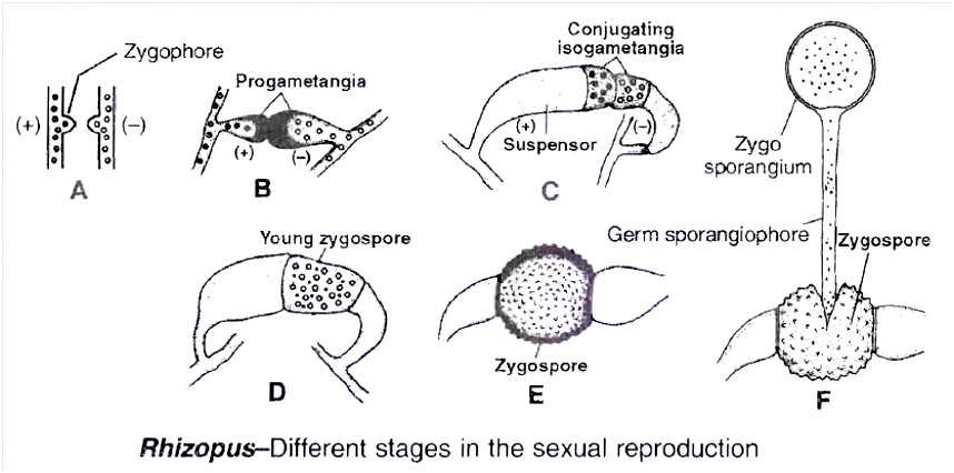
Meiosis occurs at the time of germination of zygospore.
The zygote germinates after rest period.
On germination, the exosporium cracks and endosporium produces a germsporangiophore (promycelium) that terminally develops a germ sporangium (zygosporangium) which bears large number of spores.
The meiosis produces 4 haploid nuclei where only one remain functional.
This divides repeatedly to produce coenocytic mycelium with many haploid nuclei.
Occasionally, failure of gametangial copulation results in parthenogenetic development of zygospore, which are called azygospores (parthenospores
III. Ascomycetes: The sac fungi
The mycelium consists of septate hyphae. (Yeasts are an exception in that they are basically unicellular).
They are saprophytic, decomposers, parasitic or coprophilous (growing on dung).
The septa possess central pores called septal pores. The pores allow communication and transport between adjacent cens.
Cell wall contains chitin.
Motile structures do not occur in the life cycle.
In majority of ascomycetes, the common mode of asexual reproduction is through the formation of conidia . Conidia are borne on branched or unbranched hyphae called conidiophores , e.g., Penicillium, Aspergillus.
Female sex organ is called ascogonium.
Plasmogamy occurs by means of -
- Gametangial contact (e.g., pyronema)
- Conjugation (e.g., Yeast)
- Spermatization (e.g., Ascobolus)
- Somatogamy (e.g., Peziza)
- Autogamy (e.g., Morchella).
Karyogamy is delayed after plasmogamy. A new transitional phase appears in the life cycle. It is called dikaryophase. The cells of dikaryophase are called dikaryotic cells. Each such cell possesses two different nuclei (Dikaryon). This forms a shorter phase of life cycle.
Once a cell becomes dikaryotic, it transfers the nucleus to other cells by the crozier method (method of dikaryotization) to make them dikaryotic.
Some dikaryotic cells function as ascus mother cells. This converts the cells into asci (singular ascus). Ascus is a sporangial sac peculiar to Ascomycetes. Ascus is the site of karyogamy and meiosis. 4 to 8 haploid meiospores named ascospores are produced endogenously in each ascus. In most of the cases, half the number of ascospores belong to one mating type(+) while the other half belong to the second mating type (-).
Ascospores may be arranged linearly (Neurospora) or unorderly (yeast).
The asci may occur freely or get aggregated into specific fructifications called ascocarps. Ascocarps are of many types : cup-like (apothecium, e.g., Peziza), flask-shaped (perithecium, e.g., Neurospora, Claviceps) , elongated with a slit (hysterothecium), closed (cleistothecium , e.g., Penicillium) cushion like, chambered (Ascostroma, e.g., Pleospora). The fructifications of some ascomycetes are edible, e.g., morels, truffles.
Concept Builder
Members of this class are said to be our worst fungal enemies.
(i) Morels are Ascomycetes with edible ascocarps having fleshy sponge-like conical cap or pileus and a stalk like stipe, e.g., Morchella esculenta (vern. Guchhi), M. deliciosia.
(ii) Truffles: They are edible tuber-like subterranean ascocarps, e.g., Tuber aestivum.
(iii) Claviceps purpurea causes ergot of rye and C. microcephala causes ergot of bajra in which ears are filled with sclerotia of the fungus. Eating of infected cereals produces ergotism. (It produces an alkaloid called ergotine which causes abortion, if eaten, unknowingly). This is used as a drug to promote expulsion of foetus.
(iv) Neurospora crassa (Pink bread mould), is often employed in experimental genetics, so is called Drosophila of plant kingdom.
(v) Erisyphe: The fungus produces powdery mildew (fungal disease in which pathogen results in a powdery coating of spores on the sut1ace of the host), e.g., Erysiphe graminicola.
(vi) Penicillium chrysogenum is used in commercial production of the antibiotic penicillin. The later was the first commercial antibiotic. It was discovered from P. notatum. The fungus is employed in ripening of cheese, e.g., P.roqueforti and P. camemberti.
(vii) Aspergillus: It is common green smoky mould which not only contaminates laboratory cultures (hence called weed of laboratory), but also various food stuffs including bread, butter etc. Aspergillus flavus is highly poisonous due to the presence of aflatoxins. A. oryzae is the source of diastase enzyme.
(viii) Brewing Industry: Under anaerobic conditions, sugary solutions inoculated with yeasts are converted into alcoholic beverages, e.g., beer, wine, cider, toddy. The two common yeasts used by brewing industry are Saccharomyces cerevisiae (Beer or Baker's yeast) and S. ellipsoidens (Wine yeast).
(ix) Gibberellins: They were first discovered in the extracts of Gibberella fujikuroi growing on rice (bakanae disease of rice). It is the perfect stage of fungus Fusarium moniliforme (Deuteromycete). Gibberellins are natural plant growth hormones.
Life cycle events of (A) Yeast and (B) Penicillium are described below:
(A) Yeast
Yeasts are a group of non mycelial or pseudomycelial ascomycetes which multiply asexually by budding or fission and where asci are not organised into ascocarps.
These are facultative aerobes.
Depending upon the mode of asexual reproduction, yeasts are of three types -budding yeast (e.g., Saccharomyces), fission yeast (e.g., Schizosaccharomyces) and helobial yeast (both budding and fission, e.g., Saccharomycoides).
Yeasts in which ascus formation is known are named as true yeasts.
Related forms which resemble yeasts in main characterstics, but where ascus formation is not reported, are called false yeasts, e.g., Candida, Mycoderma, Geotrichum, Cryptococcus (false yeasts belong to deuteromycetes).
Life cycle of Yeast
It is a saprophytic fungus found on substratum which is rich in sugars, e.g., sugarcane juice, fruits (banana, plums, grapes), milk etc.
Structure
This is a circular or elliptical, unicellular, colourless fungi not having a typical mycelium.
Their cell wall contains mannans, glucans, lipids, proteins and chitin.
Protoplasm of yeast can be differentiated into two regions.
Outer region called ectoplasm in the form a thin layer and endoplasm which is granular.
The protoplasm has stored foods in the form of glycogen bodies and volutin granules and fats.
Mitochondria and ribosomes are found in the cytoplasm.
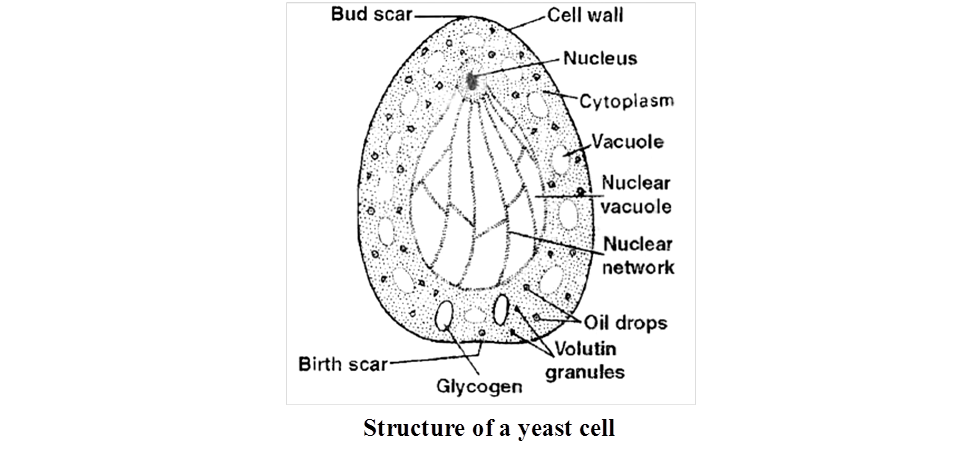
1. Vegetative Reproduction : Yeast reproduces vegetatively either by Transverse wall fission or by budding. Depending on this character, they are grouped as fission yeast (Schizosaccharomyces) and budding yeast (Saccharomyces).
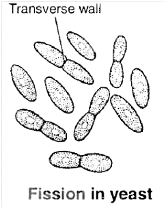
(a) By fission:
During reproduction by fission the parent cell elongates, the nucleus divides into two daughter nuclei and gradually a transverse partition wall is laid down somewhat near the middle, starting from periphery to the centre dividing the mother cell into daughter cells.
The two daughter cells so formed, may remain together for sometime and begin to divide again or they may separate soon and then divide.
(b) By budding:
Budding yeasts are rather common than the fission yeast. At the commencement of budding a small portion of the cell wall, usually near the end, softens.
The protoplast of the mother cell covered by a thin membrane bulges out in the form of a bud which ultimately develops into a daughter cell.
Meanwhile, the nucleus of the mother cell divides mitotically, (according to some, the division is amitotic).
One of the two daughter nuclei migrates into the enlarging bud.
The bud grows until it attains the size of the mother cell.
The daughter cell then becomes separated from the mother cell and the process may be repeated indefinitely.
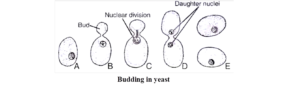
Under conditions of rapid growth, the daughter cell also starts producing buds before being detached from the mother cell and the process may be repeated several times, giving rise to chains or groups of yeast cells.
This results in the formation of branched or unbranched pseudomycelium. The cells in chains of pseudomycelium are loosely joined together.
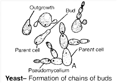
Sooner or 'A later, however, the chains break into their constituent cells.
2. Sexual Reproduction:
It takes place by the union of two cells (more often similar in size).
The copulating pair of cells may be vegetative cells or ascospores.
Yeasts may be homothallic or heterothallic and stages of fusion are extremely variable.
Three life cycle patterns are distinguishable among yeasts. These are :
(a) Haplontic life cycle: This is exhibited by Schizosaccharomyces octosporus (fission yeast).
(b) Diplontic life cycle: This is exhibited by Saccharomycoides ludwigii (Helobial yeast).
(c) Diplohaplontic life cycle: This is exhibited by Saccharomyces cerevisiae (Budding yeast).
Concept Builder
In addition to the above given methods of sexual reproduction of yeast, the following methods are also found:
- (a) Adelphogamy: Copulation between two adjoining sister cells. This is isogamous and the cells which fuse, do not separate after fission but remain united to form short chains.
- (b) Pedogamy: It is copulation between mother and the daughter cell formed by budding. The daughter remains attached to the mother and the nucleus of the bud migrates into the mother.
Saccharomyces cerevisiae = Brewer's Beer yeast or Baker's yeast
S.ellipsoidens = Wine yeast
Torulopsis utilis and Endomyces vermalis are rich in proteins. Rhodotorula is rich in vitamin A and Ashbya gossypii is rich in vitamin B2.
Yeasts are used in curing cocoa beans.
Diseases caused by yeast
(a) Candidiasis/moniliasis – Candida albicans
(b) Blastomycosis – Blastomyces dermatidis
(c) Histoplasmosis – Histoplasma capsulatus
(d) Cryptococcosis – Cryptococcus neoformans
Some yeasts reduce the yield of silk industry by attacking silkworms.
Species of Nematospora attack cotton, tomato and beans.
(B) Penicillium
Important characters
Facultative parasite and saprophytic fungi.
Mycelium is branched septate, with simple septal pore and each cell is uni or multinucleate depending upon the species.
Asexual reproduction occurs by conidia.
Conidiophores are often branched.
The first branch level is called rami and second or ultimate branches are called metulae having bottle shaped sterigmata.
Each sterigmata produces a chain of conidia.
The conidia in chain are arranged in basipetal order.
Each conidium is uninucleate, non motile, two layered, dispersed by air and germinates to form new mycelium.
Sexual reproduction: It exhibits dikaryophase and produces ascocarp.
The ascocarp is cleistothecium type.
Each ascus has 8 ascospores.
Ascospore germinates to form new mycelium.
IV. Basidiomycetes: The Club Fungi
They are the most advanced and most commonly seen fungi. Their fructifications are often large and conspicuous, e.g., mushrooms, toadstools, puff balls, bracket fungi etc.
Basidiomycetes are among the best decomposers of wood. They are able to decompose both cellulose and lignin. Lignin is not metabolised by most other fungi and even bacteria. Ganoderma species causes decay of wood even on standing trees.
Motile structures or cells are absent.
Mycelia are of two types, primary and secondary. Primary mycelium contains monokaryotic cells and is short lived.
Monokaryotic phase or primary mycelium may multiply by oidia, conidia-like spores and pycniospores.
Secondary mycelium is long lived and dominant phase of life cycle. It is represented as dikaryophase. It consists of profusely branched septate hyphae.
Septa possess dolipores or central pores with barrel-shaped outgrowths (except rusts and smuts).
Handle like outgrowths are found on the sides of septa. They are called clamp connections. Clamp connections are meant for proper distribution of dikaryons at the time of cell division.
Secondary mycelium can perennate in the soil or wood by means of sclerotia or rhizomorphs.
Dikaryophase or secondary myceium may multiply by different types of spores chlamydo-spores, aecidiospores, uredospores, teleutospores etc.
There is often differentiation of two mating types (+) and (-) in thallus.
Sexual reproduction does not involve sex organs. Instead fusion occurs between basidiospores and other monokaryotic spores, between a spore or spermatium and a hypha or between two hyphal cells of primary mycelia.
Karyogamy and meiosis occur in club-shaped structures known as basidia (singular -basidium). The name of the class is based after them.
A basidium commonly produces four meiospores or basidiospores exogenously at the tips of fine outgrowths called sterigmata or directly on the basidium.
The fungi may or may not produce fructifications called basidiocarps. The basidiocarps vary from microscopic forms to large macroscopic structures. Some puff balls and brackets can be over 50 cm in diameter.
Concept Builder
(i) Smuts : They produce thick-walled, black-coloured resting spores called smut spores. Smuts are of two types, covered and loose. In covered smut, the spore mass remains within the membranous covering of sorus, e.g., Ustilago hordei (covered smut of barley), Ustilago maydis (smut of corn). In loose smut the spores are exposed while attached to the host, e.g., Ustilago nudatritici (loose smut of wheat), U. avenae (loose smut of oat).
(ii) Mushrooms : They are edible and non edible Agaricales which possess umbrella like basidiocarp. Common examples of edible mushrooms are Agaricus campestris, A. bisporous, Volvariella volvacea (Paddy straw mushroom), Pleurotus ostreatus etc.
(iii) Toadstools: Toadstools are poisonous mushrooms which generally have white spores. Amanita caesarea (Caeser's mushroom) was used in poisoning Roman emperor Caesar. The other toadstools are-Amanita phalloides (Death cup), A. muscaria (Fly agaric) and Gynomitra esculenta (heat labile carcinogenic toxin).
(iv) Rusts : They are characterised by the formation of rusty pustules containing the spores.
(a) Puccinia graminis tritici -Black rust of wheat.
(b) Puccinia glumarum or P striiformis -Yellow rust of wheat.
(c) P. recondita -Brown rust of wheat
(v) Hallucinogens: Psilocybe mexicana (Sacred mushroom) has hallucinating properties similar to LSD. It is used by Mexican Indians during certain religious ceremonies.
(vi) Armillaria (largest fungi): A. mellea (Honey mushroom) is a serious root parasite of both hardwoods and conifers. The fungus develop rhizomorphs into the phloem of the host and hence, blocks the food supply.
(vii) Puffballs: The basidiocarp is a stalked rounded structure which, upon ripening, releases out puffs of spores. The fructification may grow above or below the substratum, e.g., Lycoperdon oblongisporum, L. giganteurn.
(viii) Bracket fungi (Shelf fungi) : They are basidiomycetes whose basidiocarps or fructifications appear on tree trunks, logs, lumber etc. just as brackets or shelves, e.g., Fomes applanatus, Polyporus sulphureus, Ganoderma.
(ix) Predator fungi : e.g., Dactylaria, Arthrobotrys.
(x) Stinkhorn. Phallus impudicus (Dead man's finger). Spore mass produces a stinking odour to attract flies.
Life History of a Mushroom
Agaricus campestris is the common field mushroom which has edible basidiocarp. The fungus is saprotrophic.
The vegetative or assimilative part of mycelium is subterranean. It is found in moist humus rich soil of open fields, grassland, piles of straw or within rotting togs.
The mycelium multiplies by fragmentation; occasionally by oidia and chlamydospores.
Life cycle of mushroom contains two types of mycelia, primary and secondary. Sex organs do not differentiate.
Primary mycelium is short lived. It consists of septate hyphae having monokaryotic cells. The mycelia are heterothallic.
The hyphae of two mating types come in contact and show somatogamy. However, only plasmogamy occurs at this time.
It gives rise to a dikaryotic cell that grows, divides and produces a long-lived and extensive dikaryotic or secondary mycelium.
The hyphae of secondary mycelium show clamp connections and dolipore septa. Its celis possess two haploid nuclei instead of single diploid nucleus.
Under favourable conditions, hyphae of secondary mycelium collect at places and give rise to rounded or pyriform compact masses of hyphae called buttons.
The buttons enlarge and produce aerial fructifications or basidiocarps.
The latter are popularly termed as mushrooms. In contrast the secondary mycelium, from which mushrooms develop, is known as spawn.
The basidiocarps or mushrooms often lie in rings. The latter are called as fairy rings, its diameter increases centrifugally every year.
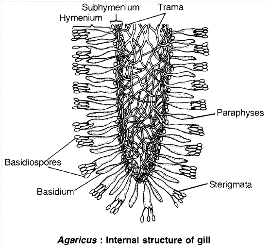
Each basidiocarp or mushroom is cream to pinkish brown in colour and consists of two parts, stipe and pileus.
The stipe or stalk is fleshy. It is slightly swollen at the base.
Pileus is umbrella-like cap of the mushroom. In the button stage, the pileus is connected to stipe by membrane called veil or velum.
It ruptures during growth of pileus. However, its remains can be seen on the upper part of stipe as annulus.
The pileus is circular in outline. Its upper surface is more or less convex. The under surface is flat or concave.
It bears 300-600 radiating rows of vertical plates named gills (lamellae).
The two sides of vertically placed gills are lined by thousands of club-shaped basidia alongwith sterile paraphyses.
The two, together constitute the fertile layer or hymenium of the gill. Hymenium is subtended by compact subhymenium.
The centre consists of interwoven hyphae called trama. Each basidium functions as the site for both karyogamy and meiosis. The two nuclei fuse to form a short-lived diploid synkaryon.
The latter, then divides meiotically giving rise to four haploid nuclei, two of (+) strain and two of (-) strain. The free end of the basidium now develops four peg-like outgrowths called sterigmata.
Each sterigmata bears an ovoid pinkish-purple meiospore termed as basidiospore. A droplet appears at the tip of sterigmata which creates tension and hanging basidiospores are carried away by air currents.
The basidiospores are liberated successively for several days. After falling on a suitable substratum, each basidiospore germinates to produce monokaryotic primary mycelium.
Life Cycle of Puccinia graminis tritici
P. graminis tritici is a macrocyclic (producing many dikaryotic spores), Heteroecious (requires more than one host i.e., primary host -wheat, secondary or alternate host -barberry).
It produces uredospores and teliospores on the wheat plant, basidiospores in soil and pycniospores and aeciospores on barberry.
It causes black or stem rust of wheat. Hence, we describe the various stages of life cycle on the basis of the host.
A. Stage of life cycle on wheat (primary host)
(i) Uredia and uredospores :
The dikaryotic aeciospores germinate on the leaves of wheat on both the surfaces and may also germinate on the stem.
They form the dikaryotic mycelium.
The germ tube protrudes out which swells up to form an elongated appressorium near the stomata.
A peg like outgrowth now arises from the appressorium and penetrates the stomata. It ramifies repeatedly to form a mass of mycelium this mycelium forms the uredospore.
As a result, some pressure is exerted on the epidermis which bursts exposing the uredospore. These clusters of uredospores have been variously referred to as uredosori or urediopustules.
A uredospore is reddish-brown, unicelled, oval or globose, stalked, dikaryotic spore. Its wall is three layered, the outer being somewhat spiny.
The uredial stage multiplies through the uredospores which germinate on fresh wheat plants (due to the red colour of spores this stage is called red rust stage).
(ii) Telia and teliospores (black rust stage) :
Towards the end of the season, the dikaryotic mycelium of the uredosorus begins to produce teliospores (or teleutospores) in the same sorus.
They are dark brown or black, stalked, thick walled, bi-celled spores. The upper cell is pointed, both the cells are binucleate.
The pustules containing teliospores are called as tella or teleutosori.
The teliospores also exert pressure on the epidermis which bursts open, exposing the spores.
It is at this stage the symptoms develop in stem also, so the disease is named black stem rust of wheat.
Karyogamy occurs inside each cell of teliospore and as a result they become diploid.
The teliospores cannot infect fresh wheat plants. They germinate in soil to form the basidiospores.
(iii) Basidia and basidiospores:
The two cells of the teliospores now act as hypobasidium. They germinate in soil and form a tube called promycelium.
The diploid nucleus migrates into epibasidium and then undergoes meiosis to form four haploid nuclei each of them develops as a basidiospore on the sterigmata.
Of these, two basidiospores belong to +ve strain and two to -ve strain.
These spores are not capable to infect a wheat plant. Each spore is unicelled, monokaryotic and unstalked.
These can infect the barberry plant (Berberis vulgaris) which is the secondary or alternate host occurring on the hills in India.
B. Stage of life cycle on barberry
(i) Spermatia or pycniospores
The basidiospores of both the strains (+ and -) can germinate on upper surface of barberry leaf.
They produce haplophase or primary mycelium of the respective strains which grows through the intercellular spaces.
Soon the mycelium forms a palisade like mat which organises like a flask shaped structure near the upper epidermis called as spermogonia or pycnidium.
They open by a single pore called ostiole.
The spermogonium is lined by palisade Ilike uninucleate cells called spermatiophores.
From the tips of spermatiophores are pinched off a large number of uninucleate cells called pycniospores or spermatia.
Besides, some sterile hyphae also arises which grow out through the ostiole.
They are called as periphysis.
In addition to these, some thin walled hyphae are also given out which become more elongated.
They are called as flexuous hyphae or receptive hyphae.
While the spermatia function as the male cells, the flexuous hyphae behave as the female hypha.
The pycniospores or spermatia protrude out of the ostiole in a nectar.
The insects feeding on this nectar transfer the spermatia from one spermogonium to the other.
Spermatization is brought about when spermatia of one strain come in contact with the trichogyne of flexuous hyphae of the other strain.
The nucleus of pycniospore or spermatium passes into the flexuous hyphae, thus bringing
(ii) Aecia and aeciospore :
An aecium or aeciosorus is produced on the lower surface of barberry leaves. It arises just beneath pycnium. The dikaryotized mycelium aggregates sub-epidermally.
This gives rise to a group of elongated dikaryotized cells which function as aecial mother cell. The mother cells differentiate a row of binucleate cells.
The alternate binucleate cells enlarge and they are identified as aeciospores. The alternate cell remaining small is disjunctor or sterile cell.
The sterile wall of the aecial cup called peridium or pseudoperianth presses the lower epidermis which eventually bursts open. Thus, the aeciospores are set free.
The aeciospores are polyhedral or ovate, binucleate, unicelled and double layered. The outer thick wall is called exine and the inner as intine.
They are set free in spring. They cannot infect the barberry bushes. These are carried from hills to the plains where they infect wheat plants. They germinate on the leaf surface from a germ tube which enters the host through stomata.
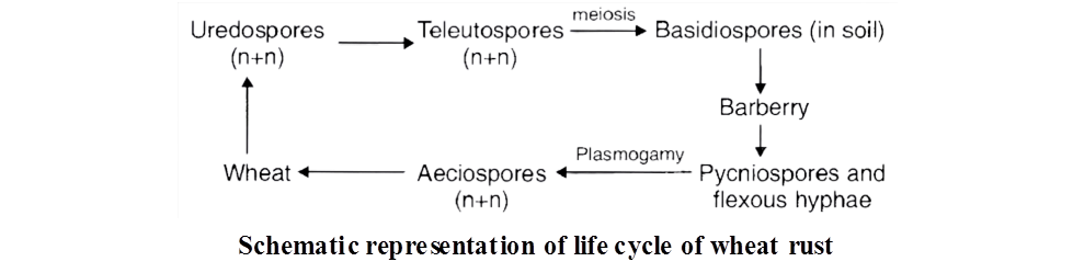
Annual Recurrence of Rust in India:
It was first studied by Prof. K.C. Mehta.
He concluded that, in India, the annual recurrence of rust on wheat takes place through uredospores.
The alternate host, Berberis vulgaris plays no role in our country. According to Mehta, if no wheat is grown on hills, the intensity of its infection in plains can be reduced.
He also suggested the involvement of certain collateral hosts like Briza minor, Bromus and Thalictrum for multiplication of uredospores during non-growing season of wheat.
V. Deuteromycetes: (The Fungi Imperfecti)
Deuteromycetes is an artificial class (form class) of fungi which has been created to include all those fungi in which sexual stage (or perfect stage) is not known (absent or not reported) .
The mycelium is septate and branched. Coenocytic forms are not known.
Asexual reproduction often occurs by conidia.
Once perfect (sexual) stages of members of Deuteromycetes were discovered they were often moved to Ascomycetes and Basidiomycetes but most members of Deuteromycetes are actually related to ascomycetes.
Some members are saprophytes or parasites while a large number of them are decomposers of litter and help in mineral cycling.
Concept Builder
(i) Leaf spot of rice : Helminthosporium oryzae causes leaf spot disease of rice commonly called brown leaf spot of rice. It caused Bengal famine in 1942-43.
(ii) Early blight: Alternaria solani causes early blight of potato. The leaves develop small oval brown spots with concentric rings (target board symptom).
(iii) Tikka disease : Circular necrotic dark brown or blackish leaf spots develop in groundnut due to Cercospora personata.
(iv) Red rot: Colletotrichum falcatum produces red rot of sugarcane.
(v) Wilts : Many economically important plants (e.g., cotton, pigeon pea) show sudden signs of wilting due to blockage of tracheary elements by growth of fungus, Fusarium especially F. oxysporum, F. udum.
(vi) Ringworm of foot/Athlete's foot is caused by Trichophyton interdigitate.
Common names of some fungi:
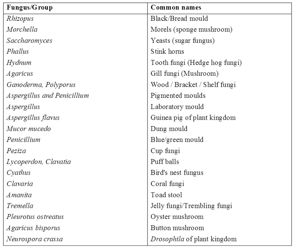
Concept Builder
Common Fungicides and their composition
- Bordeaux mixture – (CuSO4: Ca(OH)2 : H2O). First fungicide discovered by RMA Millardet Commonly known as holy water of plant pathology
- Burgandy mixture – Mixture of CuSO4 + Na2CO3 + H2O was discovered by Mass.
- Chestnut mixture – Ammonium carbonate + copper sulphate.
MYCORRHIZA (FUNGAL ROOTS)
The mutually beneficial or symbiotic association of a fungus with the roots of higher plants is termed mycorrhiza. Mycorrhizal roots differ in shape from normal roots and often show a wooly covering. These roots lack root cap and root hairs.
A fungus may get associated with roots of a number of plants and a particular plant may form association with a number of fungi. Depending upon the location of the fungus, the mycorrhiza is of two types, i.e., ectomycorrhiza and endomycorrhiza.
In ectomycorrhiza, the fungal hyphae are mainly external, forming a wooly covering on external surface of root and forms network of mycelium (Hartig net) in the intercellular spaces of the cortex. Fungal partner is commonly basidiomycetes, e.g., Pinus roots
In endomycorrhiza, the fungal hyphae enter the tissue of the root, spreading intercellularlly and intracellularly.
The fungus is able to break the cell wall in a limited way and is restricted to cortical region of the root. Some hyphae send small projections into cortical cells without destroying them.
Such fungi are termed VAM (Vesicular Arbuscular Mycorrhiza), e.g., Orchid roots.
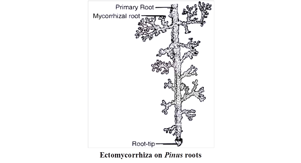
Mycorrhizal association is a symbiotic relationship as both the partners are mutually beneficial to each other. The fungal partner obtains nourishment from the cortical cells of the root and depends upon the plant for shelter.
The root cells excrete sugars and other soluble gradients which are used by fungal hyphae spreading in intercellular spaces. The hyphae may get nourishment from the cells directly and also by sending small projections into cortical cells.
The fungus seems to be essential for the growth of the plant having mycorrhiza.
The plant also gets benefit from the association as the fungal hyphae spreading in soil substantially increases the surface area of absorption, thereby enabling the plant to get enhanced supply of water, nitrogen, phosphorus and other minerals from the soil.
Orchids seldom occur without mycorrhiza. Certain forest trees like pines, birches show stunted growth if their roots are not associated with fungus.
LICHENS
Lichens are dual (composite) organisms or entities which contain a permanent association of a fungal partner or mycobiont and an algal partner or phycobiont.
Mycobiont is dominant partner and mostly belongs to ascomycetes (Ascolichens -, e.g., Graphis, CIadonia, Parmelia, Usnea, etc.) or sometimes basidiomycetes (Basidiolichens - e.g., Corella, Cora, etc.).
Phycobiont is mostly a member of Chlorophyceae (e.g., Chlorella, Trebouxia, Protococcus, Palmella, etc.) or can be a BGA (e.g., Nostoc, Chlorococcus, Scytonema, etc.).
The term lichen was coined by Theophrastus (370 -285 B.C.), also called Father of Botany.
Lichens often grow in most inhospitable and uninhabited places like barren rocks (saxicolous), soil (terricolous), icy tundra or alpines, sand dunes, roofs, walls, wood (Iignicolous), tree bark (corticolous), leaves, etc.
They commonly live under humid and exposed conditions but can tolerate extreme desiccation. However, lichens, cannot tolerate air pollution, especially due to sulphur dioxide (so are considered indicators of SO2 pollution).
Lichens are perennial. Their growth is slow. Lichens have greyish, yellowish, greenish, orange, dark brown or blackish colouration.
Structure
Based upon external morphology, the lichens are of three types :
(i) Crustose. Crust like, closely appressed to the substratum and attached to it at several places, e.g., Graphis, Lecanora, Rhizocarpon.
(ii) Foliose: The body of the lichen is flat, broad, lobed and leaf-like, which is attached to the substratum at one or a few places with the help of rhizoid like structures called rhizines, e.g., Parmelia, Peltigera.
(iii) Fruticose : The lichen is branched like a bush and attached to the substratum by means of a disc, e.g., CIadonia, Usnea, Evernia, Bryonia.
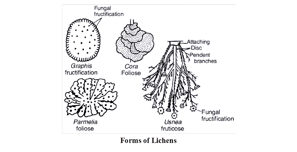
The bulk of lichen body is formed by fungal partner called mycobiont. It includes the surface, medulla (or interior) and rhizines (attaching devices).
The algal partner or phycobiont constitutes hardly 5% of the lichens body.
It is generally restricted to a narrow zone (algal zone) below the surface.
Relationship :
The fungus performs following functions:
Body structure and covering (ii) Anchoring (iii) Absorption of water and minerals. It can absorb water from wet air (atmosphere), dew and rain. Minerals are picked up both from substratum and atmosphere. Special chemicals are excreted by the fungal partner of the lichen to dissolve minerals from the substratum. (iv) Sex organs and fruitifications are of fungal origin.
The major function of alga is photosynthesis.
The cyanobacterial alga additionally takes part in nitrogen fixation. The alga picks up water and mineral salts from the fungus while the fungus obtains part of the food manufactured by the alga.
Therefore, in a lichen the association between alga and fungus is that of mutual benefit (mutualism) popularly called symbiosis.
However, at times the fungus is found to (i) send haustoria into algal cells (ii) induce alga to secrete organic substances and (iii) prevent alga to develop pectic covering. Therefore, some workers believe that the fungus is a controlled parasite over the alga. The phenomenon is called helotism.
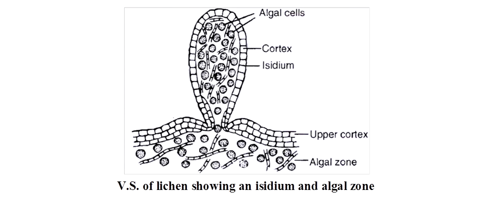
Reproduction: Lichens multiply by four methods:
(i) Progressive death and decay resulting in the separation of a lichen thallus into two or more parts.
(ii) Fragmentation caused by mechanical injury, due to wind or animal bites.
(iii) Isidia are superficial outgrowths of the lichens which are primarily meant for increasing surface area and photosynthetic activity. At time, they are broken off. Each isidium is capable of forming a new lichen because it has a core of a few algal cells surrounded by a sheath of fungal hyphae.
(iv) Soredia. These are most efficient means of asexual reproduction. They are microscopic lichen propagules which are produced in large numbers inside sori called pustules. Soredia are dispersed by air currents. After falling on a suitable substratum each soredium gives rise to a lichen, because it has a few algal cells surrounded incompletely by a weft of fungus.
Concept Builder
- Special structures in the thallus of lichen:
(i) Cyphellae help in exchange of gases, present in lower cortex.
(ii) Cephalodia help to retain moisture and its algal partner fix nitrogen also.
(iii) Breathing pores for aeration, present in upper cortex of thallus.
Early Colonisers : Lichens are early or pioneer colonisers of barren rocks, cliffs, mountains and new terrains. During their growth, lichens stick to the rocks and cliffs by secreting acids. It produces minute crevices where organic matter accumulates. It paves the way for growth of mosses.
Food: In tundra, Cladonia rangifera (Reindeer Moss) constitutes the staple food of reindeer, caribou, musk ox, etc. Cetraria islandica (Iceland Moss) is used as a food article in Iceland, Sweden and Norway Lecanora esculenta is regarded as bread of heaven by Jews. Parmelia (Rock Flower) is also a table delicacy Dermatocarpon miniatum (Stone Mushroom) is a vegetable in Japan.
Dyes : Orchil is obtained from Rocella tinctoria. The latter was also the source of litmus (R.montagnei) before the advent of synthetic products. Litmus is a pH indicator.
Perfumes: Scented incense is got from species of Ramalina and Evernia.
Medicines: Usnic acid got from Usnea (Old Man's Beard) and Cladonia has antibiotic properties. It is used in preparation of ointment for burns and wounds.
Air Pollution : Decrease in lichen population of an area is indicative of SO2 pollution.
Fires: In hot season, Usnea may produce forest fires.
VIRUS
Term virus was coined by Pasteur.
Viruses are obligate intracellular parasites. They are intermediate between living and non living entities.
Non living nature of virus
Lacking protoplast.
Ability to get crystallized, e.g., TMV, poliomyelitis virus.
Inability to live independent of a living cell. (Lack functional autonomy)
High specific gravity which is found only in non living objects
Absence of respiration.
Absence of energy storing system.
Absence of growth and division.
Living nature of virus
Being formed of organic macromolecules.
Presence of genetic material.
Ability to multiply.
Occurrence of mutations.
Occurrence of certain enzymes like, neuraminidase (first discovered), transcriptase and lysozyme in certain viruses.
Infectivity and host specificity.
Viruses can be 'killed' by autoclaving and ultraviolet rays.
They take over biosynthetic machinery of the host cell and produce chemicals required for their multiplication.
Viruses are responsible for a number of infectious disease like common cold, epidemic influenza, chicken pox, mumps, poliomyelitis, rabies, herpes, AIDS, SARS etc.
Concept Builder
Mayer described Tobacco Mosaic disease in 1886.
Iwanowsky is credited with the discovery of virus in 1892. TMV was the first virus to be discovered.
Beijerinck called virus as "Contagium vivum fluidum " (living infectious fluid).
In 1935, Stanley crystallised TMV.
Twort and d'Herelle discovered bacteriophage.
Lwoff and Wollmann discovered temperate viruses.
Shafferman and Morris discovered cyanophage, e.g., LPP-1.
Bawden and Pirie studied the chemical nature (nucleoproteins) of TMV.
Sinsheimer discovered single stranded DNA in bacteriophage f × 174
Issac and Lindemann discovered interferon
Delbruck (1938), found viruses to undergo mutations.
Reverse transcription in Retroviruses was discovered by Temin and Baltimore, so the phenomenon is called teminism. The enzyme reverse transcriptase is RNA dependent DNA polymerase.
Structural Components of Viruses
(i) Envelope is the outer thin loose covering composed of proteins (from virus), lipids and carbohydrates (both from host). It has smaller subunits known as peplomers, e.g., Herpes virus, HIV, Vaccinia virus etc. If it is not present the virus is said to be naked.
(ii) Capsid : It is the outer protein coat made up of subunits called capsomeres, their number is virus specific. These possess antigenic properties.
(iii) Nucleoid : Viruses contain either DNA or RNA. No virus contains both DNA and RNA.
(a) DNA containing viruses are called deoxyviruses.
These are of two types:
(i) Double stranded DNA (dsDNA) virus, e.g., Pox virus, Cauliflower mosaic virus.
(ii) Single stranded DNA (ssDNA) virus, e.g., Coliphage ×174, M 13 phage.
(b) RNA containing viruses or riboviruses are of two types.
(i) Double stranded RNA (ds RNA) virus, e.g., Reo virus, Wound Tumour Virus.
(ii) Single stranded RNA (ss RNA) virus, e.g., TMV, Influenza virus, Foot and Mouth disease virus, Retroviruses (HIV).
Classification of Virus
Holmes (1948) has divided viruses into three groups on the basis of specific hosts.
(a) Phytophagineae are plant viruses. They generally have ssRNA, e.g., TMV, Potato Mosaic Virus, Yellow Vein Mosaic virus, Cauliflower Mosaic virus.
(b) Zoophagineae are animal viruses. They commonly have ssRNA or dsRNA or dsDNA. e.g., Poliomyelitis virus, Influenza viruses, Small pox virus, Mumps virus, Rabies virus.
(c) Phagineae attack lower organisms
(i) Bacteriophages are bacterial viruses and they usually possess dsDNA, e.g., T2, T4, lambda ![]() phage.
phage.
(ii) Coliphages are viruses of E.coli, e.g., Coliphage fd.
(iii) Cyanophages attack blue green algae, e.g., LPP-1, SM-1 .
(iv) Phycophages attack algae.
(v) Mycophages attack fungi.
(vi) Zymophages attack yeast.
Reproduction
It is of two main types: Phagic and Pinocytic
(a) Phagic Reproduction : It is further of two types :
(i) Lytic cycle : Occurs in virulent phages, e.g., T4 bacteriophages.
(ii) Lysogenic cycle : Occurs in temperate viruses such as phage.
(b) Pinocytic Reproduction: It is found in viruses like TMV, HIV, Hepatitis B etc., in which whole of virus particle enters host cell except envelope (if present).
Structural details of some viruses
(1) Tobacco Mosaic Virus (TMV) is elongated rod like, 3000 Å long, 180 Å in diameter with molecular weight Protein 39.4 × 10⁶ dalton. 2130 capsomeres are arranged helically to form the capsid. RNA strand is helical. ssRNA consists of 6400 nucleotides. Thus, the ratio of nucleotides : capsomeres = 3 : 1
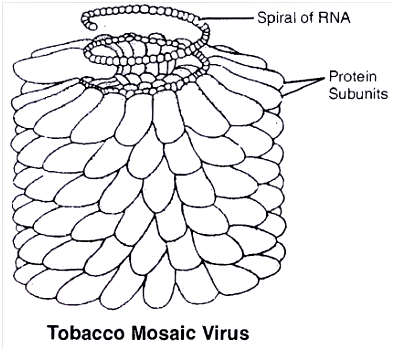
(2) Pox virus / variola is the causal agent of small pox. These are among the largest of animal viruses, are rectangular (brick shaped), 300 × 230 nm in size. Genome is dumbell shaped with central core of dsDNA. The core has two enzymes RNA polymerase and ATP phosphohydrolase.
(3) AIDS virus consists of single stranded RNA. It has 2 copies of ssRNA. Outer cover has 5 layers, i.e., outer most glycoprotein, followed by double lipid layer and the innermost has two protein layers.
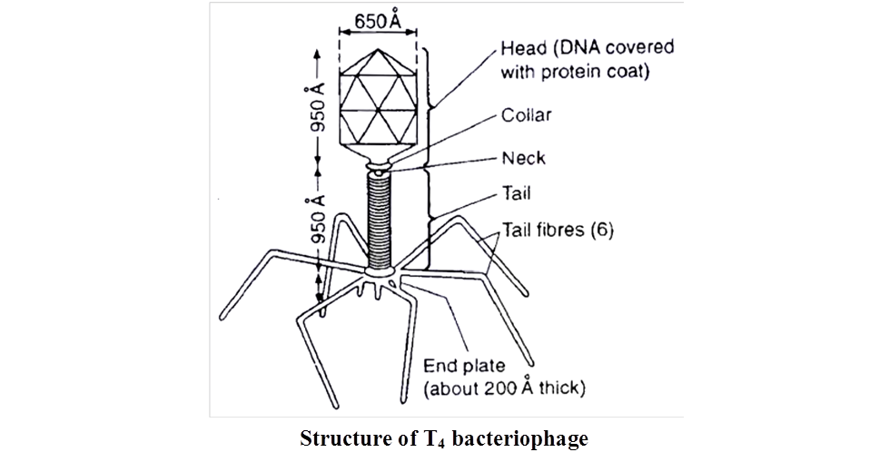
T4 Bacteriophage has a tadpole like structure with polyhedral head connected to a helical tail (binal). The head consists of nucleic acid surrounded by a protein coat or capsid. Nucleic acid is double stranded DNA. Tail is proteinaceous tube-like, core surrounded by sheath. At one end, tube is joined to the head by thin collar. At the other end, it has a hexagonal base plate with six small tail pins and six tail fibres which help in attachment of the phage to the host cell.
- Sub Viral Agents: These are viruses which lack one of the essential component, e.g., viroids, virusoids, prions
- Viroids (L. virus -poison, eidos -diminutive)
- They are the smallest self replicating particles which were discovered by Diener (1971). Viroids are infectious RNA particles which are devoid of protein coat.
- They are obligate parasites. Molecular weight of a viroid is low.
- The RNA is tightly folded to form circular or linear structures. Viroids are known to cause diseases (some 20) in plants only, e.g., Potato spindle tuber disease (PSTD), Chrysanthemum stunt and Citrus exocortis.
(2) Virusoids
- Discovered by Randle et. al., these are RNA viruses but inside the capsid of other larger virus. They replicate within the host and do not cause any infection.
(3) Prions
Discovered by Alper et al.
Proteinaceous infectious particles, causing certain diseases like
(i) Kuru disease (laughing death disease in humans)
(ii) Bovine spongiform encephalopathy (BSE or Mad cow disease)
(iii) Scrapie disease in sheep
(iv) Creutz Feldt Jakob disease
Table 5 : Viral diseases of Plants
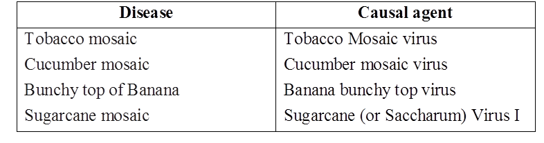
Table 6 : Viral diseases of Man
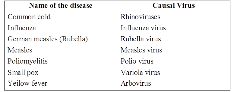
Animal viral diseases -Foot and mouth disease, Rinderpest, Ranikhet, Bird flu etc.
Concept Builder
Nomenclature of viruses :
International committee of virus nomenclature has given a system of naming the virus. The system consists of two parts. First part is common name of the virus and second part has the coded information about the virus. This is called as Cryptogram.
Kingdom: Fungi
Kingdom Fungi
Fungi are eukaryotic organisms that include yeasts, molds, mushrooms as well as other microbes.Fungi are a distinct kingdom of heterotrophic creatures. They have a wide range of forms and environments. Fungi can often be observed on stale bread and rotten fruits. Toadstools and the common mushroom that we eat are both fungi. A parasitic fungus causes white patches on mustard leaves. Some unicellular fungi, such as yeast, are utilized in the production of bread and beer. Other fungi produce illnesses in plants and animals; Puccinia, which causes wheat rust, is a good example. Penicillium, for example, is a source of antibiotics. Fungi are widely distributed and can be found in the air, water, soil, and on animals and plants. They thrive in warm, humid environments.Mycology is the branch of biology concerned with the study of fungi.
Fungi are filamentous, with the exception of yeasts, which are unicellular. Their bodies are made up of hyphae, which are long, slender thread-like structures. Mycelium refers to the network of hyphae. Coenocytic hyphae are hyphae that are continuous tubes filled with multinucleated cytoplasm. In the hyphae of others, there are septae or cross walls. Fungi's cell walls are made up of chitin and carbohydrates.
Saprophytes are fungi that are heterotrophic and absorb soluble organic materials from dead substrates. Parasites are organisms that rely on live plants and animals for their survival. They can also live as symbionts, forming lichens with algae and mycorrhiza with the roots of higher plants.
Fungi reproduce via vegetative processes such as fragmentation, fission, and budding. Spores known as conidia, sporangiospores, and zoospores are instrumental in asexual reproduction, while oospores, ascospores, and basidiospores are responsible for sexual reproduction. Fruiting bodies are structures that produce different types of spores.
The three phases of the sexual cycle are as follows:
(i) Plasmogamy, which is the fusion of protoplasms between two motile or non-motile gametes.
(ii) Karyogamy, which is the fusion of two nuclei.
(iii) In a zygote, meiosis results in haploid spores.
Two haploid hyphae of compatible mating types come together and merge when a fungus reproduces sexually. In some fungi, the fusing of two haploid cells results in the formation of diploid cells right away (2n).
Kingdom fungi can be divided into three groups based on their nutritional needs.
(i)Saprophytic fungus get their sustenance from dead organic matter. Rhizopus, Penicillium, and Aspergillus are other examples.
(ii)Parasitic fungus get their food by living on other living organisms (plants or animals) and absorbing nutrients from them. Taphrina and Puccinia are two examples.
(iii)Symbiotic fungi are those that exist in an interdependent relationship with other species in which both parties benefit. Lichens and mycorrhizae are two examples. Lichens are the result of a symbiotic relationship between algae and fungi. Algae and fungi benefit from each other in this situation because fungi give protection to algae and algae provide carbohydrates for fungi.
The morphology of the mycelium, mode of sporeformation, and fruiting bodies form the basis for thedivision of the kingdom into various classes which are discussed below.
(a) Phycomycetes:
Phycomycetes can be found in aquatic areas and on decaying wood in moist and humid environments, as well as obligatory parasites on plants. Algal fungi are sometimes known as Phycomycetes. They have aseptate coenocytic hyphae that are distinctive. Asexual reproduction occurs via zoospores (motile) or aplanospores (asexual) (non-motile). In the sporangium, these spores are produced endogenously. A zygospore is created when two gametes fuse together. The morphology of these gametes is either similar (isogamous) or distinct (heterogamous) (anisogamous or oogamous). Mucor, Rhizopus (the bread mold), and Albugo(the parasitic fungi on mustard) are other typical examples.
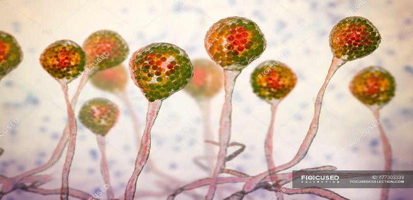
(b) Ascomycetes:
The ascomycetes, sometimes known as sac-fungi, are typically multicellular (e.g., Penicillium) or occasionally unicellular (e.g. Saccharomyces or yeast). These fungi may be Saprophytic, decomposers, parasitic, or coprophilous(growing on dung). Mycelium is septate and branched. Conidia grown exogenously on special mycelium termed conidiophores are the asexual spores. Conidia create mycelium when they germinate. Ascospores are sexual spores that are produced endogenously in sacs similar to asci (singular ascus). These asci are grouped in ascocarps, which are distinct types of fruiting structures. Aspergillus, Claviceps, and Neurospora are some examples. Neurospora is widely utilized in biochemical and genetic research. Many of the members, such as morels and truffles, are edible and regarded as delicacies.
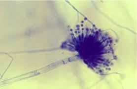
(c) Basidiomycetes:
Mushrooms, bracket fungi, and puffballs are examples of basidiomycetes. They can be found growing in soil, on logs and tree stumps, and in living things. Rusts and smuts, for example, are parasitic plant bodies. The mycelium is septate and branching. Although asexual spores are rare, vegetative reproduction by fragmentation is prevalent. Although there are no sex organs, plasmogamy is caused by the fusing of two vegetative or somatic cells of different strains or genotypes. The dikaryotic complex that results eventually gives rise to basidium. The basidium undergoes karyogamy and meiosis, resulting in four basidiospores. The basidiospores are formed exogenously on the basidium (pl.: basidia). Basidia are arranged in basidiocarps, which are fruiting bodies. Agaricus (mushroom), Ustilago (smut), and Puccinia are some of the most prevalent members.
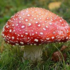
(d) Deuteromycetes:
In this category, only asexual or vegetative phases of these fungi are known, therefore they are commonly referred to as imperfect fungi. When the sexual forms of these fungi were discovered, they were placed in the appropriate groups. It's also likely that the asexual and vegetative stages were given one name (and were classified as Deuteromycetes), while the sexual stage was given another one (and placed under another class). The fungi were accurately recognized and moved out of Deuteromycetes after the links were discovered. Deuteromycetes were frequently shifted to ascomycetes and basidiomycetes after their perfect (sexual) stages were discovered. Conidia are asexual spores that Deuteromycetes use to reproduce. The mycelium is branching and septate.Some individuals are saprophytes or parasites, whereas the majority are litter decomposers who aid in mineral cycling. Alternaria, Colletotrichum, and Trichoderma are other examples.
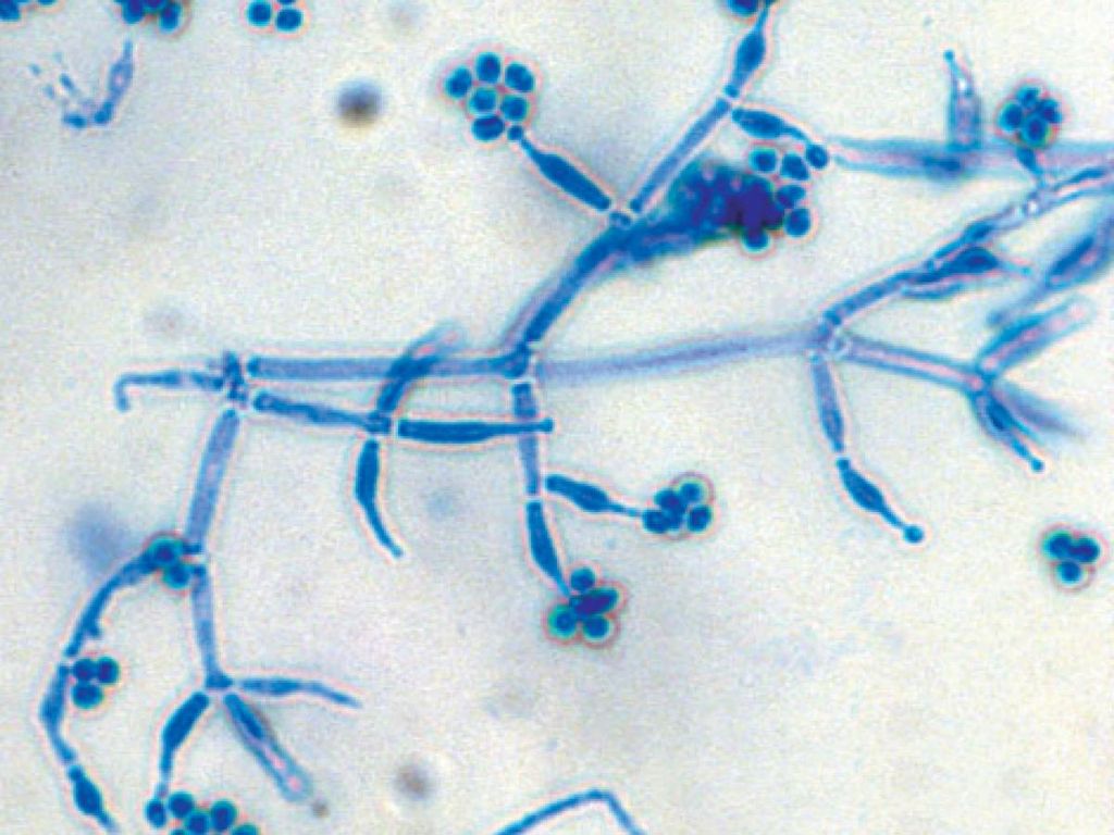
Kingdom: Plantae
- Books Name
- ACME SMART COACHING Biology Book
- Publication
- ACME SMART PUBLICATION
- Course
- CBSE Class 11
- Subject
- Biology
KINGDOM PLANTAE
Members of Plant Kingdom are multicellular, eukaryotic, photosynthetic organisms.
Main characters of the members of this kingdom are:
Cellulosic cell wall.
Non-motile, excepts some aquatic forms.
Presence of chloroplasts.
Photosynthetic mode of nutrition (oxygenic photosynthesis), e.g., different types of algae (green, brown, red algae), bryophytes, pteridophytes, gymnosperms and angiosperms.
Growth is usually indefinite.
Some of the plants are heterotrophic. They are mostly parasitic. A few are saprobes.
small group of autotrophic plants catch small animals and insects for obtaining nitrogen.They are called carnivorous or insectivorous plants.
Reproduction is both asexual and sexual. Accessory spores are present in lower plants.
An embryo stage is absent in the algal group but present in others.
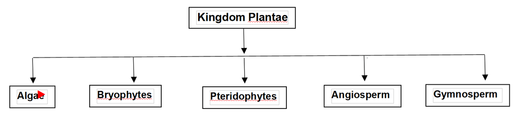
Kingdom: Plantae
Kingdom Plantae
Kingdom Plantae refers to any eukaryotic chlorophyll-containing organism that is typically referred to as a plant. Plant cells are eukaryotic in nature, with significant chloroplasts and a cellulose-based cell wall. Algae, bryophytes, Pteridophytes, gymnosperms, and angiosperms are all members of the Plantae family. A few members, such as insectivorous plants and parasites, are partially heterotrophic.
Insectivorous plants include bladderwort and Venus flytrap, while Cuscutaexists like a parasite.
Plants have two unique phases in their life cycle: diploid sporophytic and haploid gametophytic, which alternate. The lengths of the haploid and diploid phases, as well as whether they are free-living or dependent on others, varied between plant families. Alternation of generation is the term for this phenomenon.
Plants are classified according to the following criteria:
1. Plant body: Whether or not a well-differentiated plant body exists. Root, stem, and leaves, for example.
2. Vascular system: The presence or absence of a vascular system for water and other material movements. Phloem and Xylem, are two types of vascular tissues in plants.
3. The presence or absence of flowers and seeds, as well as whether the seeds are bare or wrapped in fruit, are all factors in seed production.
According to the above-mentioned criteria, the plant kingdom has been divided into five subgroups:
(A) Thallophyta:
Thallophytes have a thallus-like body structure and lack a well-differentiated body structure. Plants having a primitive and simple body structure are included. The thallus is the plant's main body, and it can be filamentous, colonial, branching, or unbranched. Algae such as green algae, red algae, and brown algae are examples. Volvox, Fucus, Spirogyra, Chara, Polysiphonia, Ulothrix, and other species are common examples.
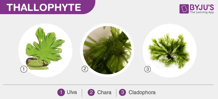
(B) Bryophytes
Bryophytes are plants that lack vascular tissues. Root-like, stem-like, and leaf-like components make up the plant's body. Bryophytes are terrestrial plants that are also known as "plant amphibians" since they require water for sexual reproduction. They thrive in wet, shaded environments. Mosses, hornworts, and liverworts are all members of the Bryophyta family. Marchantia, Funaria, Sphagnum, Antheoceros, etc. are well-known examples.
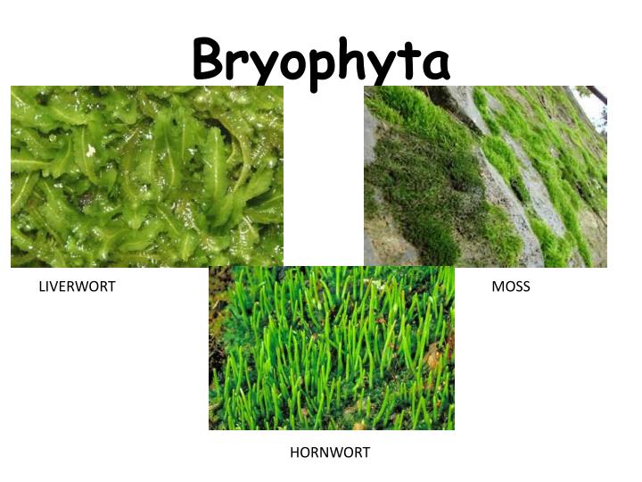
(C) Pteridophyta:
Pteridophytes have a plant body that is well-differentiated into roots, stems, and leaves. They have a circulatory system that allows water and other chemicals to be transported. Selaginella, Equisetum, Pteris, and other common examples are only a few.
Non-flowering and non-seed bearing plants. E.g. Thallophyta, Bryophyta, Pteridophyta are collectively known as Cryptogams.
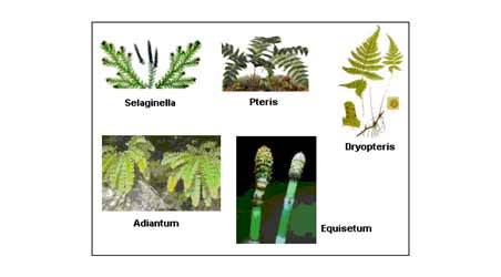
(D) Gymnosperms:
Gymnosperms have a plant body and vascular tissues that are well-differentiated. They produce naked seeds, which are seeds that are not encased in a fruit. Gymnosperms include plants such as Cycas, Pinus, Ephedra, and others.
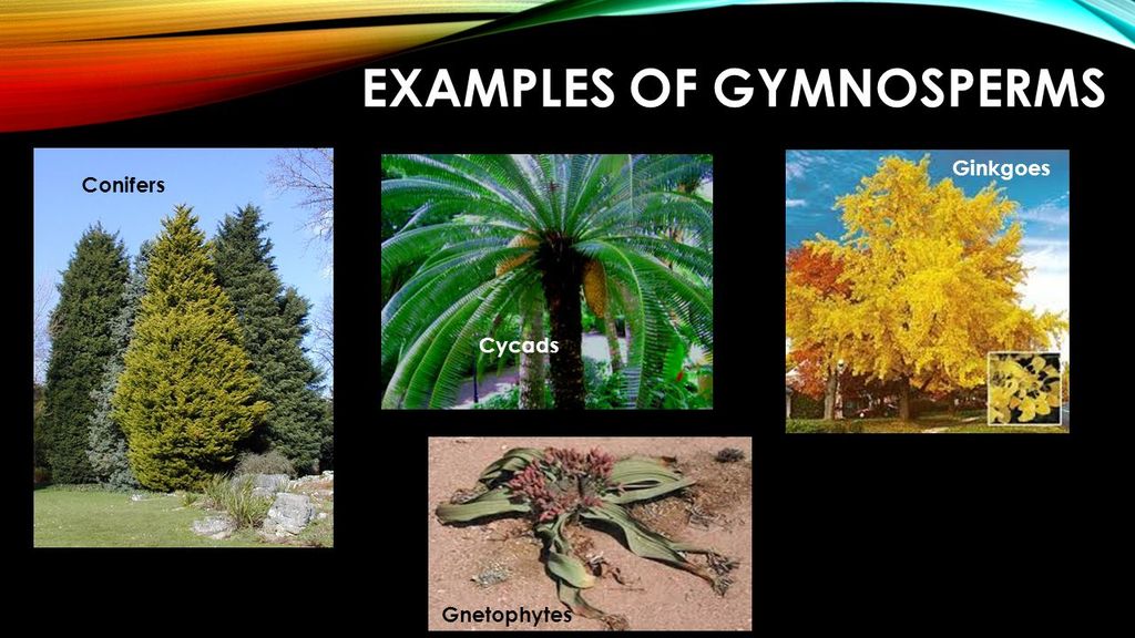
(E) Angiosperms:
Seed-bearing vascular plants with a well-differentiated plant body are known as angiosperms. Angiosperm seeds are contained within the fruits. Angiosperms are found all over the world and range in size from 0.1 cm to 100 m. For example, Wolffia is a little plant that grows to be around 0.1 cm tall, while Eucalyptus trees grow to be over 100 m tall. According to the number of cotyledons contained in the seeds, angiosperms are further split into monocotyledons and dicotyledons. Mango, rose, tomato, onion, wheat, maize, and other common examples are only a few.
Flowering and seed-bearing plants. E.g. Gymnosperms, Angiosperms are known as Phanerogams.
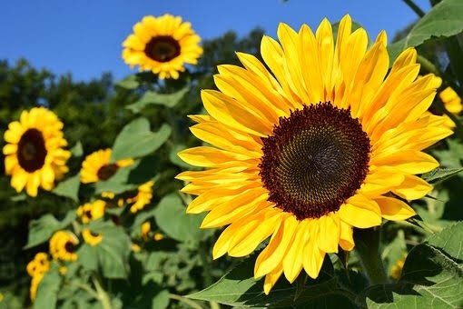
Kingdom: Animalia
- Books Name
- ACME SMART COACHING Biology Book
- Publication
- ACME SMART PUBLICATION
- Course
- CBSE Class 11
- Subject
- Biology
KINGDOM ANIMALIA
These are multicellular eukaryotic organisms.
The cells do not contain cell wall but contain only cell membrane.
They do not perform photosynthesis and have heterotrophic nutrition.
They have the power of locomotion.
They show increased sensitivity through the nervous system
Kingdom: Animalia
Kingdom Animalia
Multicellular eukaryotic organisms characterize this kingdom. Animals rely on plants for food, either directly or indirectly. Food is digested in an interior cavity, and food reserves are stored as glycogen or fat. Their feeding mode is heterotrophic and holozoic. They have a distinct growth pattern and mature into adults with distinct shapes and sizes. Higher forms have more complex sensory and motor mechanisms. The majority of them can move around. Male and female copulation is followed by embryological development in sexual reproduction. The animal kingdom is the most populous of the five kingdoms. They do not, however, have chlorophyll or a cell wall, as do plants.
Kingdom Animalia is divided into ten subphyla depending on body design or differentiation. These phyla are as follows:
(A) Phylum Porifera:
It is a phylum of organisms that have holes or perforations. Commonly known as Sponges. Poriferans are multicellular organisms with a hard outer skeleton that is non-motile. Their bodies have pores that form a canal system that aids in the passage of chemicals. They lack a well-developed organ or organ system and are not distinguished between head and tail. They primarily live in a marine environment. Spongilla and Sycon are examples of the Porifera phylum.
(B) Phylum Coelenterata (Cnidaria):
Hollow-bellied organisms are included in coelenterates. The presence of a hollow bodily cavity is one of the distinguishing characteristics of this group. The body is made up of two layers of cells: interior and exterior linings, which are differentiated into two ends. All aquatic animals are included in this phyla. Members of this phylum dwell in both colonies (corals) and solitary environments (Sea anemones). Hydra and jellyfish are examples of the phylum Coelenterata.
(C) Phylum Platyhelminthes:
Flatworms are the popular name for Platyhelminthes. Their body shape is complicated, dorsoventrally flattened, and distinct. Tissues are triploblastic and differentiated from three layers of cells. There is no actual interior cavity or coelom in them. They are symmetrical on both sides and can be free-living (Planaria) or parasitic (liver flukes). Tapeworm and Planaria are examples of Platyhelminthes phylum.
(D) Phylum Nematoda:
Nematodes, sometimes known as roundworms, belong to the Phylum Nematoda. The body of a nematode is cylindrical. They are triploblastic and bilaterally symmetrical. They have a fake bodily cavity called pseudocoelom. Elephantiasis and ascariasis are parasitic diseases caused by them. Ascaris and Wuchereria are examples of the phylum Nematoda.
(E) Phylum Annelida:
Annelids, are often known as segmented or ringed worms. They possess a head and tail that are separated by a segmented cylindrical body. The body is symmetrical on both sides and is triploblastic. They have a true cavity in their bodies. Their habitat includes both marine and freshwater environments, as well as land. Earthworms and Leeches are examples of the phylum Annelida.
(F) Phylum Arthropoda:
The term "arthropod" refers to a creature with jointed legs. This phylum includes animals with jointed appendages. This is the animal kingdom's largest phylum. They have jointed legs, an exoskeleton, and a segmented body that is bilaterally symmetrical. Organs and organ systems are well-differentiated. They have an open circulatory system, but no distinct blood vessels. Spiders, butterflies, and mosquitoes are all members of the Arthropoda phylum.
(G) Phylum Mollusca:
Molluscs are a wide group of animals that belong to the Phylum Mollusca. They are triploblastic and bilaterally symmetrical. The body is less segmented, yet organs and organ systems are well-developed. Mollusks typically have an open circulatory system. There are limbs present. Snails and octopuses are examples of the Mollusca phylum.
(H) Phylum Echinodermata:
Echinoderms are animals with spiky skin. They are triploblastic and have radial symmetry. They are true coelomates. The skeletal structure is made up of calcium carbonate and is quite hard. They are aquatic animals that live in the open ocean. Sea urchins and starfish are examples of the phylum Echinodermata.
(I) Phylum Hemichordata:
Hemichordata is a phylum of marine deuterostome organisms that is sometimes referred to as the echinoderms' sister group. The body is soft and delicate, with a proboscis. The epidermis is made up of only one layer. It is made up of worm-like marine invertebrates that are organized into organ systems. They have a circulatory system that is open. Because they are marine, they breathe through their gills. External fertilization is observed, and they have different sexes. Development is a direct process. Hemichordata includes Balanoglossus and Saccoglossus.
(J) Phylum Chordata:
Animals having a flexible cord supporting their dorsal or rear sides belong to the Chordata phylum. The majority of the species in the phylum Chordata are vertebrates or creatures with backbones. They are bilaterally symmetrical, triploblastic, and classified at the organ-system level. They have a nerve cord and a notochord. The circulatory system is one that is closed. Urochordata, Cephalochordata, and Vertebrata are the three subphyla of Chordata. Chordates include pandas, crows, sharks, salamanders, alligators, sea squirts, and many others.
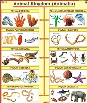
Viruses, viroids, prions and lichens:
Lichens and some acellular species like viruses, viroids, and prions are not included in Whittaker's five kingdom classification system. Here's a quick run-down of what they're all about.
Viruses were not classified since they are not actually 'alive,' as defined by organisms with a cell structure. Viruses are non-cellular organisms with a crystalline structure that is inactive outside of living cells. When they infect a cell, they use the host cell's machinery to duplicate themselves, eventually destroying the host. Viruses are classified as either living or non-living organisms. Dmitri Ivanowsky (1892), who identified specific bacteria as the causal organisms of the mosaic sickness, gave the name virus, which means venom or deadly fluid.Because they passed through bacterium-proof filters, they were discovered to be smaller than bacteria. M.W. Beijerinek (1898) discovered that an extract from sick tobacco plants could infect healthy plants, and he named the fluid Contagium vivum fluidum (infectious living fluid). W.M. Stanley (1935) demonstrated that viruses can crystallize, and that crystals are mostly made up of proteins. Outside of their respective host cell, they are inactive. Viruses are obligate parasites.Viruses include genetic material in the form of RNA or DNA, in addition to proteins. There isn't a single virus that has both RNA and DNA. A virus is a nucleoprotein with infectious genetic material. Plant viruses contain single-stranded RNA, while animal viruses have either single or double-stranded RNA or double-stranded DNA.
Bacterial viruses, also known as bacteriophages (viruses that infect bacteria), are double-stranded DNA viruses that infect bacteria. The nucleic acid is protected by a protein coat called capsid, which is made up of tiny subunits called capsomeres. The geometric forms of these capsomeres are helical or polyhedral. Mumps, smallpox, herpes, and influenza are all diseases caused by viruses. AIDS is also caused by a virus known as HIV in humans.Mosaic formation, leaf rolling and curling, yellowing and vein clearing, dwarfing, and stunted development are some of the symptoms seen in plants.
Viroids: In 1971, T.O. Diener found a novel infectious agent that caused potato spindle tuber disease and was smaller than viruses. It was discovered to be a free RNA that lacked the protein covering that viruses have, therefore the term viroid. The viroid's RNA has a low molecular weight.
Prions: In modern medicine, an agent made up of improperly folded protein has been discovered to spread some infectious brain illnesses. The agent was about the same size as viruses. Prions were the name given to these agents. The most well-known prions disorders are bovine spongiform encephalopathy (BSE), also known as mad cow disease in cattle, and its human counterpart Cr–Jacob disease (CJD).
Lichens are symbiotic (meaning mutually beneficial) interactions between algae and fungi. Phycobiont refers to the algal component, while mycobiont refers to the fungus component. Both are autotrophic and heterotrophic, respectively. Fungi offer refuge and absorb mineral nutrients and water for algae, and algae supply sustenance for fungi. Their relationship is so strong that if you saw a lichen in the wild, you'd never guess it included two different organisms. Lichens are excellent pollutant indicators since they do not develop in polluted environments.
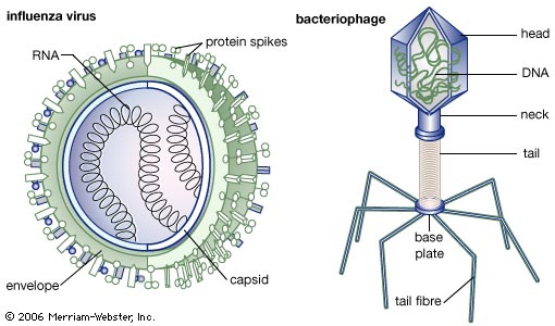
Classification Systems and taxonomy branches
- Books Name
- ACME SMART COACHING Biology Book
- Publication
- ACME SMART PUBLICATION
- Course
- CBSE Class 11
- Subject
- Biology
PLANT CLASSIFICATION SYSTEMS
Traditionally plant kingdom has been divided into two sub-kingdoms named as phanerogamae and cryptogamae.
(a) Cryptogamae (kryptos-concealed, gamos-marriage) :
All non flowering plants such as algae, fungi, lichen, mosses and ferns are included in this sub-kingdom.
The cryptogams are further classified into three divisions-thallophyta, bryophyta and pteridophyta.
(b) Phanerogamae (phaneros-visible, gamos-marriage) :
All flowering plants which bear seeds are included in this group. They are also known as spermatophytes (sperma-seed, phyton-plant), since they produce seeds.
These seed bearing plants are further divided into two divisions gymnospermae and angiospermae.
Gymnosperms have naked ovules or seeds. The angiosperms (covered seed) include all the flowering plants which produce seed and have ovules enclosed in ovary or fruit.
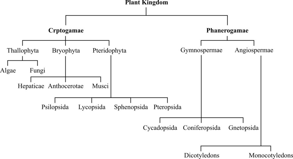
A. EARLY ATTEMPTS FOR CLASSIFICATION
Aristotle and other Greek philosophers divided living organisms into two groups: plants and animals. Aristotle also divided plants into 3 groups-herbs, shrubs and trees.
Charaka (the father of Ayurveda), in 1 st century A.D., listed 200 kinds of animals and 340 kinds of plants in his book 'Charaka Samhita'.
It is based on a few morphological characters of vegetative nature for grouping of organisms, e.g., early systems of classifications by Aristotle, Theophrastus, Pliny, Bauhin, Linnaeus etc.
Theophrastus proposed the first system of artificial classification of plants on the basis of habit and classified plants into herbs, shrubs, undershrubs and trees.
Carolus Linnaeus (1707-1778) proposed the artificial system of classification based exclusively on nature and number of stamens and carpels. It was called as Sexual System of Classification.
Classification by Linnaeus consisted of 24 classes, in which 23 classes were of flowering plants (Phanerogamia) and 24th class had flowerless plants (Cryptogamia). Details of this classification were published in Genera Plantarum (1737).
Total 24 classes given by Linnaeus were: Monandria, Diandria, Triandria, Tetrandria, Pentandria, Hexandria, Heptandria, Octandria, Enneandria, Decandria, Dodecandria, Icosandria, Polyandria, Didynamia, Tetradynamia, Monadelphia, Diadelphia, Polyadelphia, Syngenesia, Gynandria, Monoecia, Dioecia, Polygamia and Cryptogamia.
Drawbacks:
(i) This system was based on one or a few characters, hence the diverse animals or plants were placed into limited number of groups.
(ii) Natural affinities and phylogenetic relationships were not considered.
(iii) The artificial system gave equal weightage to vegetative and sexual characters; this is not acceptable and since we know that often the vegetative characters are more easily affected by environment.
C. Natural System of Classification
Also known as horizontal system of classification or 2 D system. Organisms in this system are classified on the basis of natural affinities. This system uses as many taxonomic characters as possible to group organisms.
This classification is mainly based on forms, relationship realising all information available at the time of collection of plants. This also considers internal features like ultrastructure, anatomy, embryology and phytochemistry .
Common natural systems were proposed by -John Ray, de Jussiaeu, de Candolle, Bentham and Hooker etc.
George Bentham and J.D. Hooker gave most important natural system of classification of angiosperms and published it in three volumes of 'Genera Plantarum'. They described 202 families. In this system, description of plants was based on their detailed studies and dissections. This system is followed in all British Common wealth countries including India
D. Phylogenetic System of Classification
The term phylogeny was given by Lamarck and concept of phylogeny by E. Haeckel. Phylogeny is the evolutionary history of the organism. This system is also called '3D' or vertical system.
In this system, plants are classified according to their evolutionary and genetic affinities. Organisms belonging to same taxa are believed to have a common ancestor and may be represented in the form of family tree called Cladogram.
A.W. Eichler modified Bentham and Hooker's system of classification by placing gymnosperms in the beginning. He is also called as the pioneer in phylogenetic system of classification.
Adolph Engler and Karl A.E. Prantl, two german botanists, adopted their system in "Die naturalichen Pflanzen famhien" (1887-1915). It was a German work which was later translated in English. The work had 23 volumes.
An outline of Engler and Prantl's System of Classification
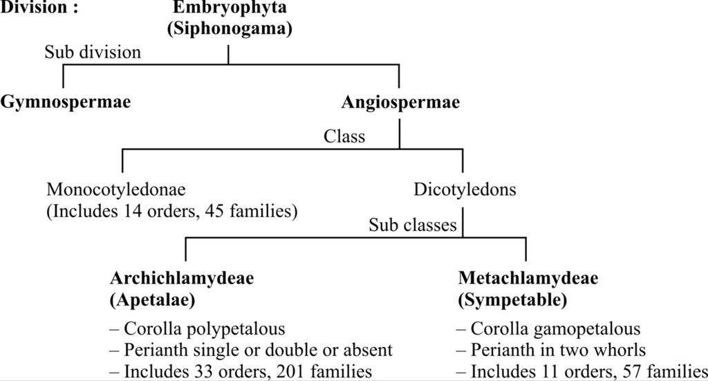
Merits: In this system families are arranged according to increasing complexity of flowers.
Demerits: Monocots were considered primitive to dicots.
According to this system, primitive forms having naked flowers were kept in the beginning. The more advanced families have distinct perianth while the highly evolved families have fused perianth.
Hutchinson, Oswald Tippo, Takhtajan and Cronquist also proposed phylogenetic systems of classification.
John Hutchinson, an English botanist, proposed phylogenetic classification in his famous book 'Famillies of Flowering plants' in 1959.
In Hutchinson's classification:
(i) Trees and shrubs are considered to be more primitive than herbs
(ii) Dicots have been considered more primitive than monocots.
(iii) Polypetalous, actinomorphic and solitary flowers are considered more primitive than gamopetalous, zygomorphic and inflorescence respectively.
An outline of Hutchinson's System of Classification
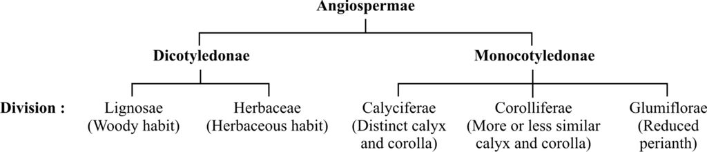
The divisions include orders that include families. Division does not include classes.
Takhtajan classified Magnoliophyta (Angiospermae) into two classes-Magnoliopsida (dicotyledons) and Liliopsida (Monocotyledons).
BRANCHES OF TAXONOMY
1. Classical Taxonomy:
In this taxonomy, organisms are classified on the basis of natural affinities. These affinities or relationships realise all informations available at the time of collection of plant, e.g., natural systems of classification.
2. Numerical Taxonomy/Phenetics/Taximetrics/Adansonian Taxonomy:
In this taxonomy, there is use of numerical methods for the evaluation of similarities and differences between the species. Firstly, number and codes are assigned to all the characters.
Then, all possible characters are compared by sophisticated calculating machines and computers without giving emphasis on a particular character.
All characters considered for analysis are given equal importance. The organization and analysis of data forms the core of this taxonomy.
The family tree prepared on the basis of phenetics is called dendrogram.
3. Cytotaxonomy/Karyotaxonom
It is based on cytological informations of the cell, chromosome number, structure and behaviour of chromosomes during meiosis
4. Biochemical (Chemotaxonomy) :
It is related with the chemical properties of plant, for example, fragrance by the presence of any aromatic compound, presence of specific crystal structures of calcium oxalate (raphides) or calcium carbonate (cystolith).
The sequencing of DNA and chemical nature of proteins have also been used in this taxonomy.
Taxonomy is divided into three types by Turril.
1. α (Alpha) Taxonomy: It deals with the collection and identification of organism on the basis of gross morphology.
2. β (Beta) Taxonomy : It deals with the collection and identification of organism on the basis of morphology and all possible evidences from cytology, anatomy, physiology and genetics.
3. ω (Omega) Taxonomy : It is based on microscopic observations and biochemical evidences
Classification Systems and taxonomy branches
Plant Kingdom
The broad classification of living organisms was proposed by Whittaker in 1969. He suggested a five-kingdomclassification, consisting of Monera, Protista, Fungi, Animalia, and Plantae. In this chapter, we will furtherstudy the classification within Kingdom Plantae or the plant kingdom. Kingdom Plantae is further classified into algae, Bryophytes, Pteridophytes, Gymnosperms, and Angiosperms.
Classification systems and taxonomy branches
Plant taxonomy can be defined as a branch of botanythat dealswith the characterization, identification, classification, and nomenclature of the plants on the basis ofsimilarities and differences.
The purpose of plant taxonomy is:
1. Identification: To identify the unknown species based on its characteristics and compare it with already existing species.
2. Characterization: To provide a description of all the characteristics of the newly identified plant species.
3. Classification: A suitable placement of the known species into different groups according to similarities and differences.
4. Nomenclature: Providing a suitable scientific name to the convention.
Only a few vegetative features were considered in the first classification system. Along with morphological traits, modern taxonomy studies have become more comprehensive and taken into account numerous morphological, cellular, and molecular factors, such as cellular and reproductive aspects, mode of nutrition, habitat, evolutionary relationships, and so on.
Plant classification systems are divided into three categories.
(I) Artificial system of classification: it was the earliest system that tried to classify organisms based on a few superficial characteristics. Ithowever did not consider morphological details and the evolutionary relationship among the organisms. This system gives equal importance to vegetative and sexual characters. Vegetative characters are greatly influenced by their surroundings. Therefore the closely related species could not be properly classified.More than 2000 years ago, Aristotlewas able to classify plants into herbs, shrubs, and trees, solely on the basis of morphology characters. Carl Linnaeus in his book Systema Naturaegives the hierarchical system of classification for the plant kingdom and animal kingdom. He proposed Binomial Nomenclature and the rules for naming every species.
(II) Natural system of classification: In this system, more traits were taken into account, in order to characterize. It was based on the natural similarities of vegetative and floral characteristics among organisms it considers various external and internal features like the anatomy of the plant, cell types of an embryo, and phytochemistry.
Bentham and Hooker’s system of classification categorized plants into cryptogams (non-flowering plants) and Phanerogams(flowering plants). It helped to determine relationships between the various groups of plants and failed to identify the phylogenetic relationship among different groups as it incorrectly placed gymnosperms between monocotyledons and dicotyledons.
(III) The phylogenetic system of classification: This system is based on evolutionary sequences and genetic links. This system was developed after the publication of Darwin's theory of evolution. Apart from the morphological characteristics found inthe fossil record, genetic components were also taken into consideration.This system of classification is most widely accepted by biologists throughout the world. According to this system, all the organisms occupying the same taxa originated from a common ancestor. Various taxonomists like Englar and Prantl, Hutchinson, Takhtajan, Robert F Thorn, etc. have contributed to the phylogenetic system of classification.
Numerical taxonomy is based on all observable characteristics present in a species. A number of codes are assigned to all the characters and the data is then processed in such a way that each character is given equal importance and at the same time, the hundreds of characters can be considered. It can be easily carried out using a computer.
Cytotaxonomy is based on cytological information like chromosome number cell structure and behavior. Chemotaxonomy exploits the chemical constituents of the plant to resolve confusion related to classification. All these techniques are widely used by taxonomistsnowadays.
Algae
- Books Name
- ACME SMART COACHING Biology Book
- Publication
- ACME SMART PUBLICATION
- Course
- CBSE Class 11
- Subject
- Biology
ALGAE
- Algae are chlorophyll containing thallophytes and are characterised by the absence of embryo stage and presence of unicelled non-jacketed gametangia. The study of algae is called algology or phycology.
- The main characteristics of algae are:
1. Algae are usually aquatic, either marine or fresh water, or may occur in a variety of habitats like moist stones, soil and wood.
2. Reserve food is mainly starch.
3. Vascular tissues are absent. Being aquatic, water conduction is not required even in giant forms.
4. Nutrition is autotrophic.
5. Vegetative reproduction by means of fragmentation.
6. Asexual modes of reproduction by means of Mitospores like -Zoospores, Aplanospores, Hypnospores, Akinetes, Palmella stage, etc.
7. Sex organs are non-jacketed. They are unicellular. In case the sex organs are multicellular (e.g., Chara), every cell is fertile.
8. Sexual reproduction involves gametic fusion, that may be isogamy, anisogamy and oogamy in different groups.
9. Life cycle is of various types such as haplontic, diplontic or diplohaplontic.
Concept Builder
1. Algae shows a wide range of forms like -unicellular (e.g., Chlamydomonas), colonial (e.g., Volvox), palmelloid (e.g., Tetraspora), dendroid (e.g., Prasinocladus), filamentous unbranched (e.g., Spirogyra) or branched (e.g, Cladophora), heterotrichous (e.g., Fritschiella, Coleochaete, Stigeoclonium), siphonaceous (e.g., Vaucheria), parenchymatous (e.g., Ulva).
2. F.E. Fritsch (1935) in his book Structure and Reproduction In Algae proposed a practical and sound classification of algae. He divided algae in 11 classes on the basis of pigmentation, reserve food, flagellation, thallus structure and mode of reproduction.
3. Kingdom Plantae of Whittaker (1969) includes mainly three types of algae -red algae, brown algae and green algae.
Comparison of Some Characteristics of Algae

Algae
Algae
Algae are a class of creatures that are mostly aquatic, photosynthetic, and nucleus-bearing but lack the real roots, stems, leaves, and specialized multicellular reproductive systems that plants have. They can be found in a range of various environments, including damp stones, soils, and wood. Some of them are also found in the presence of fungi (lichen) and animals (e.g., on sloth bears).
Algae come in a wide range of shapes and sizes, from colony forms like Volvox to filamentous forms like Ulothrixand Spirogyra. Kelps, for example, are a type of marine organism that develops gigantic plant bodies. Algae reproduce vegetatively, asexually, and sexually. Fragmentation is the mode of vegetative reproduction. A thallus forms from each fragment.Asexual reproduction occurs by the formation of spores, the most common of which are zoospores. They are flagellated (motile) and produce new plants upon germination. The fusing of two gametes occurs during sexual reproduction. These gametes can be flagellated and comparable in size (as in Ulothrix) or non-flagellated and similar in size (as in Spirogyra). Isogamous reproduction refers to this type of reproduction. Anisogamous refers to the fusion of two gametes of different sizes, such as those found in Eudorina species. Volvox and Fucus are examples of oogamous fusion that occurs between a big, nonmotile (static) female gamete and a smaller, motile male gamete.
Algae are beneficial to humans in a number of ways. Algae photosynthesis accounts for at least half of the total carbon dioxide fixing on the planet. They enhance the amount of dissolved oxygen in their immediate environment because they are photosynthetic. They are crucial as primary producers of energy-rich chemicals, which are the foundation of all aquatic species' food cycles. Among the 70 species of sea algae used as food are many Porphyra, Laminaria, and Sargassum species. Certain coastal brown and red algae, such as algin (brown algae) and carrageen (red algae), produce huge amounts of hydrocolloids (water-holding compounds) that are commercially exploited.Agar, a commercial product derived from Gelidium and Gracilaria, is used to culture bacteria as well as in ice cream and jelly preparations. Chlorella, a protein-rich unicellular alga, is utilized as a nutritional supplement by astronauts. Chlorophyceae, Phaeophyceae, and Rhodophyceae are the three major groups of algae that are described below.
A. Chlorophyceae: Green algae are members of the Chlorophyceae family. Unicellular, colonial, or filamentous plant bodies are all possible. Because chlorophyll a and b pigments predominate, they are frequently grass green. Pigments are concentrated in specific chloroplasts. In different species, the chloroplasts might be discoid, plate-like, reticulate, cup-shaped, spiral, or ribbon-shaped. In the chloroplasts, most members have one or more storage bodies called pyrenoids. Pyrenoids contain protein besides carbohydrates. Food may be stored in the form of oil droplets by some algae. Green algae have a stiff cell wall made up of an inner cellulose layer and an outside pectose layer.
Figure 1
Vegetative reproduction is usually accomplished through fragmentation or the creation of several spore types. Flagellated zoospores produced in zoosporangia are used for asexual reproduction. Sexual reproduction can be isogamous, anisogamous, or oogamous, with significant differences in the kind and development of sex cells. Chlamydomonas, Volvox, Ulothrix, Spirogyra, and Chara are some of the most common green algae.
B. Phaeophyceae: Brown algae, also known as Phaeophyceae, are mostly found in marine environments. They display tremendous variety in size and form. They range in complexity from simple branched filamentous forms (Ectocarpus) to profusely branched forms such as kelps, which can reach 100 meters in height. Chlorophyll a, c, carotenoids, and xanthophylls are all present. Depending on how much of the xanthophyll pigment fucoxanthin is present in them, they range in color from olive green to various colors of brown. Food is stored as laminarin or mannitol, both of which are complex carbs. The vegetative cells have a cellulose wall that is normally covered with a gelatinous layer of algin on the outside. In addition to plastids, the protoplast has a centrally positioned vacuole and nucleus.The plant body has a stalk, the stipe, and a leaf-like photosynthetic organ, the frond, which is normally linked to the substratum by a holdfast. Fragmentation is used in vegetative reproduction. Biflagellate zoospores, which are pear-shaped and have two unequally attached flagella, are used for asexual reproduction in most brown algae. Isogamous, anisogamous, and oogamous sexual reproduction are all possible. Gamete union can occur in water or within the oogonium (oogamous species). The gametes are pyriform (pear-shaped) and have two flagella connected to their sides. Ectocarpus, Dictyota, Laminaria, Sargassum, and Fucus are the most prevalent types.
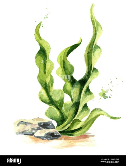
Figure 2
C.Rhodophyceae: Rhodophycea members are frequently referred to as red algae due to the preponderance of the red pigment r-phycoerythrin in their bodies. The majority of red algae are found in the sea, with higher quantities in warmer climates. They can be found in well-lit areas near the surface of the sea as well as at considerable depths in oceans where light is scarce. The majority of red algae have multicellular red thalli. Some of them have a complicated body structure. The food is stored as floridean starch, which has a structure that is extremely similar to amylopectin and glycogen. In most cases, red algae reproduce vegetatively by fragmentation. Non-motile spores and non-motile gametes are used to reproduce asexually and sexually. Sexual reproduction is oogamous, with complicated post-fertilization processes.Polysiphonia, Porphyra, Gracilaria, and Gelidium are the most prevalent members.
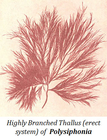
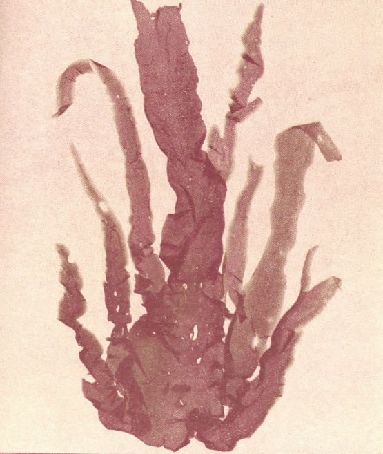
Figure 3
Bryophytes
- Books Name
- ACME SMART COACHING Biology Book
- Publication
- ACME SMART PUBLICATION
- Course
- CBSE Class 11
- Subject
- Biology
BRYOPHYTA
Bryophyta is a small group of plants having multicellular diploid sporophyte living as a parasite on an independent multicellular haploid gametophyte.
On the basis of evolution, bryophytes stand in between algae and pteridophyta. They are the first amphibians of plant kingdom, oldest bryophyte fossils are about 350 million years old.
General characters of Bryophyta :
Bryophytes live in damp, humid and shaded habitats. They may form green carpets or mats on damp soil, rocks, walls, tree trunks during rainy season.
The dominant phase or plant body is a free living gametophyte (n). It is thallus like and may be prostrate or erect.
Vascular tissues are absent.
True stem, leaf and roots are absent. Instead, rhizoids occur for attachment. The latter may be unicellular (e.g., Riccia and Anthoceros) or multicellular (e.g., mosses).
Vegetative reproduction is quite common through fragmentation, tubers, gemmae, buds, adventitious branches etc.
Asexual reproduction by mitospores is absent.
Sex organs are multicellular and jacketed, called antheridium (male) and archegonium (female). The jacket cells are sterile (Bryophytes are first archegoniates).
Antherozoid is twisted and comma shaped with two flagella.
An external layer of water is essential for the swimming of male gametes to reach the archegonia i.e., zooidogamy. So these are called amphibians of plant kingdom.
Zygotes do not undergo reduction division immediately. It develops inside archegonia and divides by mitosis to produce embryo (so these are considered as first embryophytes). The embryo develops further into a sporophyte which is parasitic over the gametophyte (may be partial pasasite as in mosses).
The sporophyte of bryophytes is also called sporogonium, it is composed of three parts viz., capsule, seta and foot. It produces meiospores or haploid spores inside the capsule part (after meiosis in spore mother cells), while attached to the gametophyte.
All bryophytes produce only one type of spores (Homosporous).
On germination each spore produces a gametophyte (either directly or through a juvenile filamentous stage called protonema, e.g., in mosses).
Bryophytes show heteromorphic or heterologous alternation of generations in the life cycle i.e., haplodiplontic life cycle.
Concept Builder
Term bryophyta was proposed by Brown. Hedwig is called father of bryology. Prof. Shiv Ram Kashyap is known as "Father of Indian Bryology".
Aquatic bryophytes are Riccia fluitans, Ricciocarpus, Fontinalis and Riella. Saprophytic bryophytes are Buxbaumia and Cryptothallus, while Frullania is an epiphytic form.
Economic importance of bryophytes
(i) Prevention of soil erosion: Bryophytes, especially mosses, form dense mats over the soil and prevent soil erosion against falling rains.
(ii) Soil formation: Mosses are an important link in plant succession on rocky areas. They take part in building soil in rock crevices formed by lichens. Growth of Sphagnum (Bog moss) ultimately fills ponds and lakes with soil.
(iii) Water retention : Dry Sphagnum has great water absorbing capacity. This characteristic is employed by gardeners to keep seedlings and cut plants moist during transportation and propagation. Sphagnum moss was used in place of absorbent cotton, so is also called cotton moss.
(iv) Peat: Sphagnum often grows in acidic marshes. The older dead parts of moss and other marshy plants got slowly carbonised, compressed and fossilised over thousands of years and have produced a dark spongy mass called peat. Peat is dried, compressed and cut to form blocks. The peat blocks are used as fuel. Peat is also a good manure. It overcomes soil alkalinity and increases its water retention as well as aeration.
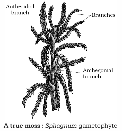
Classification of Bryophyta :
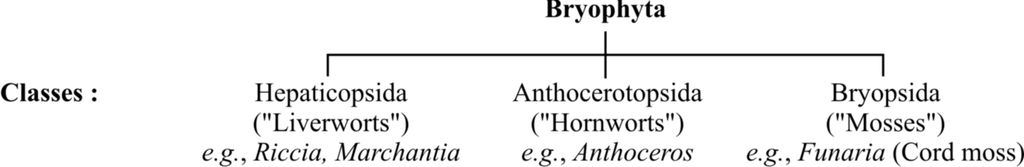
Some characteristic features of these classes are given below:
Hepaticopsida or Liverworts
They have thalloid structure (e.g., Riccia, Marchantia) with dorsiventral symmetry. On the ventral side of the thallus they have multicelled scales and unicelled rhizoids. Thallus has two distinct zones i.e., assimilatory and storage.
Leafy members like Porella have leaf like tiny appendages in two rows on the stem like structures.
Asexual reproduction occur by means of fragmentation, or by specialized structure called gemmae (e.g., Marchantia). These are '8' shaped, stalked, green and multicellular asexual buds developing in small receptacles (gemma cups) on dorsal surface of thallus. Each gemma germinates to produce two thalli in Marchantia.
Antheridia and archegonia are scattered and found embedded in dorsal side of thallus (e.g., Riccia) or may be projected from the thallus in form of stalked receptacles called antheridiophore and archegoniophore respectively (e.g., Marchantia).
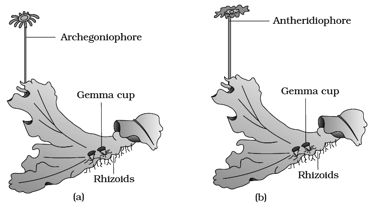
(a) Female thallus (b) Male thallus
Sporophyte may be represented by capsule only (e.g. , Riccia) or differentiated into foot, seta and capsule (e.g., Marchantia).
Capsule may contain spores only (e.g., Riccia) or spores and elaters (e.g., Marchantia). Elaters are diploid, hygroscopic structures with spiral thickenings which help in spore dispersal.
Sporophyte lacks assimilatory tissue, thus it is a complete parasite over the gametophyte.
Spores (n) germinate to form the thalloid gametophyte.
Concept Builder
Anthocerotopsida or Hornworts Anthoceros, Notothylas (Members of Anthocerotae) are called as hornworts, as they possess elongated 'horn like' sporophytes. Capsules of these members contain spores and pseudoelaters (elater like structures without spiral thicknings). Anthoceros cells have one chloroplast with one pyrenoid, so resembles algal thallus.
Bryopsida -Musci or True Mosses:
Gametophytic phase consist of two stages -first is protonema (juvenile stage) , and second is gametophore or leafy stage (mature).
Protonema is filamentous, creeping, green and branched structure developing through spore germination.
Gametophore consist of erect axis bearing spirally arranged leaves and sex organs.
Rhizoids are multicellular (with oblique septa) and branched.
Vegetative reproduction occurs by fragmentation and secondary protonema (filament developing from structure other than spore).
Sex organs develop in the axis of leaves.
The mosses have an elaborate mechanism of spore dispersal from capsule.
Mosses may be known by different names : Cord moss (Funaria), Maiden hair moss (Pogonatum), Peat/Bog moss (Sphagnum) , Hair cap moss (Polytrichum) .
Life cycle of Moss
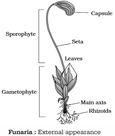
A typical moss plant like Funaria grows in moist, shady places forming dense carpet on soils, rocks, tree trunks etc., during the rainy season.
The radially symmetric plant body is differentiated into stem or axis, leaves or phylloids and rhizoids.
The rhizoids are multicellular. Moss plants multiply extensively by vegetative means.
Reproduction
1. Vegetative reproduction: It occurs by following ways:
(i) Apospory - Formation of gametophyte (i.e., the plant body) from sporophyte without meiosis.
(ii) Protonema buds - Primary protonema formed as a result of spore germination gives rise to group of cells called buds. These buds when separated give rise to new plants.
2. Sexual reproduction: Funaria is monoecious and autoecious plant i.e., male and female sex organs are borne on same plant, but on different branches, called as antheridial and archegonial branch respectively.
Antheridium :
Cluster of antheridia (male sex organ) are borne on antheridial branch, surrounded by a large number of closely arranged perigonial leaves.
An antheridium is a club-shaped structure. Paraphyses occurs mixed with antheridia. Terminal cell of paraphyses is capitate. It is borne over a small multicellular stalk.
There is a single layered jacket which encloses mass of sperm mother cells. Each sperm mother cell (androcyte) produces two biflagellate sperms having curved body.
Archegonium:
At the apex of archegonial branch, archegonia (female sex organ) intermingled with paraphyses are present, surrounded by a cluster of perichaetial leaves.
An archegonium is a flask-shaped structure having a multicellular stalk. It has a tubular neck and a swollen basal venter.
Venter has a two-layered wall which encloses two cells, a venter canal cell and an egg or oosphere. The neck has a single layered wall made of 6-spirally twisted rows of cells.
There is a cover made up of 4-cells. The neck encloses 6-10 Neck Canal Cells (NCC). At maturity, the neck canal and venter canal cells gelatinises. The lid cells separate.
The opened archegonia attract sperms by sucrose and K+ ions present in their mucilage. A sperm enters an archegonium, fuses with the oosphere and forms a diploid zygote or oospore
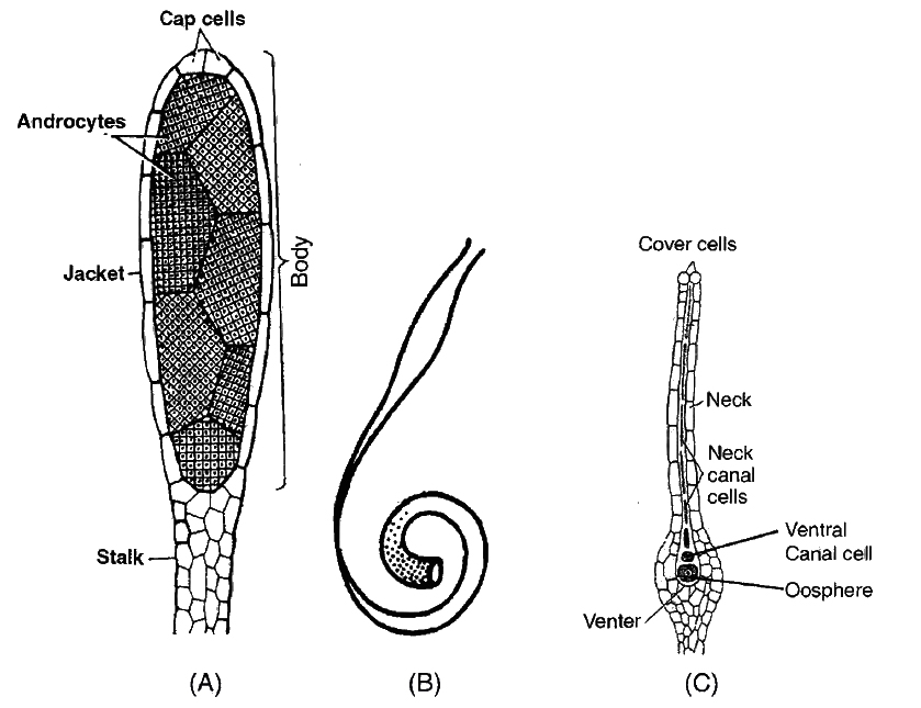
Development of sporophyte
The venter cells form a cover over zygote called calyptra (haploid -gametophytic tissue). The diploid oospore develops into a sporophyte. Sporophyte consists of three parts-foot, seta and capsule. Foot is embedded in the tip of gametophytic plant. It takes part in fixation of sporophyte and absorption of water and mineral salts from the gametophyte.
Seta is a narrow stalk which lifts the capsule in the air. The capsule is further differentiated into 3 parts-apophysis, theca and operculum. Apophysis contains assimilatory tissue and stomata (having a single circular guard cell).
Theca contains a central sterile column or columella, two spore sacs, air cavity and some assimilatory tissue. Its tip contain two rows of acellular teeth called peristome.
There are 32 peristomial teeth in each capsule arranged in two rows of 16 each. The teeth in outer row (exostome) are dark brown and hygroscopic in nature, and also have transverse bands of cellulose.
However teeth of inner row (endostome) are thin, light brown and non-hygroscopic. Inside the spore sac, diploid spore mother cells are formed.
They undergo meiosis and produce haploid spores.
The haploid spores are liberated from the capsule by removal of operculum and hygroscopic movement of peristomial teeth.
Since Funaria like mosses have some assimilatory tissue or cells with chloroplast in their sporophyte, unlike liver worts, sporophyte of mosses are often called as semiparasite over the gametophyte. Such cells are present in the apophysis, capsule wall and in the spores.
On germination, each haploid spore produces a filamentous juvenile gametophyte called protonema. Protonema has two types of branches, green and nongreen.
The nongreen branches (rhizonema) are subterranean. They function as rhizoids. Green branches (chloronema) are epiterranean.
Protonema can multiply vegetatively. It ultimately bears buds on its green branches. The buds grow to form moss plants. The latter, therefore, represent adult gametophyte.
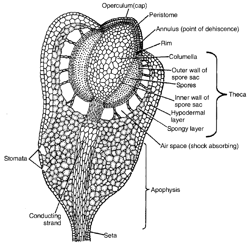
Bryophytes
Bryophytes
Mosses, liverworts, and hornworts are all classified as bryophytes. They are non-vascular plants, meaning they do not have roots or vascular tissue and instead take water and nutrients from the air through their surface. It has a variety of mosses and liverworts that are usually found growing in wet shaded regions in the hills. Bryophytes are also known as amphibians of the plant kingdom since they can live in soil but need water to reproduce sexually. They prefer moist, humid, and shady environments. On bare rocks/soil, they serve a crucial role in plant succession.Bryophytes' plant bodies are more distinct than algae's. It is thallus-like, prostrate or upright, and has unicellular or multicellular rhizoids that connect to the substratum. They are devoid of genuine roots, stems, and leaves. They may have structures that resemble roots, leaves, or stems. The bryophyte's primary plant body is haploid. It is called a gametophyte because it generates gametes. Bryophytes have multicellular sex organs. The male sex organ is referred to as the antheridium. Biflagellate antherozoids are produced by them. Archegonium, the female sex organ, is flask-shaped and produces a single egg. The antherozoids are discharged into the water, where archegonium is present. The zygote is formed when an antherozoid unites with the egg. Zygotes do not go through reduction division immediately.
They generate a sporophyte, which is a multicellular body. The sporophyte is not a free-living organism; it is linked to the photosynthetic gametophyte and feeds on it. To form haploid spores, some sporophyte cells undergo reduction division (meiosis). The gametophyte is produced when these spores germinate.
Bryophytes are not economically important in general, although some mosses supply food for herbivorous mammals, birds, and other creatures. Because of their ability to hold water, Sphagnum moss species have traditionally been utilized as fuel and as packing material for the trans-shipment of living material. Mosses and lichens were the first creatures to inhabit rocks and are hence extremely important ecologically. They break down rocks, allowing higher plants to thrive on the substrate. Mosses decrease the impact of pouring rain and prevent soil erosion by forming dense mats on the soil. Liverworts and mosses are two types of bryophytes.
A. Liverworts: Land plants with no circulatory system are known as liverworts. The liverworts like moist, shaded environments such as stream banks, marshy terrain, damp soil, tree bark, and deep in the forests. The plant body of a liverwort, such as Marchantia, is a thalloid. The thallus is dorsiventral and adheres to the substrate tightly. On the stem-like structures of the leafy members, there are two rows of small leaf-like appendages. In liverworts, asexual reproduction occurs by the fragmentation of thalli or the creation of specialized structures known as gemmae (sing. gemma). Gemmae are asexual, multicellular green buds that grow in little receptacles on the thalli called gemma cups. The gemmae separate from the parent body and germinate, resulting in the formation of new individuals.Male and female sex organs are generated on the same or distinct thalli during sexual reproduction. The sporophyte has three parts: a foot, a seta, and a capsule. Within the capsule, spores are formed after meiosis. These spores become free-living gametophytes when they germinate.
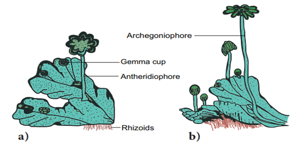
Figure 4: Marchantia (a) Male Thallus. (b) Female Thallus.
B. Mosses: The gametophyte, which has two stages, is the most important stage in a moss' life cycle. The protonema stage, which arises immediately from a spore, is the first stage. It takes the form of a creeping, green, branching, and filamentous stage. The second stage is the leafy stage, which grows as a lateral bud from the secondary protonema. They are made composed of thin, erect axes with spirally arranged leaves. They are linked to the soil by rhizoids, which are multicellular and branching. The sex organs are carried at this stage. Mosses reproduce vegetatively by fragmentation and budding in their secondary protonema. The sex organs antheridia and archegonia are generated at the top of the leafy branches during sexual reproduction.The zygote grows into a sporophyte, which has a foot, seta, and capsule after fertilization. Moss sporophytes are more complex than liverwort sporophytes. There are spores in the capsule. After meiosis, spores are generated. Mosses have a complex system for dispersing their spores. Mosses such as Funaria, Polytrichum, and Sphagnum are common examples.
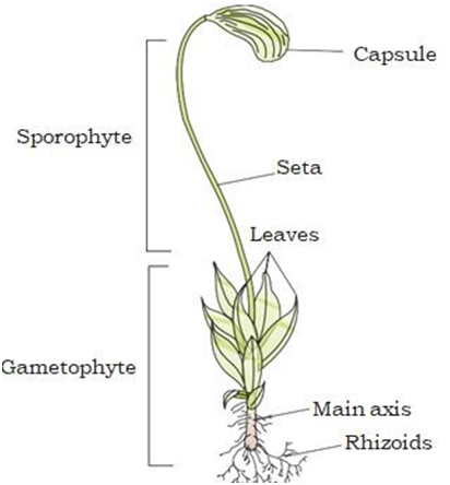
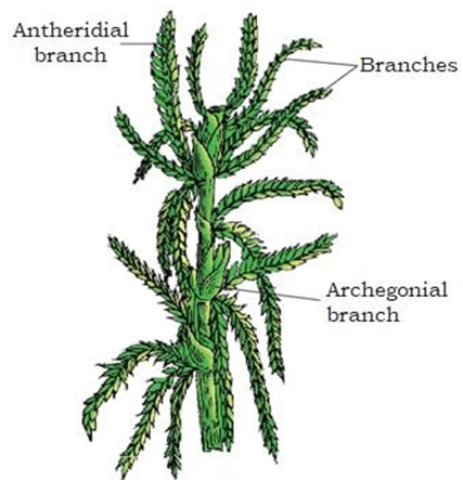
Pteridophytes
- Books Name
- ACME SMART COACHING Biology Book
- Publication
- ACME SMART PUBLICATION
- Course
- CBSE Class 11
- Subject
- Biology
PTERIDOPHYTA
General characters of pteridophyta
They are most primitive vascular, flowerless, seedless, spore producing cryptogamic land plants. Commonly called vascular amphibians "or" botanical snakes. These are first successful plants on land. Evolutionarily these are first terrestrial plants to possess vascular tissues.
Term pteridophyta was given by Haeckel.
1. Dominant phase of plant body is a sporophyte.
2. Plant body is differentiated into true stem, leaves and roots.
3. Vascular tissues are present. They are of two types, xylem and phloem. In xylem , true vessels are absent. In phloem, companion cells and sieve tubes are absent. Instead, sieve cells are present. Secondary growth is exceptionally present in Isoetes and Botrychium.
4. On the basis of leaves pteridophytes are of two types. In megaphyllous plants, leaves are large in relation to stem and leaf trace is broken by leaf gaps, e.g., ferns. In microphyllous forms, leaves are small and receive a single unbroken leaf trace, e.g., club moss and horsetails.
5. Sporangia are associated with leaves called sporophylls. Meiospores are formed inside sporangia. In some cases sporophytes may form distinct compact structures called strobili or cones (Selaginella, Equisetum).
6. Spore on germination produces small, reduced, free living', independent, multicelled, non-vascular, haploid gametophyte. This thalloid gametophyte is called prothallus. These require cool, damp shady places to grow. Because of this restricted requirement and the need of water for fertilization are reasons due to which the spread of living pteridophytes is limited and restricted to narrow geographical regions. The development of gametophyte may be endosporic (inside the spore wall) or exosporic (outside the spore wall).
7. Sex organs are multicellular and jacketed. Number of NCC in archegonia may be one uninucleate (Selaginella) or one binucleate (ferns).
8. Sperms are flagellated. They require an external supply of water for swimming to reach archegonia. The chemical stimulus is provided by malic acid.
9. Embryo stage is present.
10. Heteromorphic or heterologous alternation of generation is present in the life cycle.
11. Pteridophytes are divided in four groups, viz., Psilophyta (Psilotum), Lycophyta (Lycopodium, Selaginella), Sphenophyta (Equisetum), and Pterophytai/Filicophyta (Dryopteris, Pteris, Adiantum, Salvinia).
12. True ferns are leptosporangiate members of Filicophyta and are most conspicuous of the pteridophytes.
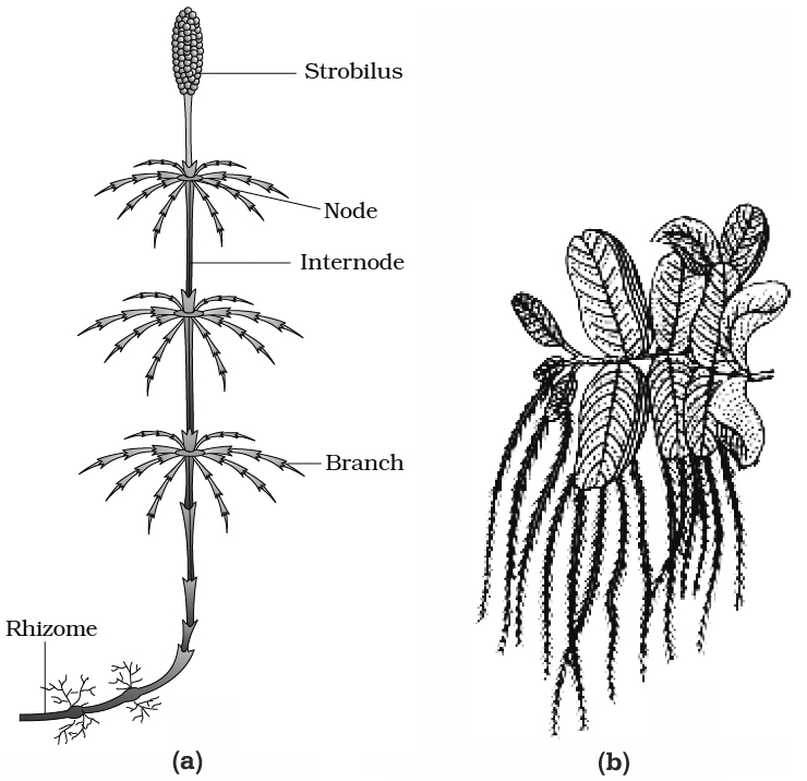
Concept Builder
1. Sporangium development is of two types in pteridophytes:
(i) Leptosporangiate -When sporangium develops from single superficial cell, e.g., Pteris, Dryopteris, Adiantum.
(ii) Eusporangiate -When sporangium develops from group of cells, e.g., Selaginella, Equisetum
2. Majority of pteridophytes produce same type of spores (homosporous, e.g., ferns like Adiantum, Pteris, Dryopteris). Only a few produce different types of spores, namely megaspores and microspores (heterosporous, e.g., Selaginella, Marsilea, Salvinia, Azolla etc.). Heterospory leads to seed habit in plants.
3. Embryogeny in pteridophytes is of two types:
(i) Exoscopic embryogeny : Axis of embryo is directed towards archegonial neck, e.g., Equisetum.
(ii) Endoscopic: Axis of embryo is directed inward towards venter, e.g., Selaginella.
4. Development of embryo may be :
(i) Holoblastic -When entire zygote is involved in embryonic development, e.g. , Ferns.
(ii) Meroblastic -When part of zygote is involved in embryonic development, e.g., Selaginella.
Stelar system
The central vascular cylinder of axis of pteridophytes is referred to as stele. It is a primary vascular tissue consisting of pith, xylem, phloem and pericycle.
It first appeared in pteridophytes like Rhynia. So lets study different types of steles proposed by Van Teigham and Douliot.
Types of stele :
1. Protostele: Simplest type, without pith
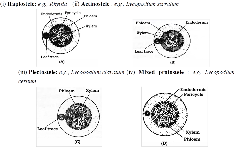
2. Siphonostele: With pith
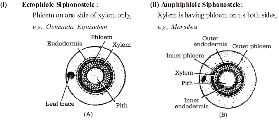
(iii) Solenostele : Stele with one or few non (iv) Dictyostele : Stele with large number of
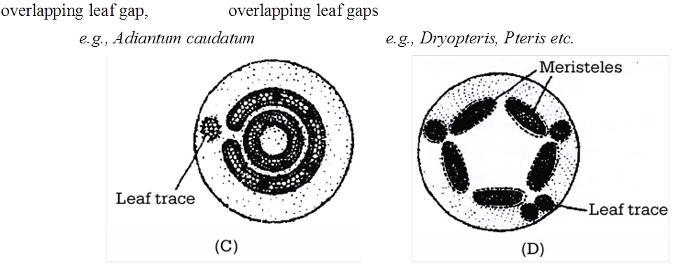
The specific features of true ferns are as follows :
i. The stem is underground rhizome (sometimes oblique, called root stock) in most of the ferns, except in tree ferns like Cyathea, Alsophyia where the stem is aerial.
ii. Leaves are large and are called fronds. Leaves may be simple or pinnately compound.
iii. Young leaves show circinate ptyxis, as they are coiled in the form of a watch spring. This coiling protects the growing point which comes to lie in the centre.
iv. Younger parts of stem, young leaves, petiole and rachis of mature leaves possess brown stiff hairs called ramenta. Ramenta protect them from mechanical injury and desiccation.
v. Sporangia occurs on the ventral surface of leaves in clusters called sori (singular-sorus). The fertile leaves are known as sporophylls. A sorus is covered by a flap-like outgrowth from its surface called indusium (true indusium, e.g., Dryopteris) or turned margin of the sporophyll (false indusium , e.g., Adiantum).
vi. Development of sporangium in true ferns is leptosporangiate.
vii. The male gamete is spirally coiled and multiciliated.
Life cycle of a fern (Dryopteris)
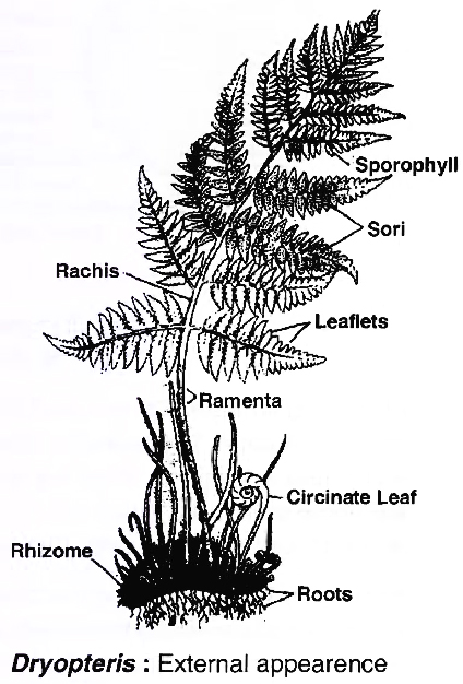
Plant body is a perennial, independently living evergreen sporophyte. It is differentiated into stem, leaves and roots. Roots are adventitious.
Stem is an underground dark brown rhizome.
The large aerial leaves or fronds develop acropetally in spirals from the upper surface of the rhizome.
The young leaves show circinate ptyxis. Persistent leaf bases of the dead leaves are found in older parts of rhizome.
Venation is open furcate in leaflets. Ramenta or paleae are present.
Reproduction
The fern multiplies vegetatively by fragmentation of rhizome and development of adventitious buds.
In Dryopteris, the adventitious buds present at leaf base separate and grow into a new plant.
In Adiantum caudatum and a few other species, adventitious buds develop at leaf tips.
When such leaf tips happen to touch the soil, they form new plants.
The process helps in spreading the fern over a large area. Because of this reason A. caudatum is also called walking fern.
Ventral surface of leaflets of a mature leaf bear yellowish brown clusters of sporangia called sori. The leaves having sori or sporangia are called sporophylls.
In Dryopteris, the sori develop in two rows, one on either side of the veins. Each row contains 4-6 sori. Each sorus is covered by a membranous sheath of its own.
This covering is called true indusium. The covered sori of Dryopteris are kidney-shaped in outline. This has given the name of male shield fern to Dryopteris.
A sorus consists of a parenchymatous cushion of placenta. The placenta bears a number of stalked biconvex sporangia. In each sporangium, there is a single layered jacket that encloses 12-16 diploid spore mother cells.
A marginal row of jacket cells are differentially thickened to form annulus. The remaining marginal cells constitute stomium. The diploid spore mother cells divide meiotically to form haploid spores.
With the maturity of spores, the indusium shrivels. The exposed sporangia dehisce in the region of stomium due to differential contraction of annulus. The spores are thrown away. They are dispersed by air currents.
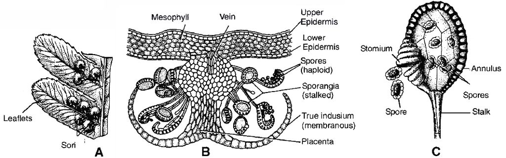
A. Fern leaflet (Sporophyll) showing sori, B. V.S. Sporophyll, C. A Sporangium

B. One archegonium C. One antherozoid
After falling on a suitable soil, each spore germinates to form a flat, heart shaped, green, thalloid gametophyte called prothallus.
The prothallus is monoecious, that is, it bears both the types of sex organs, male antheridia and female archegonia. They are borne ventrally.
Antheridia occur in the area of rhizoids, while archegonia are borne behind the apical notch in an area called apical cushion. Antheridium has a 3 celled jacket and about 32 sperm mother cells. The sperms are multiflagellate (= multiciliate) and spirally twisted.
Archegonium is flask-shaped in outline. It has an embedded venter and a projecting neck. Neck has 4-rowed wall which encloses a single binucleate neck canal cell.
The venter contains a single venter canal cell and an oosphere. In the mature state, the venter canal cell and the neck canal cells gelatinise. The neck opens.
Sperms are attracted to the opened archegonia by malic acid (chemotactic stimulus) present in their mucilage. A sperm fuses with an oosphere to form a diploid oospore.
The oospore gives rise to an embryo which grows in size to form the fern plant.
Life Cycle of Selaginella :
Salient features
Plant body is an evergreen sporophyte. It is differentiated into stem, leaves and roots. The roots often develop at the tips of special structures called rhizophores.
Plant body of Selaginella chrysocaulos and S.selaginoides is erect. Its leaves are isophyllous and arranged in two rows. In S.kraussiana the plant body is prostrate. Its leaves are anisophyllous or dimorphic and arranged in four rows.
Bower and Goebel named rhizophore of Selaginella as an organ sui-generis, i.e., an organ having the characters of both stem as well as root, but independent in origin.
The stem in Selaginella is distelic. 'Rarely stem is monostelic.
The plant body can multiply vegetatively by fragmentation, bulbils and tubers. Bulbils and tubers help in perennation as well.
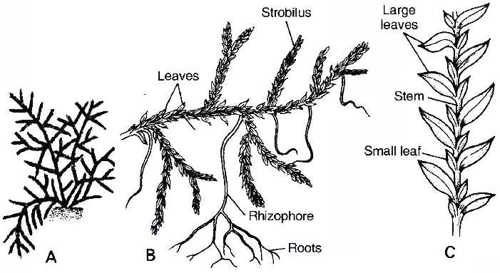
C. Arrangement of leaves
Sporangia develop in the axil of fertile leaves or sporophylls at the tips of small branches called spikes. There are two types of sporangia, microsporangia and megasporangia, hence this genus is heterosporous.
Each microsporangium produces a large number of small haploid microspores. A megasporangium develops only four haploid megaspores. Growth of the gametophytes is precocious.
Microspore produces an endosporic 13-celled male gametophyte. The latter has one prothallial cell, 8 jacket cells and four androgonial cells. The androgonial cells form 128-256 androcytes or sperm mother cells. A sperm mother cell gives rise to a biflagellate sperm.
Megaspore produces a partially exosporic female gametophyte. The female gametophyte contains an exposed generative apical cushion and a storage tissue. The two are separated by a
diaphragm
The apical cushion produces archegonia and rhizoids. Each archeogonium has a single female gamete or oosphere. The mucilage oozing out of neck of archegonium in Selaginella and fern contains malic acid.
The neck of archegonium in Selaginella and fern contains only one neck canal cell. Fertilization requires the help of external water for swimming of sperm. It produces diploid oospore.
Oospore divides to form a suspensor and embryo. The development of embryo is meroblastic. Suspensor helps in pushing the embryo into food laden storage tissue. Embryo has a foot for absorbing nourishment, a shoot tip and a root tip. The latter two elongate and produce an independent sporophytic plant body.
Selaginella shows two distinct generations, sporophyte and gametophyte, one producing the other. They are morphologically different. The phenomenon is called heteromorphic or heterologous alternation of generations.
Concept Builder
1. Common names Spike moss (Selaginella)
Bird's nest moss (Selaginella rupestris)
Club moss (Lycopodium)
2. Smallest pteridophyte =Azolla pinnata (an aquatic fern). It is commonly used as biofertilizer.
3. Largest pteridophyte = Cyathea (tree fern)
4. Apogamy : It is development of haploid sporophyte from gametophyte without fertilisation. It was reported by Farlow in Pteris cretica.
5. Apospory : It is development of diploid gametophyte from sporophyte without meiosis. It was reported by Druery in Athyrium. Due to apospory, polyploidy is common in ferns.
6. Psilotales like Rhynia (fossil) were first tracheophytes.
7. Psilotum shows presence of rhizoids and dichotomous branching of stem.
8. Under dry conditions, the xerophytic species of Selaginella roll into brown balls. The phenomenon is called cespitose habit. In this state, it may be uprooted. The brown balls become green and unroll again under moist conditions. Because of this characteristic, these plants are known as resurrection plants, e.g., S.lepidophylla, S.bryopteris.
9. Selaginella rupestris shows a near approach to seed habit.
Pteridophytes
Pteridophytes
A pteridophyte is a vascular plant that disperses spores and has xylem and phloem. Pteridophytes are frequently referred to as "cryptogams," meaning that their mechanisms of reproduction are hidden since they do not produce blooms or seeds.Horsetails and ferns are examples of Pteridophytes. Pteridophytes are utilized as soil binders and for therapeutic purposes. They're also commonly used as ornamentals. They are the first terrestrial plants to have vascular tissues – xylem and phloem – according to evolution. Pteridophytes prefer chilly, damp, shady environments, yet some can thrive in sandy soil.
Pteridophytes, have a sporophyte that is divided into real roots, stems, and leaves. The vascular tissues in these organs are well-differentiated. Pteridophyta leaves can be microscopic (microphylls) like Selaginella or huge (macrophylls) like ferns. Sporophytes have sporangia that are cared for by sporophylls, which are leaf-like appendages. Sporophylls can sometimes develop unique compact structures known as strobili or cones (Selaginella, Equisetum).In spore mother cells, the sporangia produce spores by meiosis. The spores germinate to form prothallus, which isa small, multicellular, free-living, primarily photosynthetic thalloid gametophyte. These gametophytes can only thrive in chilly, wet, shaded environments. The proliferation of live pteridophytes is limited and restricted to certain geographical locations due to this specific constrained requirement and the necessity for water for fertilization. Male and female sex organs, known as antheridia and archegonia, are found in gametophytes. The transmission of antherozoids — male gametes released from antheridia – to the mouth of archegonium requires water. The zygote is formed when the male gamete fuses with the egg contained in the archegonium.The zygote then creates a multicellular, well-differentiated sporophyte, which is the pteridophyte's dominant phase. The majority of pteridophytes have homosporous plants, which means that all of their spores are of the same type (e.g. Psilotum, Lycopodium, and Equisetum). Heterosporous genera include Selaginella and Salvinia, which generate two types of spores: macro (big) and micro (small). Megaspores and microspores, respectively, germinate and produce female and male gametophytes. Female gametophytes are kept on the parent sporophytes for varying amounts of time in these plants. The female gametophytes are responsible for the development of the zygotes into newborn embryos. This occurrence is a prelude to the seed habit, which is considered a crucial evolutionary step.
Pteridophytes are divided into four groups: Psilopsida (Psilotum), Lycopsida (Selaginella, Lycopodium), Sphenopsida (Equisetum), and Pteropsida (Dryopteris, Pteris, Adiantum).
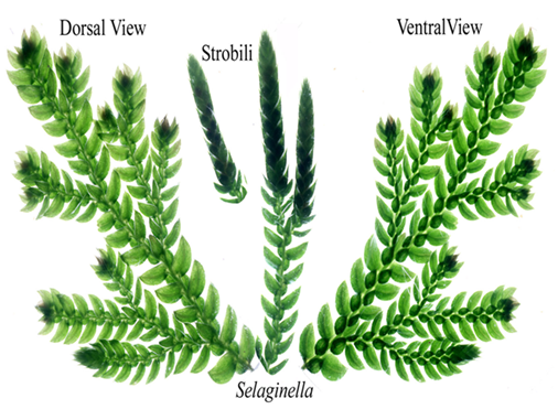
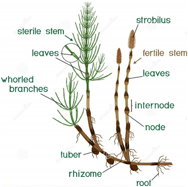
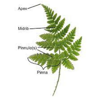
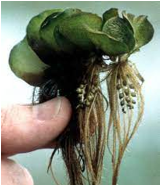
Figure 6
Gymnosperms
- Books Name
- ACME SMART COACHING Biology Book
- Publication
- ACME SMART PUBLICATION
- Course
- CBSE Class 11
- Subject
- Biology
GYMNOSPERMS
The term gymnosperm was used by Theophrastus (300 Be) in his book Enquiry into plants.
They are popularly called naked seeded vascular plants. Goebel called them as phanerogams without ovary.
It is smallest group of plant kingdom.
General characters of gymnosperms:
Majority of gymnosperms are perennial woody plants forming either shrubs or trees. Ephedra is a climber. Some of these are very large and live for thousands of years, e.g., Sequoia.
Unlike bryophytes and pteridophytes, in gymnosperms the male and female gametophytes do not have an independent free living existence.
Tap root system is present. Cycas also has special azeotropic N2 fixing coralloid roots having association with Anabaena cycadae (BGA). Pinus root shows symbiotic relation with ectomycorrhiza.
The leaves may be simple (Pinus) or compound (Cycas). These leaves are well adapted to withstand extremes of temperature, humidity and wind, e.g., Needle leaf of Pinus. Scale leaves may also be present.
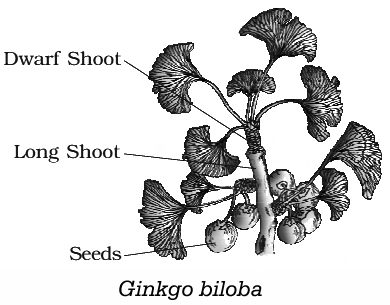
Stem is woody and branched (Pinus, Cedrus) or unbranched (Cycas).
These are heterosporous. There occurs two types of sporophylls; microsporophylls and megasporophylls. The two types of sporophylls are usually aggregated to form compact cones (strobili) i.e., pollen cones (male cones) and seed cones (female cones).
Microsporophylls do not show distinction of filament and anther.
The micros pores develop into a male gametophyte which is highly reduced and is confined to only a limited number of cells. This reduced male gametophyte is called pollen grain. The development of pollen grains takes place within the microsporangium.
Megasporophylls are not rolled like carpels. A distinction of ovary, style and stigma is absent.
Ovules (integumented. megasporangium) lie exposed on the megasporophyll. Each ovule is surrounded by a 3-layered single integument (Bitegmic in Gnetum).
Female gametophyte contains archegonia (Absent in certain members of order Gnetales)
Pollination is direct as a stigma is absent and the pollen grains directly reach the micropylar end of ovules. Pollination is usually accomplished by wind (anemophily).
Male gametophyte produces only two male gametes or sperms. Generally, one of them is functional.
External water is not required for transport of male gametes. Instead, a pollen tube is formed by the male gametophyte for effecting fertilization (siphonogamy).
Seeds contain a food laden tissue or endosperm for future growth of embryo into seedling. This tissue represents the female gametophyte, hence haploid.
Like pteridophytes, xylem does not possess vessels, except in some Gnetales. Phloem is without companion cells. Albuminous cells perform the function of companion cells. Sieve tubes are also not formed as the sieve cells are not arranged end to end in rows.
Vascular tissues are arranged into vascular bundles just like angiosperms.
Foliage leaves do not have lateral veins. Transfusion tissue (hydrostereom) occurs internally for lateral transport.
Wood is soft and homoxylous but members of Gnetales bear vessels in xylem.
Types of wood :
(i) Manoxylic :
Soft wood, vascular tissues with medullary rays, commercially less important, e.g., Cycas.
(ii) Pycnoxylic :
Compact wood without or with narrow medullary rays, commercially more important, e.g., Pinus.
(iii) Monoxylic :
With single persistent cambium ring and bundles, e.g., Pinus
(iv) Polyxylic :
With many persistent cambium rings and bundles, e.g., Cycas
Gymnosperms are, therefore, those seed plants in which the ovule remains exposed over the surface of the megasporophylls both before and after fertilisation.
Concept Builder
1. Smallest gymnosperm = Zamia
Largest gymnosperm = Sequoia
2. The three generations in seed are:
- (i) Testa, tegmen and perisperm represent parental sporophyte
- (ii) Endosperm represents female gametophyte;
- (iii) Plumule, radicle, suspensor and cotyledons (embryo) represent future sporophyte.
3. Polyembryony: It is the formation of more than one embryo inside a single seed. It was reported by Leeuwenhoek in oranges. Simple polyembryony is due to fertilisation of many eggs, e.g. Pinus ovule has 2-8 archegonia. Cleavage polyembryony is true polyembryony and very common. It is due to splitting of embryo tissue. Adventive polyembryony is the formation of extra embryos directly from diploid cells (e.g., rosette cells) other than embryonal cells.
4. Order Gnetales consists of Gnetum, Ephedra and Welwitschia. These are nearer to flowering plants in having flower like arrangement of sporophyll and possessing primitive vessels in xylem so wood is called heteroxylous. Plants of Cycadales and Coniferales are commonly called Cycads and Conifers respectively.
5. Cycas, Ginkgo and Metasequoia are living fossils.
6. Ginkgo biloba (Pagoda tree or Maiden hair tree) is oldest living fossil and it is connecting link between cycades and conifers.
7. Largest ovule (found in Cycas revoluta)
8. Largest male cone (in C. circinalis)
9. Largest male gamete (sperm) in Cycas
10. Largest female gamete (egg) in Cycas
11. Independent, free living, photosynthetic gametophyte is not found in gymnosperms and angiospersm.
12. Gymnosperms are divided into four orders-Ginkgoales, Gnetales, (e.g. Ephedra, Gnetum, Welwitschia), Cycadales and Coniferales. Ginkgoales is represented by maiden hair tree (Ginkgo biloba) where Ginkgo biloba happens to be only living member, rest all are extinct. So it is also called as living fossil.
Economic Importance
i. Edible Seeds. Seeds of Pinus gerardiana called chilgoza are edible.
ii. Timber. Gymnosperms possess soft wood. The same is used in preparation of light furniture , plywood, packing cases, match sticks, railways sleepers, etc, e.g., Cedrus deodara.
iii. Resin. Resin is a semifluid secreted by special resin canals. It solidifies on exposure to air. Therefore, it plugs the places of injury. It helps in sealing female cones after pollination. Resin is commercially extracted and distilled to obtain turpentine and resin. Resin is used in water proofing, sealing joints and preparation of writing paper. Turpentine is used as solvent in paints, polishes and wax, e.g., Pinus.
iv. Ephedrine. Drug ephedrine is obtained from Ephedra. The drug is used in curing respiratory elements including asthma.
v. Sago. A starchy food sago is obtained from stem of Cycas revoluta which is thus also called as sago palm.
vi. Canada balsam. A mounting agent used in preparation of permanent slides is obtained from Abies balsamaea.
vii. Cedar wood oil. Useful in microscopy is obtained from Juniperus virginiana.
viii. Taxol. Anticancerous chemical obtained from Taxus.
Life Cycle of Pinus
Pinus is a coniferous gymnosperm.
It is a large tree having pyramidal or excurrent shape like a "Christmas tree". The plant body is a sporophyte having stem, leaves and roots.
The main stem is straight. Stem branches are dimorphic i.e., of two types, long and dwarf. Long shoots show unlimited growth and bear only scale leaves. The dwarf branches possess 1-5 needle-like foliage leaves which are surrounded at the base by a sheath of scare leaves.
Pinus has a horizontally spreading tap root system with peg-like downwardly directed roots present at intervals.
Finer roots are of two types-normal (with root hairs and root cap) and mycorrhizal. The mycorrhizal roots occur near the soil surface.
They are devoid of root hairs and root cap. Mostly Boletus (fungus) forms an ectomycorrhiza with roots of Pinus.
The sporophytic plant body does not multiply vegetatively. It is a monoecious tree. It produces microsporophylls and megasporophylls in two types of cones, male and female.
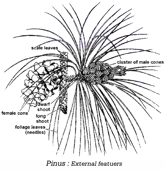
Male or Pollen Cones
These are non-woody structures occurring sub-terminally in clusters on lower long branches.
These cones are homologous to dwarf shoots. Each male cone has a short stalk, a central axis and a number of spirally arranged microsporophylls.
A microsporophyll bears two oblong, parallel microsporangia on its lower surface. In a microsporangium, the diploid microspore mother cells undergo meiosis and form haploid microspores or pollen grains.
The pollen grains are released from the male cones at 4 celled stage, having two prothallial cells, one generative cell and one vegetative cell and dispersed by air currents.
They form yellow clouds in the pine forests called as 'sulphur shower'. A pollen grain has two air sacs or wings for making it light, which are formed by its exine
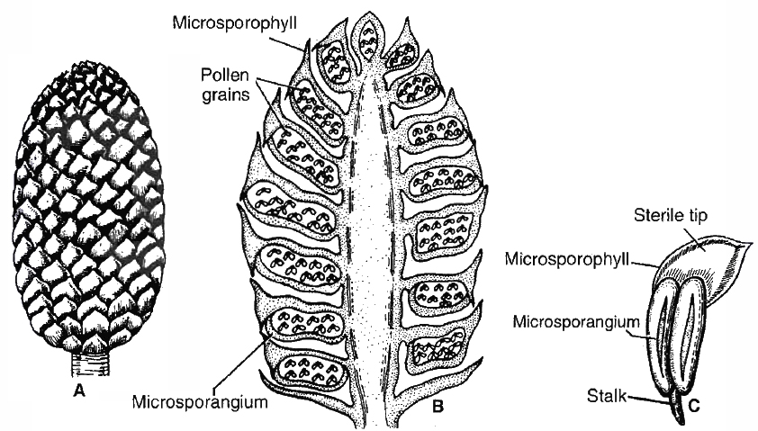
C. microsporophyll ventral view
Female or Seed Cones
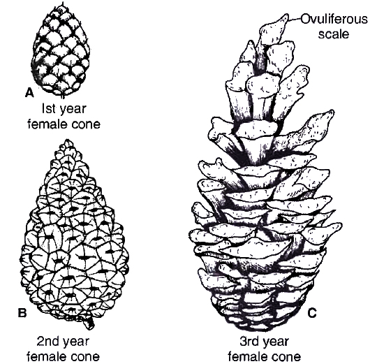
C. Third year female cone
These are initially green but become woody and brown at maturity.
They develop in groups of 2-6 on upper long branches of the tree. These are homologous to long shoots.
Each female cone has a long stalk and a central axis on which are borne a number of spirally arranged paired scales. The lower of the pair is called bract scale while the upper scale is ovuliferous scale.
The ovuliferous scale bears two ovules towards the basal region on the upper side.
Each ovule has a 3-layered integument with a terminal wide, oblique pore or micropyle and nucellus (= megasporangium). In the middle of nucellus, a megaspore mother cell differentiates.
It forms four haploid megaspores by meiosis but only one survives.
The functional megaspore gives rise to female gametophyte called endosperm.
The female gametophyte bears 1-8 archegonia. An archegonium has a short neck and a large venter.
Neck canal cell is absent. Venter canal cell is ephemeral. The venter contains a large egg or oosphere.
Pollination is anemophilous (by air) and direct. The formation of male gametes occur after pollination.
The pollen grains pass into the opened micropyle and rest on the tip of nucellus, where they are caught in the mucilagenous pollination drop oozed out by terminal part of nucellus.
Here each pollen grain germinates and forms a pollen tube (siphonogamy) but further growth is stopped due to arrival of winter in the first year.
Fertilization occurs after 13 months from the time of pollination. In the spring of next year, fertilization occurs.
Pollen tube grows and pierces an archegonium.
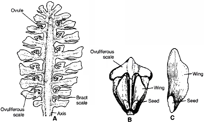
One male gamete or male nucleus fuses with the oosphere to form diploid zygote or oospore. The oospore forms an embryo while the ovule matures into a seed.
Part of the upper surface of the ovuliferous scale is peeled off alongwith the seed to form its wing.
A female cone takes about 26 months for reaching maturity. It then opens to release winged seeds which are dispersed by air. After falling on a suitable soil, each seed gives rise to a new plant.
Concept Builder
(a) Development of embryo is meroblastic in Pinus.
(b) The number of cotyledons in Pinus ranges from 3 -18.
(c) There are 4 tiers in proembryo, namely basal embryo tier, suspensor tier, rosette tier and upper tier.
(d) Development of sporangium in Pinus is eusporangiate.
(e) Pinus shows polyembryony by three methods -simple, cleavage and adventive.
Salient Features of Cycas (Sago Palm)
Cycas is called Palm-fern. It bears crown of leaves on unbranched stem called caudex like palms. It also shows circinate ptyxis and presence of ramenta like ferns.
Roots are of two types-normal tap root and ageotropic coralloid roots. Latter are formed in form of coralline masses which bear blue green algae in their cortical cells, e.g., Anabaena, etc. They help in nitrogen fixation.
Leaves are of two types-scale and foliage, both arising in whorls. Every year almost one is added.
Old leaves fall and leave characteristic leaf scars on the stem. Foliage leaves are leathery and pinnately compound. Scale leaves are brown, and membranous.
Stem of Cycas shows manoxylic and polyxylic wood.
Rachis shows presence of diploxylic vascular bundles arranged in shape of inverted omega (w).
Transfusion tissue replaces lateral veins in the leaves for lateral transport of food.
Plants are dioecious. Male cones are found but female cones are absent (loose megasporophylls). Largest egg, sperm and ovule are found in Cycas. Ovules are orthotropous.
Sperms in Cycas are multiflageliate (= multiciliate). Pollen tube is formed in Cycas, but is haustorial in nature. Thus, Cycas shows both siphonogamy and zooidogamy.
Gymnosperms
Gymnosperm
Any vascular plant that reproduces by means of an exposed seed, or ovule, is referred to as a gymnosperm. Many gymnosperm seeds are carried in cones and are not visible until they reach maturity. Both before and after fertilization, the ovules are not contained by any ovarian wall and remain visible. The seeds that form after fertilization are unprotected, or naked. Gymnosperms are medium- to tall-sized trees and shrubs. The gigantic redwood tree Sequoia, a gymnosperm, is one of the world's tallest tree species. Taproots are the most common type of root. Some genera have mycorrhizal fungi connected with their roots (Pinus), while others have small specialized roots (Cycas) termed coralloid roots that are coupled with Nitrogen-fixing cyanobacteria.Cycas has unbranched stems, whilePinus andCedrushave branched stems. Simple or complicated leaves are possible. The pinnate leaves of Cycas last for a few years. Gymnosperm leaves are well-adapted to tolerate temperature, humidity, and wind extremes. The needle-like leaves of conifers minimize the surface area. Their thick cuticle and recessed stomata also aid in water conservation.
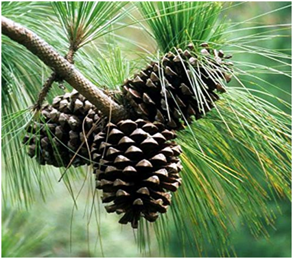
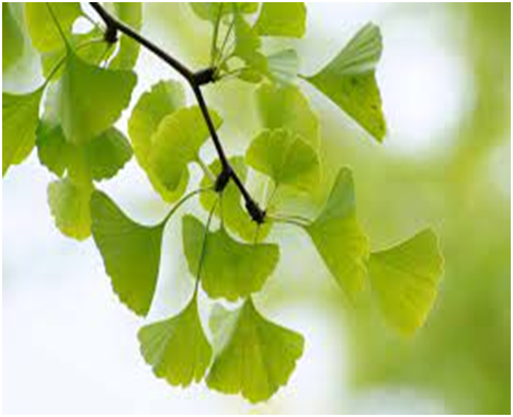
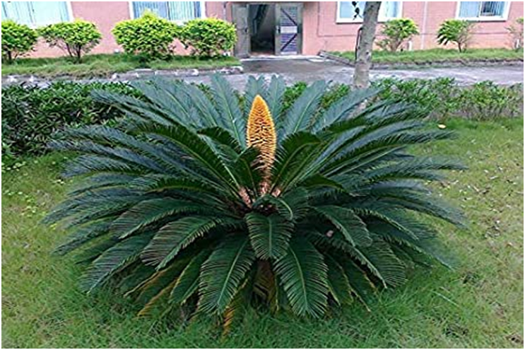
Figure 7
Gymnosperms generate haploid microspores and megaspores and are heterosporous. Both types of spores are formed in sporangia, which are carried on sporophylls that are spirally arranged along an axis to create lax or compact strobili or cones. Microsporangiate or male strobili contain microsporophylls and microsporangia. The microspores mature into a male gametophyte that is extremely small and restricted to a small number of cells. A pollen grain is a gametophyte that has been reduced in size. The microsporangia is where pollen grains grow. Macrosporangiate or female strobili are cones carrying megasporophylls with ovules or megasporangia. On the same tree, male and female cones or strobili can be found (Pinus).
Male cones and megasporophylls, on the other hand, are produced on distinct trees in Cycas. One of the nucellus cells differentiates into the megaspore mother cell. The nucellus is shielded by envelopes, and the resulting structure is known as an ovule. Megasporophylls carry the ovules, which can be grouped to create female cones. The megaspore mother cell splits into four megaspores by meiosis. One of the megaspores within the megasporangium matures into a multicellular female gametophyte with two or more archegonia or female sex organs. Megasporangium also retains the multicellular female gametophyte.
Unlike bryophytes and pteridophytes, male and female gametophytes in gymnosperms do not have their own free-living existence. They stay in the sporangia that are left on the sporophytes. The microsporangium releases the pollen grain. They are carried by the wind and come into touch with the opening of megasporophyll ovules. In the ovules, the pollen tube containing the male gametes develops towards archegonia and discharges its contents near the archegonia's mouth. The zygote develops into an embryo after fertilization, and the ovules mature into seeds. These seeds aren't protected in any way.
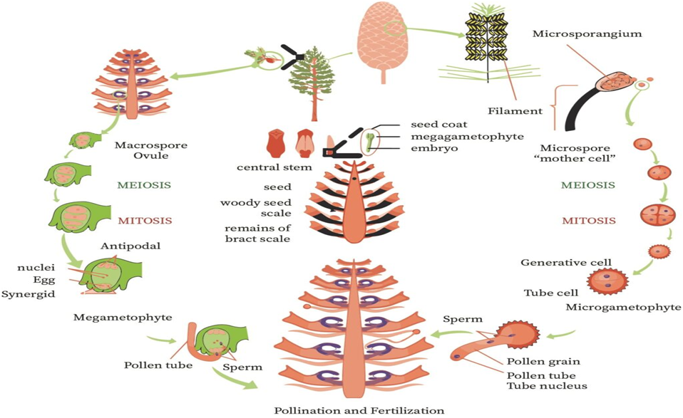
Angiosperms
- Books Name
- ACME SMART COACHING Biology Book
- Publication
- ACME SMART PUBLICATION
- Course
- CBSE Class 11
- Subject
- Biology
ANGIOSPERMS (FLOWERING PLANTS OR ANTHOPHYTES)
Angiosperms-Classification up to Class
After Linnaeus work on classification of plants, the taxonomists realised the necessity of information on natural history of vegetation and the affinities at various levels of hierarchy.
George Bentham and J.D. Hooker gave most important natural system of classification of angiosperms and published it in three volumes of Genera Plantarum.
They described 202 families. In this system, description of plants was based on detailed studies and dissections.
This system is followed in all British Commonwealth countries including India.
The system being handy, it is preferred and used by the students in practical classes. This system covered about ninety seven thousand species of seed plants.
In Bentham and Hooker system of classification, phanerogams were divided into three classes on the basis of morphological characters, such as leaf arrangement and venation pattern, number of members in floral whorls like calyx, corolla, androecium and gynoecium, number of cotyledons in the seed and seeds with or without cover.
In this classification, Class is like division, series is like class, cohort is like order and order is like family.
An outline of the Bentham and Hooker classification of phanerogamia is given below:
Class 1 : Dicotyledonae
General characters
Pentamerous flower, reticulate venation in leaves, two cotyledons in seed, open vascular bundles (with cambium), secondary growth present, wood formation occurs.
It is divided into 3 sub-classes
Sub-class 1 : Polypetalae -Petals free.
It includes three series:
Series 1 : Thalamiflorae -Flower hypogynous, stamens and pistils many (indefinite), petals free, distinct sepals are free from ovary. It has 6 orders, e.g., Ranales, Parietales, Malvales etc.
Series 2 : Disciflorae -Flower hypogynous, Calyx consists of free or united sepals, Petals free, a prominent cushion shaped disc is present below ovary. It has 4 orders, e.g., Sapindales, Geraniales etc.
Series 3 : Calyciflorae -Flower perigynous or epigynous, calyx contains united sepals (rarely free), ovary inferior. It has 5 orders, e.g., Rosales, Umbellates etc.
Sub-class 2 : Gamopetalae -Petals united or fused.
It includes 3 series:
Series 1 : Inferae -Flower epigynous, ovary inferior, stamens as many as corolla lobes or fewer. It has 3 orders, e.g., Asterales, Campanulates etc.
Series 2 : Heteromerae -Ovary usually superior, carpels more than two. It has 3 orders , e.g., Ericales, Primulales, Ebenales.
Series 3 : Bicarpellatae -Ovary usually superior, two carpels (rarely one or three) . It has 4 orders, e.g., Lamiales, Polemoniales etc.
Sub-class 3 : Monochlamydeae -Flower incomplete, no distinction between calyx and corolla, pe¬rianth present which is usually sepaloid and may be absent.
It includes 8 series:
Series 1 : Curvembryeae -Embryo curved, generally one ovule.
Series 2 : Multiovulatae Aquaticae -Plants are aquatic, submerged herbs, syncarpous ovary.
Series 3 : Multiovulatae Terrestres -Plants are terrestrial, syncarpous ovary.
Series 4 : Microembryae -Very minute or small embryo.
Series 5 : Daphnales -Ovary with one carpel and one ovule.
Series 6 : Achlamydosporeae -Ovary unilocular with one to three ovules. Ovary inferior.
Series 7 : Unisexuales -Flower unisexual.
Series 8 : Ordines Anomali -Families having plants with anomalous (abnormal) characters.
Class 2 : Gymnospermae
Sex organs are cones, perianth absent, ovule naked (not found inside ovary), seeds are also naked, haploid endosperm.
It has three families -Gnetaceae, Coniferae and Cycadaceae.
Class 3 : Monocotyledonae
Parallel venation in leaves, embryo with one cotyledon, flower usually trimerous, wood absent, no secondary growth.
It include 7 series:
Series 1 : Microspermae -Ovary inferior, seed minute.
Series 2 : Epigynae -Ovary inferior, seeds larger.
Series 3 : Coronarieae -Ovary superior, perianth coloured.
Series 4 : Calycineae -Ovary superior, perianth green.
Series 5 : Nudiflorae -Perianth absent, ovary superior.
Series 6 : Apocarpae -Carpels free (apocarpous).
Series 7 : Glumaceae -Flowers arranged in spikelets with bracts, perianth reduced, bracts large and scaly.
Merits of Bentham and Hooker's System
1. It is useful for practical value.
2. Ranales have been given the most primitive position among dicots.
3. Glumaceae is considered most advanced among monocots.
Demerits of Bentham and Hooker's System.
1. Gymnosperms were placed between dicots and monocots.
2. Many important floral characters have been neglected.
3. It is not a phylogenetic system and believes in the fixity of species.
4. Some closely related families were separated and placed under different cohorts (order) and unrelated families have been put nearer.
5. Advanced family like Orchidaceae (Microspermae) have been considered as primitive
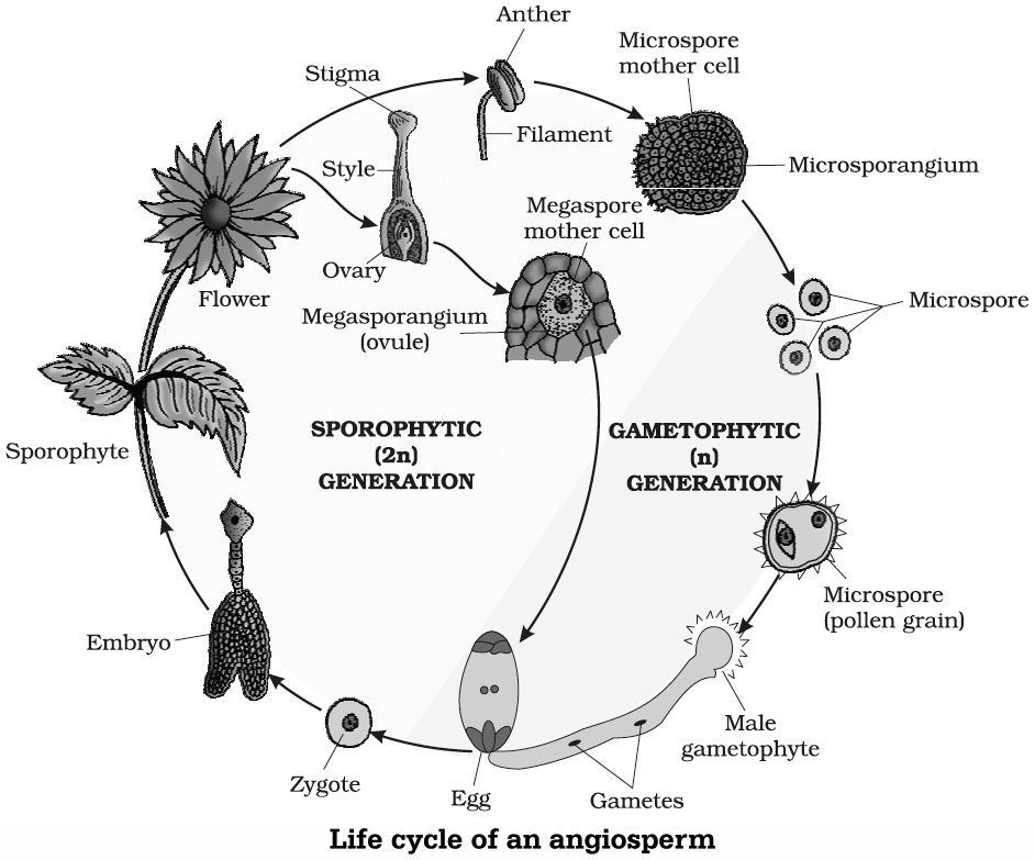
Angiosperms are those seed plants in which ovules and seeds are covered (inside fruits) and the sporophylls are organised into flowers. These are found in almost all the possible habitats. Tillandsia (Spanish moss) is an epiphytic angiosperm.
General characters
Flowering plants or angiosperms are the most recent and highly evolved plants.
Sporophylls are aggregated in flowers. It is their most striking feature. Therefore, angiosperms are also called flowering plants.
Stamen (male sex organ) consists of a filament and an anther. Carpel (female sex organ) is rolled and partly sterilised to produce a stigma, style and ovary, containing ovules.
Pollination occurs through several agencies, but most prominent amongst them is by animals, especially insects.
Pollen grains or microspores reach stigmatic surface found at the tip of carpel or megasporophyll (indirect pollination).
Female gametophyte or embryo sac is highly reduced and generally develops upto 8-nucleate stage prior to fertilization.
Archegonia are absent. Instead, there is one egg surrounded by two specialised synergid cells that attract the pollen tube. The latter brings two naked non-flagellate male gametes.
There occurs double fertilization . One produces zygote, hence embryo. The other forms primary endosperm cell.
Endosperm is formed through triple fusion and is generally triploid.
Fertilized ovules ripen into seeds. The seeds are covered by ovary wall. A fruit is technically a ripened ovary. Fruits not only protect the seeds but also help in their dispersal.
Xylem contains vessels.
Phloem possesses sieve tubes and companion cells.
Angiosperms are divided into two sub-groups-dicotyledons and monocotyledons, mainly on the basis of number of embryonic leaves or cotyledons. The two are commonly called as dicots and monocots.
Differences between dicots and monocots
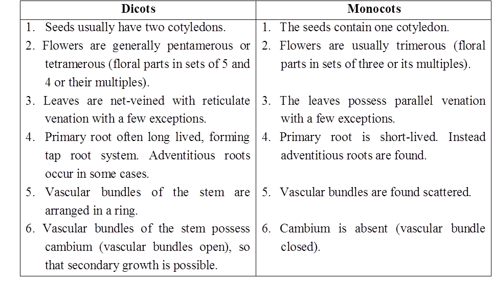
Differences between various plant groups having embryo
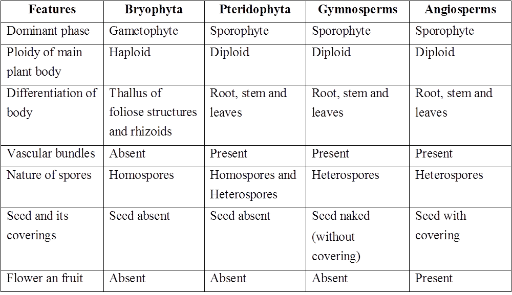
Angiosperms
Angiosperms
Angiosperms (flowering plants) generate pollen grains and ovules in specialized structures known as flowers. The seeds of angiosperms are encased in fruits. The angiosperms are a diverse group of plants that can be found in a variety of environments. They range in size from the tiniest Wolffia to massive Eucalyptus trees (over 100 meters). Food, fodder, fuel, medicines, and a variety of other commercially significant things are all provided by them. There are of two types: monocotyledons and dicotyledons. Seeds with two cotyledons, reticulate venations in leaves, and tetramerous or pentamerous flowers (four or five members in each floral whorl) distinguish dicotyledons.
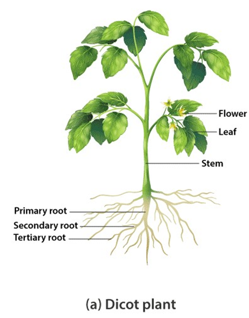
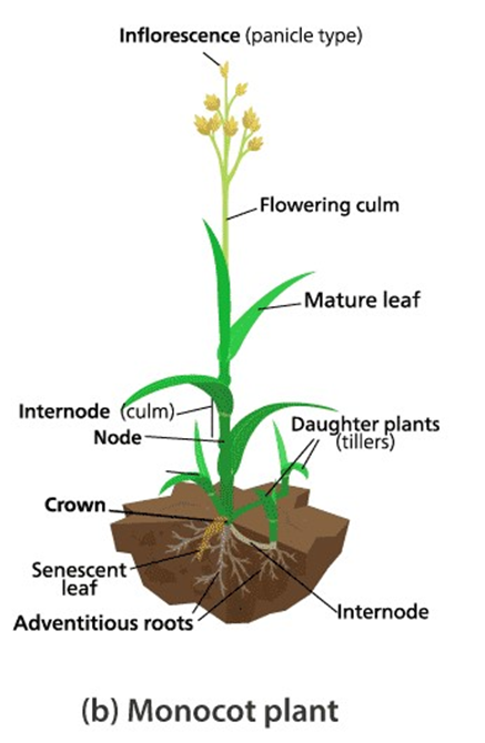
Figure 9: A dicot and monocot plant.
Monocotyledons, on the other hand, have single cotyledonous seeds, leaves with parallel venation, and trimerous flowers with three members in each floral whorl. The stamen is a flower's male sex organ. A short filament with an anther at the tip makes up each stamen. The pollen mother cell divides through meiosis within the anthers to produce microspores, which mature into pollen grains. The pistil is a flower's female sex organ. A large ovary, a long slender style, and the stigma make up the pistil. Ovules can be found inside the ovary. Each ovule has a megaspore mother cell, which goes through meiosis to produce four haploid megaspores. To produce the embryo sac, three of them degenerate and one divides.
A three-celled egg apparatus – one egg cell and two synergids, three antipodal cells, and two polar nuclei – is found in each embryo sac. Eventually, the polar nuclei combine to form a diploid secondary nucleus. Pollen grains are dispersed from the anthers and carried to the stigma of a pistil by wind or other means. Pollination is the word for this process. The pollen grains germinate on the stigma, and the pollen tubes grow through the stigma and style tissues to reach the ovule. Two male gametes are released from the pollen tubes into the embryo sac. Syngamy occurs when one of the male gametes combines with an egg cell to generate a zygote. The triploid primary endosperm nucleus (PEN) is formed when the other male gamete combines with the diploid secondary nucleus.
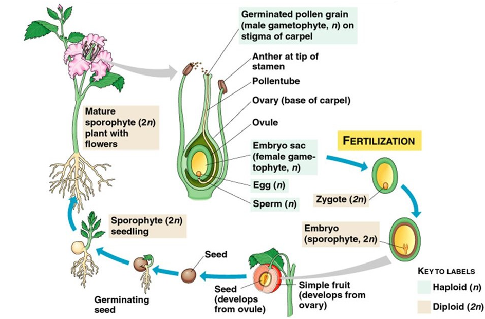
This phenomenon is known as double fertilization because it involves two fusions, syngamy and triple fusion, which is unique to angiosperms. The zygote matures into an embryo (with one or two cotyledons), and the PEN matures into endosperm, which feeds the growing embryo. After fertilization, the synergids and antipodals disintegrate. The ovules become seeds, and the ovaries become fruit, during these activities.
Plant Life Cycles and Alteration of Generations
- Books Name
- ACME SMART COACHING Biology Book
- Publication
- ACME SMART PUBLICATION
- Course
- CBSE Class 11
- Subject
- Biology
Plant Life Cycles and Alternation of Generations
Life cycle of a plant is called alternation of generations in plants, both haploid and diploid cells can divide by mitosis.
This ability leads to the formation of different plant bodies -haploid gamete producing plant (gametophyte) and diploid spore producing plant (sporophyte).
The haploid plant body produce, gametes by mitosis. Following fertilization, the zygote may divide by mitosis to produce sporophyte or may divide by meiosis to produce haploid spores.
Haploid spores in turn divide mitotically to produce gametophyte. Thus, during the life cycle of a sexually reproducing plant there occurs an alternation of generations between the haploid and diploid phases. Syngamy and meiosis seems to be instrumental for this event.
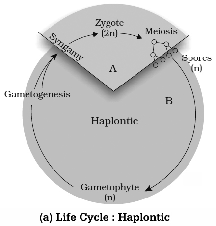
There are three chief patterns of life cycle. They fundamentally differ in the duration of the haploid and diploid phases. These are:
(i) Haplontic : It is characterized by the dominance of haplophase. The plant body is gametophyte and independent. The sporophyte is dependent on gametophyte, and is not a free living body. The major part of life is dominated by gametophyte, e.g., majority of green algae viz. Chlamydomonas, Ulothrix, Spirogyra etc.
(ii) Diplo-haplontic or Haplo diplontic: It is characterized by the existence of both sporophyte as well as gametophyte. Both of them are photosynthetic multicelled bodies and often free living with two nearly equal phases -the diplophase and haplophase. If sporophytes and gametophytes are morphologically different then the life cycle is termed as Diplo-haplontic-heteromorphic, e.g., all pteridophytes; all bryophytes some brown algae viz., Laminaria and other Kelps. If the two phases are morphologically identical, then life cycle is termed as Diplo-haplontic¬isomorphic, e.g., green algae viz., Ulva and Cladophora; brown algae, such as Ectocarpus
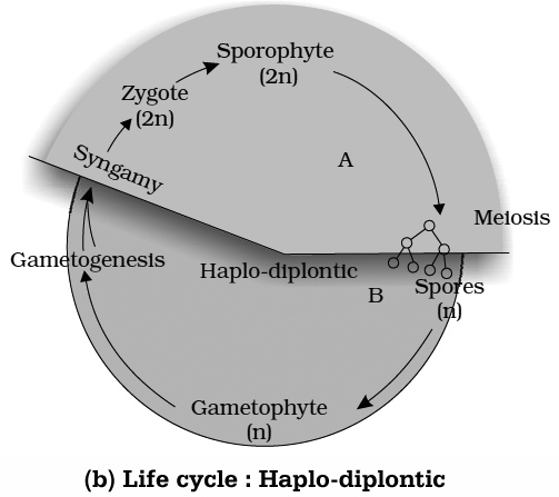
(iii) Diplontic : It is characterized by the dominance of diplophase. The plant body is sporophyte which is independent and free, living. The gametophytes are extremely reduced and are dependent physically as well as nutritionally on the sporophyte. The major part of the life is enjoyed by the sporophyte, e.g., all gymnosperms and angiosperms; Diatoms; some brown algae, viz., Fucus and Sargassum.
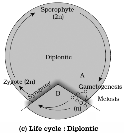
Plant Life Cycles and Alteration of Generations
Plant Life Cycles and Alternation of generations
The alternation of generations, also known as metagenesis or heterogenesis, is the transition between a sexual and asexual phase in an organism's life cycle. Both physically and chromosomally, the two phases, or generations, are often unique.
Mitosis allows haploid and diploid cells in plants to divide. This ability results in the development of haploid and diploid plant bodies. Mitosis creates gametes in the haploid plant body. A gametophyte is represented by this plant body. The zygote divides by mitosis after fertilization to generate a diploid sporophytic plant body. Meiosis produces haploid spores in this plant body. These, in turn, divide by mitosis to produce a haploid plant body.During the life cycle of any sexually reproducing plant, the gamete-producing haploid gametophyte, and the spore-producing diploid sporophyte alternate generations.
Individuals from diverse plant groups differ in the following patterns:
1. The one-celled zygote is the sole representative of a sporophytic generation. Sporophytes do not exist as free-living organisms. The zygote undergoes meiosis, which leads tothe creation of haploid spores. The gametophyte is formed when haploid spores divide mitotically. The free-living gametophyte is the major photosynthetic phase in such plants. The term "haplontic" refers to this type of life cycle. This pattern is found in many algae, including Volvox, Spirogyra, and some Chlamydomonas species.
2. The kind in which the diploid sporophyte is the dominant, photosynthetic, and independent phase of the plant is the other extreme. The single to few-celled haploid gametophyte represents the gametophytic phase. This type of life cycle is known as diplontic.This pattern is shown by the alga Fucus sp. Furthermore, all seed-bearing plants, such as gymnosperms and angiosperms, follow this pattern with some modifications, with the gametophytic phase ranging from single to multicellular.
3. Bryophytes and pteridophytes, for example, have an intermediate state (Haplo-diplontic), in which both phases are multicellular. Their dominating periods, however, are distinct.
A haploid gametophyte represents a dominant, autonomous, photosynthetic, thalloid, or erect phase that alternates with the short-livedmulticellular sporophyte that is entirely or partially dependent on the gametophyte for anchoring and nourishment. This pattern is found in all bryophytes. A dominant, autonomous, photosynthetic, vascular plant body represents the diploid sporophyte. It alternates with a haploid gametophyte that is multicellular, saprophytic/autotrophic, and independent but short-lived. The haplo-diplontic life cycle is a pattern like this.
All pteridophytes exhibit this pattern. Interestingly, while most algal genera are haplontic, some of them such as Ectocarpus, Polysiphonia, and kelps are haplo-diplontic. Fucus, an alga is diplontic.
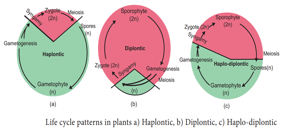
Basis of Classification
- Books Name
- ACME SMART COACHING Biology Book
- Publication
- ACME SMART PUBLICATION
- Course
- CBSE Class 11
- Subject
- Biology
Animal Kingdom covers about 35 phyla, of which 11 are considered to be major phyla. In major phyla, 10 are from Non-chordates and 1 from the chordates.
HABITAT
The place on the earth, where an animal finds optimum conditions for food and shelter is called its habitat. Habitat can be defined as the physical and geometrical space where an organism lives.
The habitat may be water, soil or earth’s surface. On the basis of their habitat, animals can be of following types :
1. Terrestrial habitat :
Animals which live on land or dry earth’s surface, are said to have terrestrial habitats.
Terrestrial animals are adapted for following types of habitat modes :
a. Aerial or flying : These animals possess wings and therefore can fly. e.g., Birds, bats.
b. Arboreal : These animals mainly live on trees. e.g., Bat, Monkey.
c. Fussorial : These animals live in burrows or underground. e.g., Rabbits, rat, earthworm etc.
d. Scansorial : These animals can climb on walls, rocks etc. e.g., Wall lizard, flying squirrel.
e. Cursorial : These animals are adapted to run very fast. e.g., Tiger, horse, dog etc.
2. Aquatic animals :
These animals live in water. They may be fresh water or marine water.
The aquatic animals may further be of following types :
a. Zooplanktons : The animals which flow passively, with water current, on the surface of water, are called zooplanktons. e.g., many aquatic protozoans, larvae, crustaceans etc.
b. Nektons : These animals can float actively in water body even against water current. e.g., many fishes such as sharks, many skates and rays.
c. Benthos : These animals live at the bottom of the water body. e.g., Deep sea fishes, many echinodermates, sponges and corals.
d. Pelagic : These animals usually live at the surface or water body. These include both zooplanktons and Nektons.
3. Amphibian
These animals living on land as well as in water.
HABITS
Nature of living is called habit. Animals lead various types of life and on this basis, they are of following types:
1. Solitary animals : Animals which have singly (not in groups). e.g., Taenia, Earthworm etc.
2. Colonial animals : Animals which live in groups and form colony. e.g., Volvox, honeybee, termites, wasp etc.
3. Gregarious animals : Animals which live in groups, but do not interact with each other. e.g., locust.
4. Free living animals : Animals, which do not depends on other organisms for food and shelter and live independently. e.g., cat, dog etc.
5. Parasitic animals : Animals which depends on other living organisms for food are called parasitic organisms. e.g., Entamoeba, Taenia, Fasciola, Ascaris, Louse etc.
6. Saprophagous animals : Animals which obtain their food from dead and decaying organisms and organic matter are called saprophagous animals. e.g., house fly etc.
7. Sanguivorous animals : Animals which feed on the blood of other organisms. e.g., Mosquitoes, Leech etc.
8. Sessile (sedentary) animals : Animals which do not move here and there are remain immotile, are called sessile organisms. e.g., Herdmania.
9. Motile animals : Animals, which have locomotory organs and can move here and there, are called motile organisms. e.g., man horse etc.
10. Diurnal animals : Animals, which are active during day time for obtaining their food, are called diurnal organisms. e.g., man, horse, dog etc.
11. Nocturnal animals : Animals, which are active at night for obtaining food are called nocturnal animals. e.g., Cockroach etc.
12. Crepuscular animals : Animals which are active at dusk (evening) for obtaining their food are called crepuscular animals. e.g., rabbit.
13. Vasperal animals : Animals which are active early morning. e.g., Birds.
14. Caprophagous animals : Animals which can eat their own faecal matter to re-digest it. e.g., Rabbit.
Table - Main Phyla of Animal Kingdom
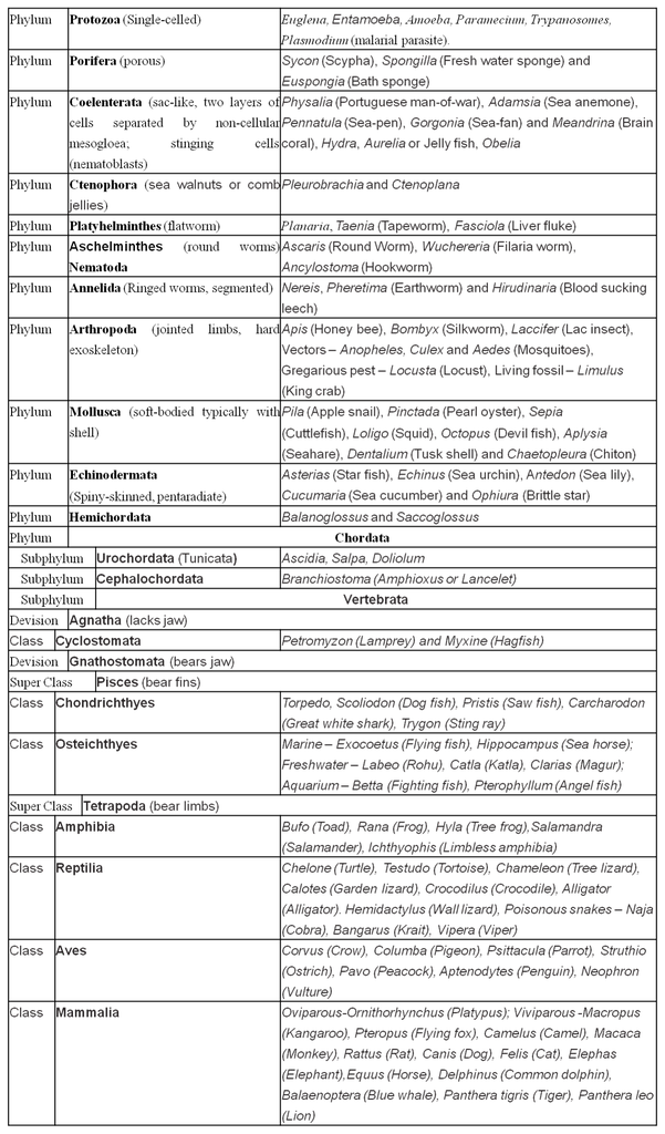
METAZOA
Animal groups are characterised by mobility and the presence of a sensory or a nervous system. These systems receive stimuli from the environment and animals respond by exhibiting some behaviour.
The only exception is of poriferans (pore-bearers) or the sponges. They have no cell that can be termed as nerve cell.
Like plant life, early animal life also arose in sea. The animals which live on the sea floor are called Benthonic (e.g., echinoderms, corals and deep sea fishes), whereas those which swim about actively in sea are called Nektons.
The multicellular eukaryotic organisms with holozoic mode of nutrition are called metazoans. Based on complexity of organisation, metazoans are further sub-divided into two sub-kingdoms, Parazoa and Eumetazoa.
(a) Parazoa: Parazoa include the sponges in which the cells are loosely aggregated and do not form tissues or organs.
(b) Eumetazoa: Eumetazoa includes the rest of animals in which the cells are organised into structural and functional units called tissues, organs and organ systems.
LEVELS OF ORGANISATION
1. Cellular Level :
Though all members of Animalia are multicellular, all of them do not exhibit the same pattern of organisation of cells.
For example, in sponges, the cells are arranged as loose cell aggregates, i.e., they exhibit cellular level of organisation.
Some division of labour (activities) occurs among the cells.
2. Tissue Level :
In coelenterates and ctenophores, the arrangement of cells is more complex.
Here the cells performing the same function are arranged into tissues, hence it is called tissue level of organisation.
3. Organ Level :
A still higher level of organisation, i.e., organ level is exhibited by members of Platyhelminthes and other higher phyla where tissues are grouped together to form organs, each specialised for a particular function.
In animals like Annelids, Arthropods, Molluscs, Echinoderms and Chordates, organs have associated to form functional systems, each system concerned with a specific physiological function.
This pattern is called organ system level of organisation. Organ systems in different groups of animals exhibit various patterns of complexities.
For example, the digestive system in Platyhelminthes has only a single opening to the outside of the body that serves as both mouth and anus, and is hence called incomplete.
A complete digestive system has two openings, mouth and anus.
Similarly, the circulatory system may be of two types:
(i) Open type
(ii) Closed type
(i) Open type:
In this, the blood is pumped out of the heart and the cells and tissues are directly bathed in it. The blood is pumped by the heart into the blood vessels that open into blood spaces (sinuses).
There is no capillary system (e.g., in most arthropods, non-cephalopod molluscs and tunicates). These sinuses are actually the body cavities and are called the haemocoel.
The pressure of the blood is low; it moves slowly between the tissues, and finally, returns to the heart via the open-ended blood vessel
(ii) Closed type :
Many invertebrates and all the vertebrates, including humans, have a closed circulatory system. In closed type, the blood flows inside the body through a series of blood vessels of varying diameter (arteries, veins and capillaries).
In this system, the same blood regularly circulates in the body under high pressure and returns back to the heart without leaving the system of tubes.
SYMMETRY
The animals can be categorised on the basis of their body symmetry:
(i) Radial symmetry: When any plane passing through the central axis of the body divides the organism into two identical halves, it is called radial symmetry. The animals with radial symmetry are put in the group Radiata. For example, cnidarians (hydra and jelly fish). Biradial symmetry is present in sea anemone, ctenophores.
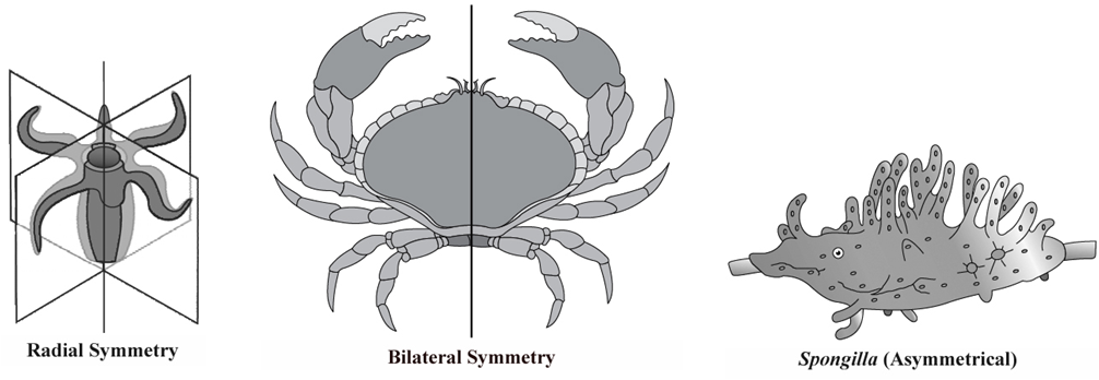
(ii) Bilateral symmetry: The animals with bilateral symmetry are put in group Bilateria. The body can be divided into identical right and left halves in only one plane. For example, platyhelminthes, annelids, arthropods etc. (platyhelminthes to chordates).
(iii) Asymmetry: Asymmetric organisms cannot be divided along any plane to produce two equivalent halves. Sponges are mostly asymmetrical.
GERM LAYERS
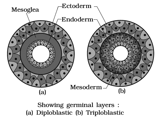
Germ layers give rise to all the tissues/organs of the fully formed individuals. On the basis of number of germ layers animals can be
1. Diploblastic: In diploblastic animals, the body cells are arranged in two layers-an outer ectoderm and an inner endoderm with an intervening undifferentiated mesoglea. e.g. Coelenterates.
2. Triploblastic: If the body wall in animals is made of three germ layers i.e. ectoderm, mesoderm and endoderm, they are called triploblastic animals. e.g., Platyhelminthes to chordates.
BODY PLAN
Though diverse in shape and size, animals have body that fits in one of the three basic plans:
1. Cell aggregate : Cell aggregate type of body plan is present in sponges. They are clusters of cells with a rudimentary division of labour among them. There are no tissues, or organs.
2. Blind sac : Blind sac type of body plan is present in platyhelminthes and coelenterates where the alimentary canal has only one opening.
3. Tube within tube : Tube-within tube type of body plan is present in Nemathelminthes, Annelida, Arthropoda, Mollusca, Echinodermata and Chordata. The digestive system is a continuous tube with an opening at both end.
BODY CAVITY OR COELOM
Presence or absence of cavity between the body wall and the gut wall is very important for classification
1. Acoelomate : The animals in which the coelom is absent are called acoelomates, for example poriferans, coelenterates, ctenophores, flatworms. In flatworms, the spaces between various organs are filled with special tissue called parenchyma.
2. Pseudocoelomate : The body cavity is not completely lined with mesoderm. Instead, the mesoderm is present as scattered pouches in between the ectoderm and endoderm. Such a body cavity is called pseudocoelom e.g. in roundworm.
3. Eucoelomate : The true coelom is a body cavity which arises as a cavity in embryonic mesoderm. In this case, the mesoderm of the embryo provides a cellular lining, called coelomic epithelium or peritoneum, to the cavity. The coelom is filled with coelomic fluid secreted by the peritoneum. True coelom is found in annelids, echinoderms and chordates. True coelom is of two types:
- Schizocoelom : It develops by the splitting up of mesoderm. It is found in annelids, arthropods and molluscs. Body cavity of arthropods and non-cephalopod molluscs is called haemocoel.
- Enterocoelom : The mesoderm arises from the wall of the embryonic gut or enteron as hollow outgrowths or enterocoelomic pouches. It occurs in echinoderms and chordates.
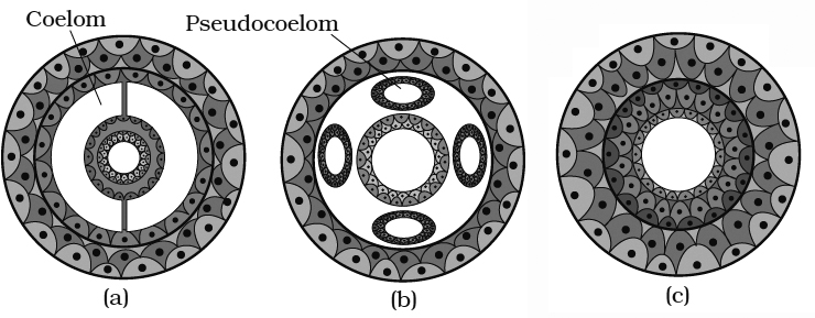
Segmentation
In some animals, the body is externally and internally divided into segments with a serial repetition of at least some organs.
For example, in earthworm, the body shows this pattern called metameric segmentation and the phenomenon is known as metamerism.
Metameric segmentation is present in annelids only.
Notochord
Notochord is a mesodermally derived rod-like structure formed on the dorsal side during embryonic development in some animals. Animals with notochord are called chordates and those animals which do not have this structure are called non-chordates, e.g., porifera to echinoderms.
Basis of Classification
Basis of classification in Animals
Despite differences in structure and shape across animals, there are essential similarities in cell organization, body symmetry, coelom type, and digestive, circulatory, and reproductive system patterns. These characteristics are used to classify animals, and some of them are covered here. The requirement for classification grows in importance as more than a million species of animals have been described to date. Newly described species can also be assigned a systematic position thanks to categorization.
A. Level of organization: Even though all members of Animalia are multicellular, they do not all have the same cell organization pattern. Sponge cells, for example, are grouped as loose cell aggregates, indicating that they have a cellular level of organization. Among the cells, there is some division of labour (activities). The cell arrangement in coelenterates is more complicated. The cells that perform the same function are grouped together ies, which is referred to as the tissue level of organization. Members of the Platyhelminthes and other higher phyla have an even higher degree of organization, namely the organ level, in which tissues are gathered together to form organs, each specialized for a certain function. Organs have come together to form functional systems in creatures including Annelids, Arthropods, Molluscs, Echinoderms, and Chordates.
B. Symmetry: The symmetry of animals can be used to classify them. Sponges are generally asymmetrical, which means that any plane passing through the centre split them in half evenly. Radial symmetry occurs when any plane traveling through the body's central axis divides the organism into two identical halves. This is the body plan of coelenterates, ctenophores, and echinoderms. Bilateral symmetry is found in animals such as annelids, arthropods, and others whose bodies can be divided into identical left and right halves in just one plane.
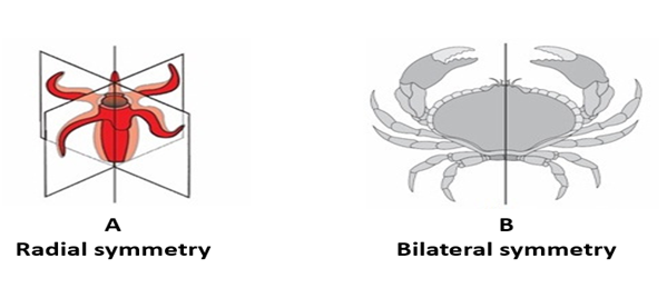
C. Germinal layers: During embryonic development, a germ layer is the first layer of cells that arises. Gastrulation is the process through which an early embryo develops three germ layers. Ectoderm, endoderm, and mesoderm are the three types of cellular layers that make up the body. Diploblastic creatures, such as coelenterates, have their cells organized in two embryonic layers, external ectoderm, and an internal endoderm. Between the ectoderm and the endoderm, there is an undifferentiated layer called mesoglea. Triploblastic animals are those in which the growing embryo has a third germinal layer, mesoderm, between the ectoderm and endoderm.
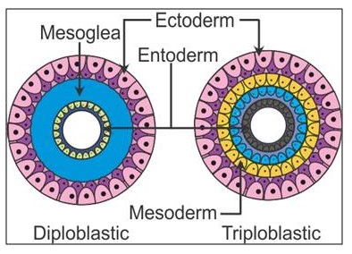
D.Coelom: When it comes to classification, the presence or absence of a cavity between the body wall and the gut wall is crucial. The coelom is the name for the bodily cavity that is lined by mesoderm. Coelomates are animals that have a coelom, such as annelids, mollusks, arthropods, echinoderms, hemichordates, and chordates. Mesoderm does not line the body cavity of some animals; instead, it appears as scattered pouches in between the ectoderm and endoderm. Pseudocoelom is the name for such a bodily cavity, and animals with them are known as pseudocoelomates, such as aschelminthes. Acoelomates, such as Platyhelminthes, are animals that do not have a body cavity.
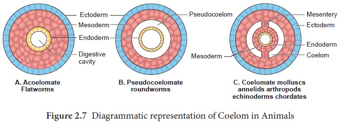
E. Segmentation: The body of several animals is divided into segments both visibly and internally, with at least some organs repeating serially. The body of an earthworm, for example, displays a pattern known as metameric segmentation, and the phenomenon is known as metamerism.
F. Notochord: In some animals, the notochord is a mesodermally derived rod-like structure that forms on the dorsal side during embryonic development. Chordates are creatures that have a notochord, while non-chordates are animals that do not have one, such as Porifera and Echinoderms.
The broad classification of Animalia is based on common fundamentalfeatures as discussed above. A schematic diagram for the same is given below.
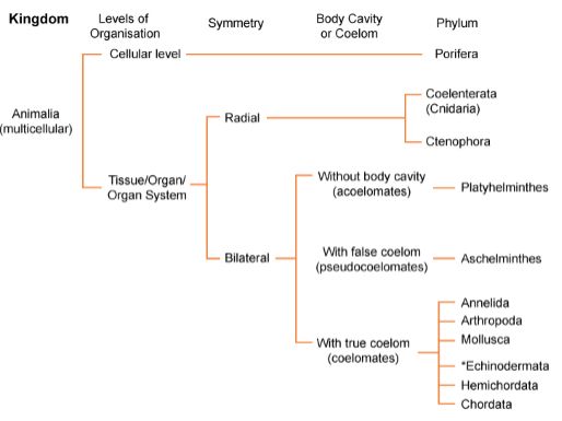
Classification of Animals
- Books Name
- ACME SMART COACHING Biology Book
- Publication
- ACME SMART PUBLICATION
- Course
- CBSE Class 11
- Subject
- Biology
TABLE : Salient Features of Different Phyla in the Animal Kingdom

Phylum : Porifera
- Books Name
- ACME SMART COACHING Biology Book
- Publication
- ACME SMART PUBLICATION
- Course
- CBSE Class 11
- Subject
- Biology
PHYLUM: PORIFERA (Sponges)
These are primitive, multicellular, asymmetrical (except Leucosolenia, Scypha) organisms having cellular level of organisation.
Most of them are marine and remain attached to rocks (sessile). A few live in fresh water e.g., Spongilla. This is the only phylum in animal kingdom without any nerve cells.
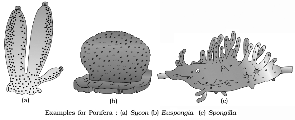
Important characters of phylum porifera
1. Body wall: The body wall of a common sponge consists of following layers:
(a) Pinacoderm (dermal layer)
It is outer cellular layer which consists of (i) flattened pinacocytes and (ii) oval porocytes
(b) Choanoderm (gastral layer) :
It is inner cellular layer which consists of highly specialized flagellated cells called choanocytes (collar cells).
They are the characteristic cells of this phylum responsible for ingestion of food, secretion of mesohyal and differentiation of sex cells.
(c) Mesohyl layer (mesenchyme) :
Basically, it is a noncellular layer found between pinacoderm and choanoderm. It has fine dispersed spongin fibres and numerous spicules.
It also contains amoebocytes (amoeba-like cells) of both pinacoderm and choanoderm.
Amoebocytes are modified into the following types :
- Archaeocytes. They may be converted into other types of cells and are also called undifferentiated "totipotent" cells.
- Trophocytes. They provide food to developing cells and are called nurse cells.
- Thesocytes. They store food granules.
- Gland cells. They secrete a slimy substance.
- Spongioblasts. They secrete spongin fibres of the mesohyllayer.
- Scleroblasts. They secrete spicules. In calcareous sponges, they are called calcoblasts and in silicious sponges, they are called silicoblasts.
- Collencytes. They secrete collagen fibers and form connective tissue.
- Myocytes. They form a circular ring around the osculum and help in closing and opening of the osculum.
- Germ cells (Sex cells). They form sperms and ova and develop during breeding season.
- Chromocytes. They contain pigment granules and excretory substances.
(xi) Phagocytes. They collect food from choanocytes through their pseudopodia and also engulf excreta and damaged tissues.
2. The central body cavity of a sponge is called spongocoel or paragastric cavity.
3. The continuous water current flowing through the canal system is very important for the life of a sponge. It brings in food and oxygen and carries away carbon dioxide, nitrogenous wastes and reproductive bodies. Thus the canal system helps the sponge in nutrition, respiration, excretion and reproduction.
4. Skeleton: Almost all sponges possess an internal skeleton. It may consist of calcareous or siliceous spicules or of fine spongin fibres or both, located in the mesohyl layer.
5. Digestion: It is intracellular and takes place inside food vacuoles as in protozoans.
6. Circulation: Distribution of food from the ingesting cells to others is brought about by wandering amoebocytes of mesohyl layer.
7. In case of Sycon, pinacoderm is divided into exopinacoderm and endopinacoderm. Exopinacoderm lines the incurrent canal and the spongocoel. Radial canals are lined by flagellated choanocytes.
Concept Builder
What is luffa sponge?
A luffa sponge isn't a sponge at all but a gourd. When dried, the fibrous material found in gourd forms a "skeleton" similar to that of some sponges. It can be used for the same purposes e.g., scrubbing.
8. Canal System: The body of a sponge is organized in such a manner as to form a complex system of pores and canals. This system is called canal system. It is meant for food gathering, respiration and removal of waste.
- Three types of canal systems are found in sponges:
(i) Ascon type : It is the simplest type of canal system which is found in Leucosolenia and few other sponges.

(ii) Sycon type : It is more complex than the ascon type. It is found in Sycon and some other sponges.
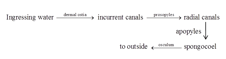
(iii) Leucon type : It is most complex canal system which is found in Spongilla and some other sponges.
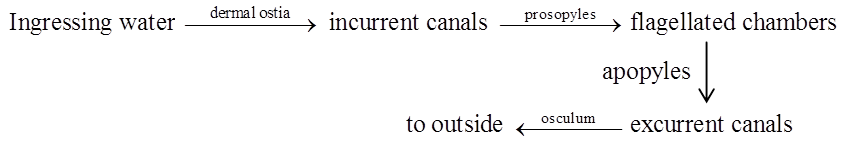
In Demospongiae, the leuconoid condition is derived from a larval stage, called the rhagon. It is a conical organism with a flattened base. The canal system of rhagon larva does not occur in any adult sponge. Because of its derivation from rhagon stage in Demospongiae, the leucon type of canal system is also called the rhagon type.
9. Respiration: Exchange of gases occurs by diffusion through the plasma membranes of the cells as in protozoans.
10. Excretion: Removal of metabolic wastes also occurs by diffusion through the plasma membranes of the cells as in protozoans. Ammonia is chief excretory waste.
11. Reproduction: Both asexual and sexual reproduction occur in sponges. Asexual reproduction occurs by fragmentation, budding and gemmules. They are hermaphrodite and show internal fertilization.
12. Development: Zygote undergoes holoblastic cleavage (complete division). The development is indirect and includes a free swimming larva, the amphiblastula (in Sycon) or parenchymula (in Leucosolenia and other Porifers) for dispersal of the species.
CLASSIFICATION
Based on the type of skeleton, phylum Porifera is divided into three classes.
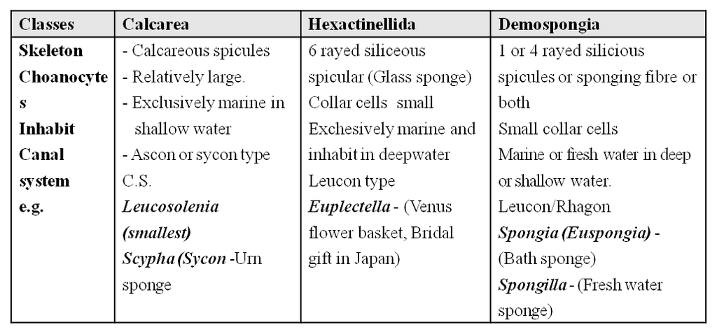
Concept Builder
Cultural Awarness
1. Using sponges: For centuries, people around the world have used natural sponges with spongin skeletons for cleaning, bathing by taking advantage of these sponges' soft flexible and highly porous bodies.
e.g., Euspongia. The ancient Greeks also used sponges as padding inside helmets.
2. Spongin fibres are elongated protein fibres which form a fibrous network.
3. Digestion in sponges is intracellular.
4. Regeneration in sponges was demonstrated by H.V.Wilson (1907). It is brought about by archaeocyte cells.
5. Sponge cells, seperated by straining pieces of sponge through a fine net, can reaggregate and grow into a sponge. So, a sponge is a republic of cells which identify one another, aggregate and grow together.
6. Sponge reproduces asexually by fragmentation. During sexual reproduction, some cells become egg or sperm cells. After fertilisation, the zygote develops into a flagellated larva which swims, settles in a new place and grows into a sponge.
7. Proterospongia is a connecting link between protozoa and porifera.
8. In Hyalonema, root tuft consists of bundle of long anchoring spicules. These may pass through the columella (body axis) as gastral cone. It is commonly known as glass rope sponge.
9. Euspongia is commonly found is Mediterranean sea. It is commonly known as Bath sponge.
10. Amphiblastula is the hollow larva of Sycon etc. whereas parenchymula is the solid larva of most of sponges, e.g., Leucosolenia.
Phylum : Porifera
Phylum Porifera
This animal group comprises Sponges and is thought to be the oldest on the planet. Sponges are the most basic multicellular creatures. They don't have any tissues or organs, despite being multicellular. Sponges need to be near water, thus they dwell in an aquatic environment. Water is essential for nutrient absorption, gas exchange, and excretion. The sponges' bodies have numerous holes or pores known as Ostia. Sponge bodies are built so that water can circulate through them, filtering out food and absorbing dissolved oxygen while also removing waste.This phylum of organisms lacks a specialized digestive, neurological, or circulatory system. Instead, they have a canal or water transport system that performs the activities of digesting, excretion, and gas exchange.
Their bodies are asymmetrical, and their design has been optimized to facilitate the most efficient flow of water via the core hollow that is present inside. Bacteria and other food particles found in the water are their main sources of nutrition. The spongocoel is a huge central hollow canal in their bodies. Water enters the spongocoel through the Ostia and exits through the osculum. Choanocytes or collar cells, with their flagellum jutting out, line up the spongocoel and canals.The water is moved throughout the sponge's body by the beating of this flagellum from all choanocytes. Common examples areSycon (Scypha), Spongilla (Freshwatersponge), and Euspongia(Bath sponge).
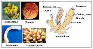
Phylum : Coelenterata (Cnidaria)
- Books Name
- ACME SMART COACHING Biology Book
- Publication
- ACME SMART PUBLICATION
- Course
- CBSE Class 11
- Subject
- Biology
PHYLUM: CNIDARIA (Coelenterata
- The phylum Cnidaria (the old name is Coelenterata) includes about 9,000 species, mainly marine. They are sessile, free swimming, radially symmetrical invertebrates and more complex than sponges
- General characters of this phylum are as follows:
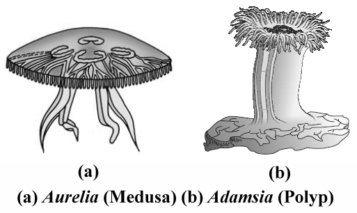
1. Cnidarians exhibit a blind sac body plan and are radially symmetric. They are more advanced than sponges in having true tissues. They are, however, acoelomate.
2. Body wall consists of only two cell layers, the ectoderm and the endoderm, separated by a jelly-like mesoglea. These animals are therefore diploblastic, that is, arising from two embryonic cell layers.
Concept Builder
Body wall in coelenterates (with reference to Hydra).
They are diploblastic animals i.e., they are derived from two layers-ectoderm and endoderm. These germ layers form the epidermis and gastrodermis.
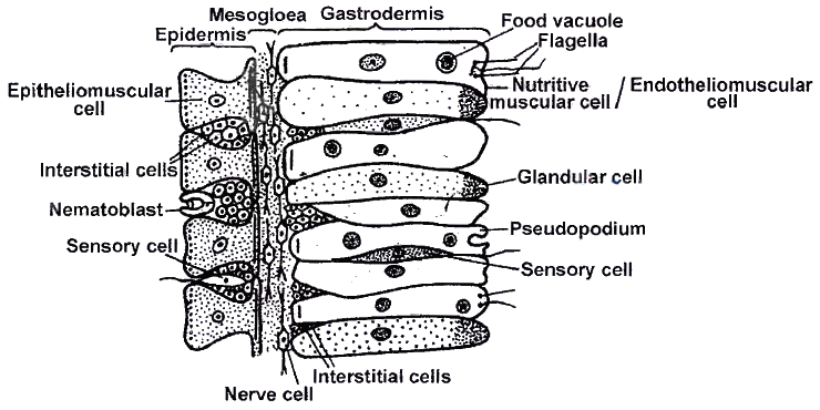
(1) Epidermis: The various epidermal cells are:
(a) Epithelio-muscular cells: Each cell has two functional parts, the outer epithelial part, extending to the body surface and the basal muscular part drawn out into two muscle processes along the longitudinal axis of the body. The muscle processes contain a contractile fibril myoneme.
(b) Glandulo-muscular cells : The epithelio-muscle cells, chiefly in the region of the pedal disc, are especially modified to secrete sticky material for attachment of animal to substratum.
(c) Interstitial cells: They are totipotent cells which give rise to all the different cells of the body.
(d) Sensory cells: They are scattered throughout the epidermis. They are most numerous on the tentacles, hypostome and basal disc. They receive and transmit impulses.
(e) Nerve cells: They occur for the first time in coelentrates. They are present at the base of epithelio-muscle cells. They conduct impulses in all the directions.
(f) Germ cells : They remain in restricted regions. They proliferate to form gonads. They are ectodermal in hydrozoa and endodermal in Scyphozoa and Anthozoa.
(g) Cnidoblasts : Many of the interstitial cells of the epidermis become specialized to form the stinging cells, called the cnidoblasts. They migrate to the tentacles through the mesoglea by means of amoeboid movements. Projecting cnidoblasts act as organs for offence and defence.
Structure:
- The cytoplasm of a cnidoblast contains a conspicuous nucleus lying to one side, and a peculiar oval or pyriform sac filled with a poisonous fluid, hypnotoxin. The sac is a part of stinging apparatus, known as the nematocyst (stinging structure).
Occurence of nematocysts :
- The nematocysts occur scattered mostly singly, throughout the epidermis of the body but remain absent on the basal disc.
- They are abundant in the epidermis of the oral region and the tentacles where they cluster as wart-like "nematocyst batteries".
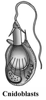
Difference between four nematocyst

(2) Gastrodermis: It is the inner layer of the body. The cells are of following types :
(a) Endothelio-muscle or nutritive muscle cells : They help in contraction of the body and nutrition.
(b) Endothelio-gland cells : They secrete digestive enzymes in coelenteron. In the region of hypostome and mouth are found mucous gland cells. Gland cells are absent in the tentacles and basal disc.
(c) It also contains interstitial cells, sensory cells and nerve cells but no cnidoblasts.
(3) They enclose gastrovascular cavity (coelenteron), having single opening hypostome. Their digestion is intra and extracellular.
Concept Builder
1. The green colour of Chlorohydra viridissima comes from alga Chlorella vulgaris (Zoochlorella), that lives inside the gastrodermal cells of Hydra in symbiotic relationship.
2. Corallium rubrum (red coral) has been used widely in jewellery and known as red moonga.
HYDRA

- Some important characters of Hydra are :
(i) Hydra flourishes well in cool, clean and stagnant water.
(ii) Number of tentacles surrounding mouth in Hydra is 6-10. Tentacles of Hydra are hollow while that of obelia are solid.
(iii) Food of Hydra is water fleas (Daphnia and Cyclops). Thus, it is carnivorous. It cannot digest starch.
(iv) No free larval stage in development of Hydra. Hence, development is direct.
(v) Most species of Hydra are dioecious/unisexual.
(vi) Male Hydra bears 1-8 conical testes towards distal end whereas female Hydra bears 1-2 rounded ovaries towards proximal end of the body.
(viii) Locomotion in Hydra occurs by looping, somersaulting, gliding, walking and floating etc.
METAGENESIS AND POLYMORPHISM
- Colenentrata members shows polymorphism most common is polyp & medusa forms.
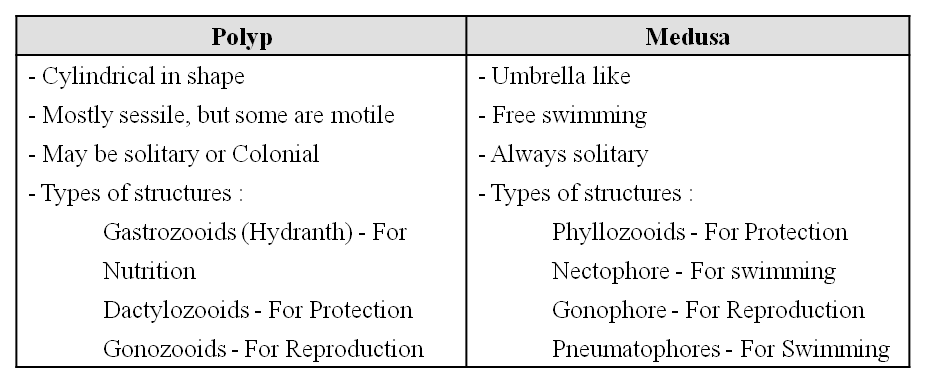
- In the life-cycle there is an alternation of the asexual polyp phase and the sexual medusa phase and it is termed as Metagenesis.
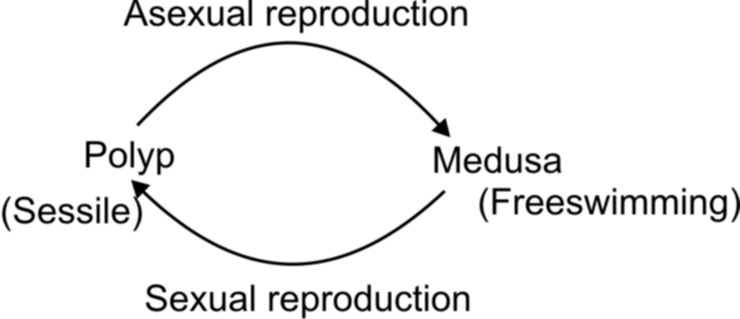
- During sexual reproduction, medusae liberate gametes into water. Following fertilization, the zygote forms a ciliated larva called Planula, which swims, settles and grows into a sessile polyp. (Some cnidarians, like Hydra, do not have a medusa stage. Hydra has no larval form, no metagenesis).
- Polyps reproduce asexually by budding where as, medusa liberates gametes into water during 'Sexual reproduction. Both asexual and sexual forms are free living.
CLASSIFICATION
Chiefly on the basis of the dominance of medusoid or polypoid phase in the life cycle, the phylum Cnidaria is divided into three classes

Concept Builder
Coral Reefs :
- A coral reef is a ridge or mound in a shallow, tropical sea, has its upper surface near the surface of water, supports a variety of animals and plants. It is formed of calcium carbonate produced by some of its inhabitant, chiefly medreporarian corals. Coral reefs form stable marine ecosystems. The coral reefs are of three kinds :
(i) Fringing Reefs : The coral reefs lying close to the shores of some volcanic island or part of some continent are called the fringing reefs. Hawaiian is lands are an ideal example of permanent fringing reefs.
(ii) Barrier Reefs : They are like the fringing reefs but are located some distance from the shore. The stretch of water of lagoon separates the barrier reefs from land. Australia's Great Barrier Reef is an example of such a reef which is about 900 nautical miles long and 90 miles from sea shore.
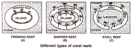
(iii) Atoll reefs : An atoll reef is also called coral island or lagoon island which is a circular or horse shoe shaped reef that encircles a lagoon but not an island. It may be complete or broken by a number of channels of which only a few are navigable. The Lakshadweep and Maldive islands in the Indian Ocean are composed of Atoll reef.
Phylum : Coelenterata (Cnidaria)
Phylum Coelenterata
They are also known by the name Cnidaria. They are radially symmetrical aquatic, primarily marine, sessile, or free-swimming organisms. Typically, these are invertebrate species with a very basic tissue arrangement. Coelenterates can be found in colonies or alone. Coelenterates are the simplest animal group with genuine tissues and the coelenteron or gastrovascular cavity as a distinguishing feature. The word Cnidaria comes from the stinging capsules or nematocysts found on the tentacles and body, which are called Cnidoblasts or Cnidocytes. Cnidoblasts are the phylum Cnidaria's distinguishing trait.They contain stinging capsules called nematocysts. It aids in both prey capture and defence. Cnidoblasts are employed for prey capture, defence, and anchoring. Cnidarians are diploblastic and have tissue-level organization. They have a single aperture in the center of their gastro-vascular cavity, and their mouth is hypostome. Extracellular and intracellular digestion are both involved.The skeleton of some cnidarians, such as corals, is made of calcium carbonate. Polyp and medusa are the two primary body types of cnidarians. The former, like Hydra, Adamsia, and others, are sessile and cylindrical, whereas the latter, like Aurelia or jellyfish, is umbrella-shaped and free-swimming. Cnidarians often show alternation of generations (Metagenesis). When both polyps and medusae exist within a Cnidarian life cycle, polyps reproduce asexually by budding off medusae, and medusae form the polyps sexually (e.g. Obelia). Examples: Physalia (Portuguese man-of-war), Adamsia (Sea anemone), Pennatula (Sea-pen), Gorgonia (Sea-fan), and Meandrina (Brain coral).
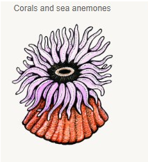
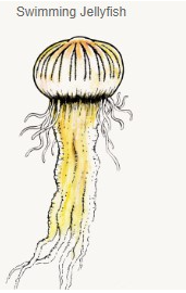
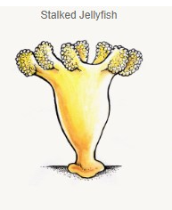
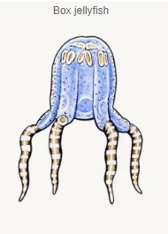
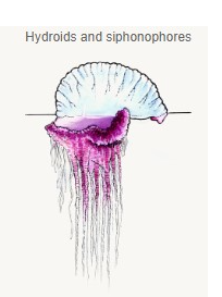
Figure 6: Phylum Cnidaria.
Phylum : Ctenophora
- Books Name
- ACME SMART COACHING Biology Book
- Publication
- ACME SMART PUBLICATION
- Course
- CBSE Class 11
- Subject
- Biology
PHYLUM: CTENOPHORA / SEA WALNUT / COMB JELLIES
Ctenophores are exclusively marine, solitary, free swimming or pelagic, very active animals with transparent and flat or oval body shape and have following important characters :
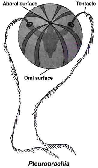
1. Body soft, delicate, transparent and gelatinous without segmentation.
2. Polyp phase is absent in their life cycle, shape is typically spherical, pear shaped or cylindrical, flat in some.
3. They are radially symmetrical, diploblastic, tissue level of organisation and devoid of cnidoblast cells.
4. Tentacles may be present or absent. When present, the number of tentacles is 2. They are solid and possess adhesive cells called colloblasts (lasso cells). Digestion is both extracellular and intracellular. Bioluminescence (the property of a living organism to emit light) is well-marked in ctenophores.
5. The animals move by cilia, which join together to form comb plates. There are eight median combplates forming locomotor organs, hence organisms are called comb-jellies or sea-walnuts.
6. Gastrovascular cavity is branched and opens to the exterior by stomodaeum.
7. They are diploblastic animals but the mesoglea is different from that of cnidaria; it contains amoebocytes and smooth muscle cells and is comparable to a loose layer of cells. From this viewpoint, ctenophores may be considered as triploblastic.
8. Skeletal, circulatory, respiratory and excretory systems are absent. Nervous system is diffuse type.
9. The presence of a special sense organ 'Statocyst' at the opposite end of the mouth (aboral end) is the characteristic of the members of this phylum.
10. All are hermaphrodite. Testes and ovary formed side by side from endoderm of digestive canals.
11. Asexual reproduction doesn't occur. They reproduce only by sexual means. Fertilization is external. Development is indirect and an immature ciliated stage called 'cydippid larva' is found in some forms.
Examples: Hormiphora (The sea walnut), Pleurobrachia (The sea gooseberry), Ctenoplana, Cestum (The venus girdle)
Concept Builder
Word roots and Origins :
(a) Colloblasts from the Greek kolla meaning "glue" and blastos meaning "bud".
(b) Ctenophore from the Greek ktene meaning "comb" and phors meaning "bearing".
Phylum : Ctenophora
Phylum Ctenophora
Ctenophores, often known as sea walnuts or comb jellies, are biradially symmetrical, diploblastic animals with a tissue-level organization that are entirely marine. The body has eight rows of ciliated comb plates on the outside that aid in movement. The existence of comb plates containing cilia, which are employed for propulsion, distinguishes the phylum. Ctenophores have a more complex body than sponges, although it is less complicated than bilaterians. They have tissue-level organization, just like cnidarians. Extracellular and intracellular digestion both are observed. Ctenophores exhibit a lot of bioluminescence (the ability of a living creature to emit light). They do not have stinging cells. In order to capture prey, ctenophores possess sticky cells called Colloblasts. The sexes are not divided. Sexual reproduction is the only way to reproduce.
External fertilization is used in conjunction with indirect development.Examples arePleurobrachia and Ctenoplana.
Phylum: Platyhelminthes
- Books Name
- ACME SMART COACHING Biology Book
- Publication
- ACME SMART PUBLICATION
- Course
- CBSE Class 11
- Subject
- Biology
PHYLUM : PLATYHELMINTHES (Flatworms)
Phylum Platyhelminthes contains about 13,000 species, mostly parasites that live in other animals including man.
The important characters of this phylum are:
1. The flat worms are mostly parasites but some are free living e.g., Planaria.
2. They are acoelomate, triploblastic, bilaterally symmetrical and dorsoventrally flattened animals.
3. They have organ system level of organisation.
4. Body is not segmented except in class Cestoda (false segmentation).
5. Body is covered with a cellular, syncytial, one layered, partly ciliated epidermis; while in parasitic trematodes and cestodes, epidermis is lacking and the body is covered with cuticle.
6. Exoskeleton and Endoskeleton are completely absent. However hooks, spines, suckers (in parasitic form), teeth or thorns may be present which act as adhesive organs.
7. The space between the body wall, alimentary canal and other organs is filled with a peculiar connective tissue called the parenchyma. It helps in transportation of food materials.
8. Digestive system is totally absent in tapeworms, so they absorb food directly through body surface. In Trematoda and Turbellaria, it consists of mouth, pharynx and blind intestine (anus absent).
9. Respiratory and circulatory systems are absent.
10. Excretory system consists of single or paired protonephridium with flame cells.
11. Nervous system is primitive. The main nervous system consists of a pair of cerebral ganglia or brain and one to three pairs of longitudinal nerve cords connected to each other by transverse commissures. This type of nervous system is called ladder like nervous system e.g., Planaria (Ougesia).
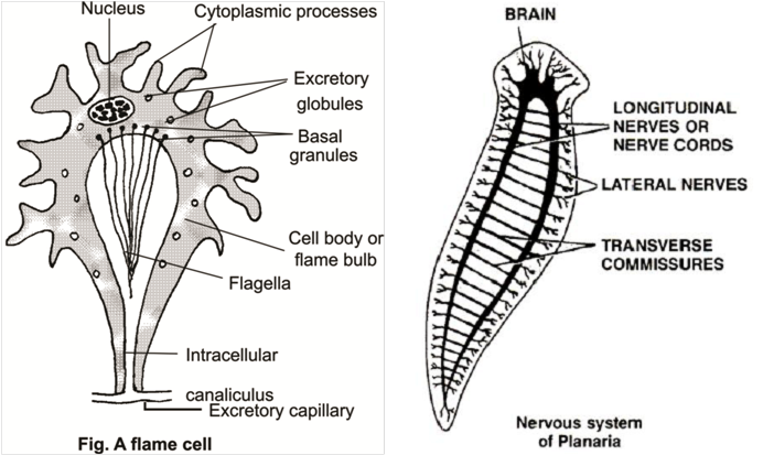
12. Sense organs are of common occurrence in Turbellaria but these are greatly reduced in parasitic forms.
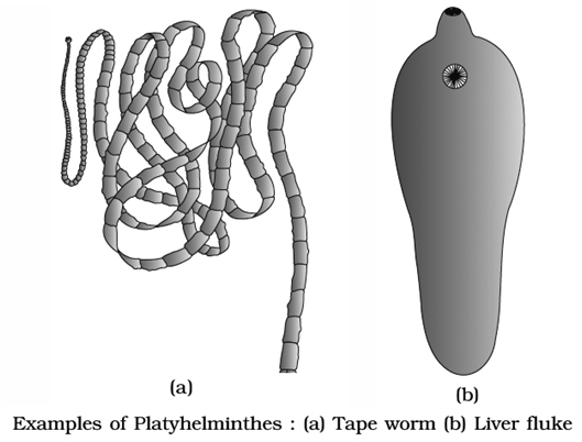
13. Sexes are united, i.e., hermaphrodite with very few exceptions like Schistosoma (Blood fluke).
14. Asexual reproduction by fission occurs in many fresh water forms like Turbellarians.
15. In majority of forms, eggs are devoid of yolk but provided with special yolk cells and are covered by egg shell.
16. Cross fertilization in trematodes and turbelleria and self-fertilization in cestodes is very common. Fertilization is internal. Development indirect with many larval stages.
17. Life cycle complicated, involves one or more hosts.
18. Regeneration. It is well marked in some flat worms like Planaria.
CLASSIFICATION
- Phylum platyhelminthes is divided into three classes.

Phylum: Platyhelminthes
Phylum Platyhelminthes
Flatworms and tapeworms are popular names for Platyhelminthes. They are a type of invertebrate with a soft body. In reality, there are over 20,000 different species of these animals. A few of these parasites live on humans and other animals as parasites. Furthermore, because of their parasitic nature, they do cause some discomfort to the host animal. Schistosomiasis, often known asbilharziasis, is a parasitic flatworm disease. A few species in this phylum are known to be major causes of disease. They are triploblastic, acoelomate, and bilaterally symmetrical. Their body is dorsoventrally flattened which is its most noticeable feature. They are flat because the body has no cavities. They also lack specialized systems and do not have segmented bodies. They have no specialized circulatory and respiratory organs, which allows oxygen and nutrients to pass through their bodies by diffusion.Around 80% of the flatworms are parasitic, with a few free-form flatworms thrown in for good measure. Scavengers or predators are the free-living forms. The parasitic species feed on the tissues of the organism they dwell in. Planaria, for example, has a high potential for regeneration. This phylum's animals come in a wide range of sizes. Some are minuscule, while others can reach lengths of up to two feet. They're also hermaphrodites, which means they have both sexes living in the same body.They typically reproduce both sexually and asexually. For instance, Taenia (Tapeworm) and Fasciola (Liver fluke).
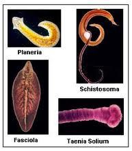
Phylum: Aschelminthes / Nemathelminthes
- Books Name
- ACME SMART COACHING Biology Book
- Publication
- ACME SMART PUBLICATION
- Course
- CBSE Class 11
- Subject
- Biology
PHYLUM: NEMATHELMINTHES OR ASCHELMINTHES
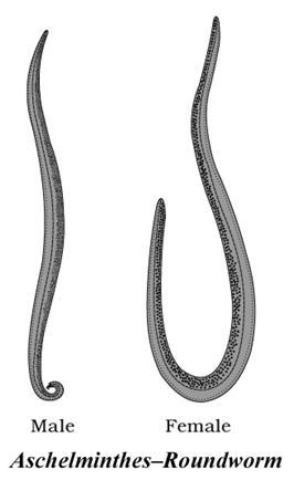
The phylum includes bilaterally symmetrical, triploblastic, pseudocoelomate animals with organ system grade of organisation. They are circular in cross-section.
They are free living, aquatic or terrestrial or parasitic (on plants or animals).
Excretory system involves an excretory cell (a large giant H-shaped cell) called Renette cell (multinucleated).
They have tube-within tube type of body plan evolved along Protostomic evolutionary line.
Sexes separate, show sexual dimorphism, fertilisation internal, development direct or indirect.
CLASSIFICATION
On the basis of caudal receptor or phasmids, Aschelminthes can be divided into 2 classes-


Important classes of this phylum are:
Class : Nematoda.
Examples: Ascaris, Wuchereria, Enterobius, Trichuris. Rhabditis is a free living roundworm.
Class : Rotifera.
Examples: Wheel worms
Phylum: Aschelminthes / Nemathelminthes
Phylum Aschelminthes
Roundworms are the popular name for Aschelminthes. They are triploblastic, bilaterally symmetrical, and pseudocoelomates creatures. Aschelminthes can be parasitic or free-living. The free-living creatures, which feed on bacteria, are abundant in soils and sediments. Others are plant parasites that can cause disease in economically significant crops. The remaining parasites can be found in both animals and humans. Hookworms, pinworms (Enterobius), Guinea worms (Dracunculus), and intestinal roundworms are examples of parasitic worms. A well-developed muscular pharynx completes the alimentary canal. Through the excretory hole, an excretory tube transfers body wastes from the body cavity. In most of the Aschelminthes, the respiratory and circulatory systems are absent. They utilize the process of diffusion for the circulation of oxygen. Aschelminthes are dioecious, which means they are separate. Females are frequently taller than males.Internal fertilization occurs, and development can be either direct (the young resemble the adult) or indirect. The Giant Intestinal Roundworm (Ascaris lumbricoides) is an endoparasite that lives in the human gut. They're quite prevalent in kids. Ascariasis is a disease caused by these worms. Many adult roundworms dwell inside the intestine, obstructing the digestive tract.Examples: Ascaris (Roundworm), Wuchereria (Filaria worm), Ancylostoma (Hookworm).
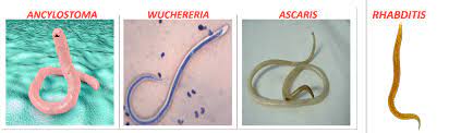
Phylum : Annelida
- Books Name
- ACME SMART COACHING Biology Book
- Publication
- ACME SMART PUBLICATION
- Course
- CBSE Class 11
- Subject
- Biology
PHYLUM : ANNELIDA
- The phylum includes over 9,000 species of metamerically segmented animals with a true coelom called the Schizocoelom.
General characters of Phylum Annelida
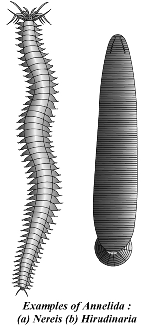
1. The organisms are triploblastic, bilaterally symmetrical, coelomates with organ system level of body organization and are metamerically segmented.
2. Body wall has an epidermis of columnar epithelium coated externally by moist albuminous cuticle and with circular and longitudinal muscle fibres.
3. Chitinous setae, aiding in locomotion, mayor may not be present on fleshy parapodia; absent in leech.
4. Annelids are the first animals to have a true schizocoelic coelom. Coelom is divided by septa into compartments.
5. The coelomic fluid act as a hydrostatic skeleton.
6. Digestive system is complete and digestion is extracellular.
7. Respiration by moist skin (cutaneous respiration) or through gills (Branchial respiration).
8. Blood vascular system is usually closed. Respiratory pigments, either haemoglobin or erythrocruorin, are dissolved in blood plasma. Free amoeboid blood corpuscles are present, butthere are no RBCs. ln leech, there is no true blood vascular system.
9. Nephridia are the excretory organs. Ammonia is chief excretory waste.
10. The nervous system consists of a nerve ring and asolid, double, mid-ventral nerve cord with ganglia and lateral nerves in each segment.
11. Sensory organs include tactile organs, taste buds, statocysts, photoreceptor cells and eyes with lenses.
12. The sexes may be separate (e.g., Nereis) or united (e.g., earthworm, leech).
13. Development is mostly direct (e.g., Earthworm). There is indirect development in Nereis. Larva, when present is trochophore.
CLASSIFICATION OF ANNELIDA
Based on presence or absence/of Parapodia, Setae and Sense organs

NEREIS
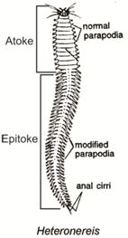
Inhabits in sea - shore between tide mark, burrower, nocturnal, carnivorous, gregarious, fertilization - in sea.
Parapodia in each segment except first & last. During breeding body divides in two parts. Anterior asexual part - Atoke and posterior sexual portion Epitoke. This change is known as epitoky.
Phylum : Annelida
Phylum Annelida
Phylum Annelida contains around 17000 species. Ringworms and segmented worms are other names for annelids. They can be found in a variety of habitats, including coastal waterways, freshwater, and wet terrestrial places. The annelids can be as small as a few centimeters long or as large as three meters. In addition, some species in this phylum have unusual forms and vibrant colors. Annelids are invertebrate creatures with bilateral symmetry. They are triploblastic and coelomate. Their body surface is divided by segments called metameres. They have longitudinal and circular muscles that let them move around. Nereis and other aquatic annelids have lateral appendages called parapodia that aid in swimming.There is a closed circulatory system in place. Osmoregulation and excretion are aided by nephridia (sing. nephridium). The neural system is made up of paired ganglia (sing. ganglion) coupled to a double ventral nerve cord via lateral nerves. Earthworms and leeches are monoecious, but Nereis, an aquatic variety, is dioecious. Sexual reproduction is prevalent. Nereis,Pheretima (Earthworm), and Hirudinaria are other examples (Bloodsucking leech).
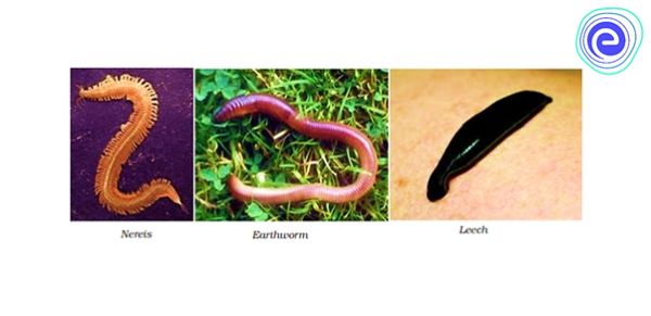
Phylum : Arthropoda
- Books Name
- ACME SMART COACHING Biology Book
- Publication
- ACME SMART PUBLICATION
- Course
- CBSE Class 11
- Subject
- Biology
PHYLUM . ARTHROPODA
- It is the largest phylum in the animal kingdom, including 900,000 species. The largest class is insecta with 750,000 species.
- General chacters are :
1. They are triploblastic, coelomate and bilaterally symmetrical animals. The body cavity is full of haemolymph (blood) and it's known as haemocoel. The true coelom is restricted to gonads.
2. The body is covered by chitinous cuticle, which forms the exoskeleton which is shed at intervals i.e., undergo moulting/ecdysis.
3. They have a segmented body, each segment bearing a pair of jointed appendages covered by a jointed exoskeleton. Chitinous exoskeleton is secreted by the underlying epidermis.
4. The body is divided into head, thorax and abdomen. In some cases, the head and thorax is fused to form cephalothorax. In insects, the thoracic segments have legs and wings, the abdomen has no legs in insects.
5. Respiratory organs are gills, book gills, book lungs or tracheal system.
6. Excretion takes place through green glands or malpighian tubules since nephridia are absent.
7. Sensory structures in arthropods are antennae for perceiving odour, eyes, statocysts or balance organs and sound receptors (in chirping crickets and cicadas). Eyes are simple or compound. In honey bees, butterflies, moths and some other insects, the gustatory receptors are present on their feet.
8. The heart is dorsal and circulatory system is open.
9. The central nervous system consists of paired pre-oral ganglia connected by commissures to a solid double ventral nerve cord.
10. In land arthropods, the fertilization is always internal.
11. Arthropods are mostly dioecious, oviparous. In some like the scorpion, the eggs hatch within the female body. They bring forth the young ones alive. They are viviparous. Development is direct or indirect.
Advancement Over Annelida
1. Distinct-head in all species.
2. Jointed appendages serving a variety of functions.
3. Jointed exoskeleton for protection and muscle attachment.
4. Striped muscles arranged in bundles for moving particular body parts.
5. Special respiratory organs such as gills, trachea, book lungs in majority of cases.
6. Well developed sense organs such as compound eyes, statocysts, auditory organs, taste receptors etc.
7. Endocrine glands and pheromone secretion for communication.
CLASSIFICATION
- The phylum arthropoda is divided into five classes.
Class 1. Crustacea
(i) The body is divisible into cephalothorax (head + thorax) and abdomen.
(ii) Dorsally, the cephalothorax is covered by a thick exoskeletal carapace.
(iii) There are present two pairs ofantennae and a pair ofstalked compound eyes and biramous appendages.
(iv) Respiration is carried out either by body surface or by giliis.
(v) Excretion takes place usually by antennary glands (green glands).
(vi) Sexes are usually separate. Sexual dimorphism is also seen.
(vii) Development is usually indirect.
Examples: Palaemon (Prawn), Macrobrachium (Prawn), Astacus (Crayfish), Palinurus (Lobster), Cancer(Crab), Lucifer(Shrimp), Eupagurus (Hermit Crab), Oniscus (Wood louse, terrestrial), Oaphnia (Water-flea), Cyclops, Balanus (barnacle). Tiny crustaceans such as Oaphnia and Cyclops act as zooplanktons which form important link in the food chain in water.
Class 2. Chilopoda
(i) Body is long, segmented and divisible into head and trunk.
(ii) Each trunk segment bears a pair of legs. The first pair of legs are modified into poison claws.
(iii) There is a single pair of antennae and ocelli.
(iv) Many legs are present.
(v) Respiration occurs by tracheae.
(vi) Excretion takes place by Malpighian tubules.
(vii) Development is direct
Examples: Scolopendra (Centipede).
Class 3. Diplopoda
(i) Body is divisible into head, thorax and abdomen.
(ii) There is a single pair of antennae and ocelli.
(iii) Except first thoracic segment, (if does not have legs) each thoracic segment bears a pair of legs, however each abdominal segment has two pairs of legs.
(iv) Respiration occurs by tracheae.
(v) Excretion takes place by Malpighian tubules.
(vi) Development is generally direct e.g., Julus (Millipede).
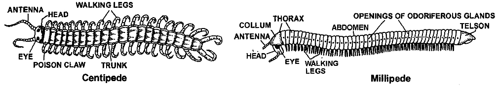
Class 4. Insecta (Hexapoda) [Largest number of species]
(i) Body is divisible into head, thorax and abdomen.
(ii) There is a pair of antennae and a pair of compound eyes.
(iii) The thorax consists of three segments with three pairs of legs and usually two pairs of wings. For example, mesothoracic wings which are thick and leathery are called tegmina and metathoracic wings are membranous (in cockroach). Wings may be absent in same insects.
(iv) The abdomen may consists of ten segments.
(v) Respiration usually takes place by tracheae. (No respiratory pigments).
(vi) Heart is tubular and divided into chambers.
(vii) Malpighian tubules are the excretory organs. Uric acid is chief excretory waste. (viii) Sexes are separate.
(ix) Development may be direct or indirect e.g., silver fish, cockroach, bedbug, locust, termites, butter flies, rat flea, beetle, wasp, aphid, glow worm etc. Maggot is the larva of Housefly.
- Insects communicate with each other by ectohormones called Pheromones. Pheromones are chemicals secreted outside the body and perceived (as smell by other individuals of same species). They help in communication amongst the organisms of the same species.
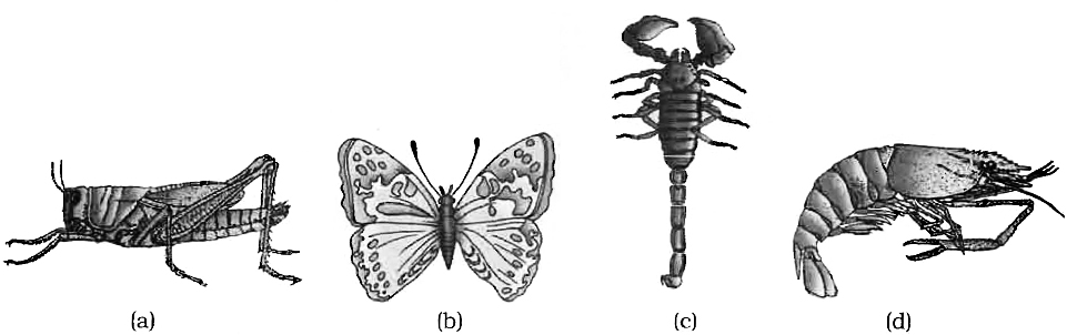
Class 5. Arachnida
(i) The body is usually divisible into cephalothorax and abdomen.
(ii) The cephalothorax bears simple eyes and six pairs of appendages (one pair of chelicerae, one pair of pedipalpi and four pairs of legs).
(iii) Antennae are absent.
(iv) Respiratory organs are book lungs or tracheae or both.
(v) Excretion takes place by Malpighian tubules or coxal glands or both.
(vi) Development is generally direct.
Examples: Tick, Mite, Aranaeus (garden spider), Palamneus (Scorpion), Buthus (Scorpion).
(vii) Spiders spin the web by means of proteinaceous secretions of abdominal glands known as "spinnerets".
- There are two more classes in the phylum Arthropoda. These classes are Onychophora and Merostomata. Peripatus is an important example of the class onychophora which has characters of phylum Annelida and Arthropoda.
- Hence it is called "connecting link". Umulus, (the king crab or Horseshoe crab) is an example of class Merostomata which respires with book gills.
- The king crab is called "living fossil". A living fossil is a living animal of ancient origin with many primitive characters.
Concept Builder
1. Peripatus is considered as connecting link between annelida and arthropoda as it has unjointed legs and breathes by trachea.
2. Larvae of different Arthropods
(a) Bombyx(Silkworm) Caterpillar/Silkworm.
(b) Beetles, honey bee Grub
(c) Musca (Housefly) Maggot
(d) Culex, Anopheles Wriggler
(e) Pennaeus(marine prawn) Mysis, nauplius, protozoea
(f) Cancer(Crab) Megalopa, metanauplius, zoea
3. Terga are dorsal plates whereas sterna are ventral plates of exoskeleton.
4. Arthrodial membranes join the different sclerites.
5. Halters are drumstick shaped, second pair of reduced wings of housefly and mosquito which helps in balancing.
6. Eggs of Culex are cigar shaped; they are laid vertically on the surface of water in clusters; airfloats are absent; whereas in Anopheles eggs are boat shaped, laid singly and horizontally; they have airfloat.
7. Larva of Culex is bottom feeder whereas larva of Anopheles is surface feeder.
8. Adult of Culex lies parallel to the surface and both ends of body deflexed whereas in Anopheles, the body is inclined at an angle of 45° to the surface. So, they can be distinguished with the help of sitting posture.
9. In Spider, Spinnerets are present anterior to the terminal anus. They produce silken thread.
10. Von Frisch described the process of communication of food source in honey bees.
11. Insecticides sprayed over the mosquitoes desensitize their nervous system as well as chemoreceptors and mechanoreceptors ofthe antennae.
12. Johnston's organs are present on antennae of mosquitoes.
13. The life cycle is generally accompanied with metamorphosis. Larvae and adults may show different feeding habits and occupy different habitats.
14. Insects like bees, wasps, beetles, moths and butterflies are good pollinators for important crops.
15. Ants, termites and locusts are eaten by Chinese and Indians; Eggs of aquatic bugs are used as food by Mexicans; Ox-warbles are eaten by Red Indians.
16. Dragon flies feed on the larvae of mosquitoes
17. Glow worm (Firefly) shows bioluminescence.
18. Adult Culex and Anopheles can be distinguished with the help of Sitting posture.
19. The insects may be divided into five groups on the basis of their mode of development.
1. Ametabola insects – metamorphosis absent. The young ones resemble adult.
![]()
ex. Lepisma (Silver fish)
2. Paurometabola insects – gradual metamorphosis
![]()
- ex. Cockroach, Grasshopper, Locusts etc.
3. Hemimetabola insects – incomplete metamorphosis. Habitat of young ones is different from adults.

ex. Dragon flies (Naiads aquatic but adults aerial)
4. Holometabola insects – complete metamorphosis
![]()
ex. House flies, Butterflies, Mosquitoes etc
5. Hypermetabola insects – various forms of larvae

ex. Blister beetle.
Phylum : Arthropoda
Phylum Arthropoda
Arthropods are animals with joint legs. Spiders, ants, bees, crabs, shrimps, millipedes, centipedes, and other insects belong to this phylum of the kingdom Animalia. Arthropods make up more than two-thirds of all identified species on the planet. The presence of an exoskeleton, which makes arthropods adaptable, isprotective and allows flexibility and mobility.It is largely responsible for their success and survival. They are organized at the organ-system level. They are coelomate animals that are triploblastic, segmented, and bilaterally symmetrical. Arthropods have a chitinous exoskeleton covering their bodies. The head, thorax, and abdomen make up the arthropod body. Their appendages are joined. Gills, book gills, book lungs, and the tracheal system are examples of respiratory organs.The circulatory system is open. There are sensory organs like antennae, eyes (complex and simple), and statocysts for balance and orientation. Malpighian tubules are used for excretion. The majority of them are dioecious. Internal fertilization is common. The majority of them are oviparous. Direct or indirect development is possible. Apis (Honey bee), Bombyx (Silkworm), and Laccifer(Lac insect)are some examples of commercially important insects. Anopheles, Culex, and Aedes are examples of vectors (Mosquitoes). Limulus (King crab) is known as a living fossil, while Locusta(Locust) is a gregarious pest.
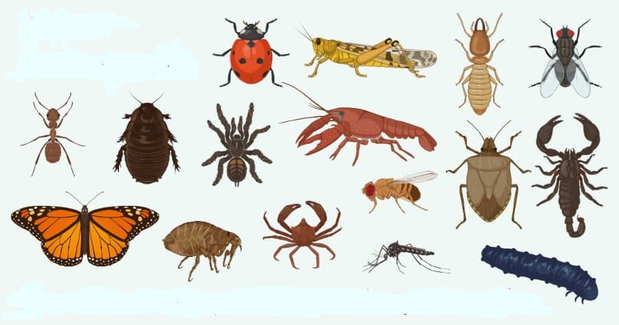
Phylum : Mollusca
- Books Name
- ACME SMART COACHING Biology Book
- Publication
- ACME SMART PUBLICATION
- Course
- CBSE Class 11
- Subject
- Biology
PHYLUM MOLLUSCA
Phylum mollusca is the second largest phylum in animal kingdom which includes over 60,000 species. General characters are :
- Molluscs are terrestrial, aquatic (fresh water or marine) triploblastic, bilaterally symmetrical, schizocoelic and unsegmented animals.
- Most molluscs secrete a shell of calcium carbonate that protects and supports their soft tissues.
- The body is organised into three general regions: head, foot and visceral hump.
- The visceral hump contains the digestive tract and other visceral organs.
- The body is covered by a soft and spongy skin fold called mantle which secretes the shell.
- The space between the hump and the mantle is called mantle cavity in which feather like gills are present.
- Molluscs typically employ a feeding organ called radula which is armed with rows of chitinous teeth. The radula is protruded from the mouth and worked back and forth to rasp the food into fine particles.
- Circulatory system is mainly of open type but some reduced sinuses are present. Respiratory pigment is haemocyanin.
- Respiration occurs by feather like gills, pulmonary sac or both, or through general body surface.
- Excretion occurs by paired Organ of Bojanus. Another excretory organ called Keber's organ (Pericardial gland) is also present in Unioalongwith paired organ of Bojanus. It pours the waste into pericardium from where the waste is carried to the organ of Bojanus that opens out through mantle cavity.
- Sense organs include eyes, statocysts and osphradia (chemoreceptor to test chemical nature of water).
- Reproduction sexual, adults can be dioecious (unisexual) or monoecious (bisexual or hermaphrodite) Fertilization is generally external, development is direct or through free larval forms like trochophore, veliger (in Pila), glochidium (in Unio) -an ectoparasite on fishes.
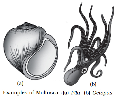
Pila produces 2 types of sperms :
(i) Eupyrene -Motile and functional sperms, 25 µm long, thread like with a single cilium.
(ii) Oligopyrene -Non-motile and non-functional sperms, 32.5 µm long, spindle shaped with 4-5 cilia.
CLASSIFICATION
Molluscs' classification is mainly on the basis of shell and foot.

Concept Builder
(i) Architeuthis (Giant AtJantic squid) is the largest and heaviest among invertebrates, 55 feet length.
(ii) Nautilus is the only cephalopod with external shell, so ink glands are absent.
(iii) The colour change in cephalopods occurs due to chromatophores.
Phylum : Mollusca
Phylum Mollusca
Molluscs are a diverse group of organisms that play a vital role in the global ecology. A calciferous shell is found on many creatures in this phylum. The bodies are usually soft and protected by a hard exoskeleton. They can be found both on land and in the deepest parts of the ocean. This is the second-largest phylum of animals. Molluscs are organ-system-organized terrestrial or aquatic (marine or freshwater) animals. They are animals that are bilaterally symmetrical, triploblastic, and coelomate. A calcareous shell covers the body, which is unsegmented with a distinct head, muscular foot, and visceral hump. Over the visceral hump, a soft and spongy layer of skin forms a mantle.The mantle cavity is the region between the hump and the mantle, and it contains feather-like gills. They have breathing and excretory systems. Sensory tentacles can be found in the anterior head region. The radula, a file-like rasping organ in the mouth, is used for feeding. They are both a food and a jewellery source. The firm shells are used to create stunning jewellery. Even though it sounds strange, these are also raised as pets in some parts of the world. Bivalve and gastropod pearls are particularly valuable because they are lined with nacre. Natural pearls are generated when a small foreign object becomes lodged between the mollusc's mantle and shell.Bivalve molluscs are useful as freshwater and marine bioindicators. However, not all molluscs are beneficial to humans. Snails and slugs, for example, are considered pests.They are usually dioecious and oviparous with indirect development. Examples: Pila (Apple snail), Pinctada (Pearl oyster), Sepia (Cuttlefish), Loligo (Squid), Octopus (Devilfish), Aplysia (Seahare), Dentalium (Tusk shell), and Chaetopleura (Chiton).
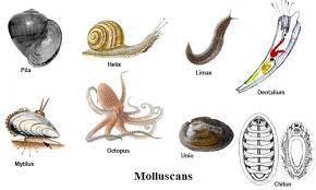
Phylum : Echinodermata
- Books Name
- ACME SMART COACHING Biology Book
- Publication
- ACME SMART PUBLICATION
- Course
- CBSE Class 11
- Subject
- Biology
10. PHYLUM: ECHINODERMATA
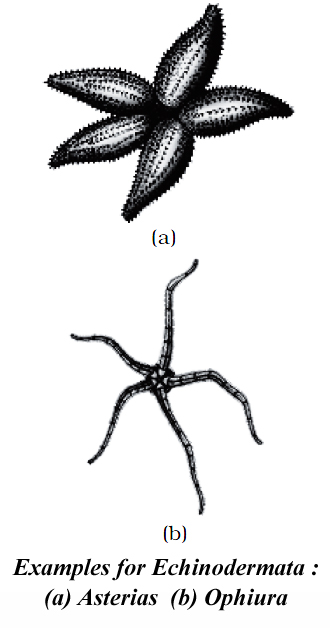
They are exclusively present in marine water and there are no parasitic forms. Similarities with chordates :
(i) They have tube within tube type of body plan which has evolved along deuterostomic evolutionary line.
(ii) They possess a true coelom called enterocoelom.
(iii) They have mesodermal skeleton made of calcareous plates or ossicles.
All these characters make the echinoderms closer to chordates. The other characters are :
1. The symmetry is bilateral in larvae but pentamerous radial in adults.
2. Many echinoderms bear a number of calcareous spines on their body surfaces called tubercles. Between the spines, there are pincer like structures called pedicellariae to keep the surface clean. Pedicellariae are made of three calcareous plates. Two calcareous valve like structures in the form of jaws resting upon a basal calcareous plate.
3. Between the spines, there are finger like processes called dermal branchiae which help in respiration.
4. Mouth is on ventral/oral side and aniss is on dorsal or aboral side.
5. There is no distinct anterior and posterior end i.e., no cephalisation.
6. Haemal and Perihaemal Systems: Instead of blood vascular system, there are present haemal and perihaemal systems which are of coelomic origin. Thus the so called circulatory system is open type. The so called blood is often without a respiratory pigment. There is no heart.
7. Respiratory Organs : Gaseous exchange occurs by dermal branchiae or papulae in star fishes, peristominal gills in sea urchins, genital bursae in brittle stars and cloacal respiratory trees in holothurians. Exchange of gases also takes place through tube feet.
8. Excretory organs : Specialized excretory organs are absent. Nitrogenous wastes are diffused out via gills or dermal branchiae. Ammonia is chief excretory matter.
9. Sexes are separate. Reproduction is sexual. Fertilization is usually external. Development is indirect with free-swimming larva.
CLASSIFICATION
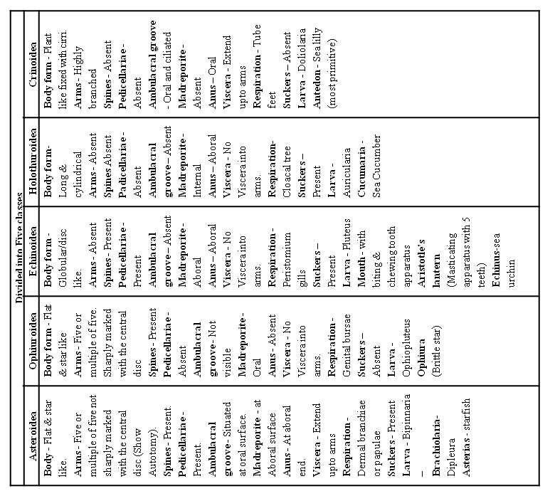
Concept Builder
1. The starfish uses the suction cup-like ends of the tube feet of one arm to hold on to rocks, as it moves the other arms. It can open bivalves (molluscs) by attaching two arms to either sides of a bivalve and pulling them apart. The bivalve opens out, the stomach of starfish is then everted through its mouth and introduced into the open mollusc which is partly digested before being ingested. Echinoderms predate on coral polyps also.
2. Word roots and origin : ampulla from Latin meaning ''flask''.
Phylum : Echinodermata
Phylum Echinodermata
Invertebrates with spiny skin are known as echinoderms. The endoskeleton of these creatures is made up of calcareous ossicles. All of them are marine, having organ-system organization. There are no freshwater or terrestrial organisms in this phylum. Adult echinoderms have radial symmetry, whereas larvae have bilateral symmetry. They are coelomate and triploblastic creatures. With the mouth on the bottom (ventral) side and the anus on the upper (dorsal) side, the digestive system is complete. Echinoderms are distinguished by the presence of a water vascular system that aids motility, food capture and transport, and breathing. This system has a central ring canal and radial canals that extend along each arm. Through these structures, water circulates. The madreporite is a structure present on top of the body. This is responsible for the regulation of the water in the water vascular system. There is no excretory system. Sexes are distinct. Reproduce sexually. External fertilization is common. The development of free-swimming larva is indirect. Asterias (starfish), Echinus (sea urchin), Antedon (sea lily), Cucumaria (sea cucumber), and Ophiura (Brittle star) are among examples.
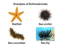
Phylum : Hemichordata / Stomachordata
- Books Name
- ACME SMART COACHING Biology Book
- Publication
- ACME SMART PUBLICATION
- Course
- CBSE Class 11
- Subject
- Biology
PHYLUM: HEMICHORDATA / STOMOCHORDATA
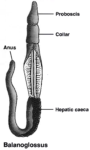
Hemichordata was earlier considered as a sub-phylum under chordata. But now it is placed as a separate phylum under non-chordata.
The phylum consists of a small group of worms like marine animals with organ system level of organisation.
Its characteristics are :
(i) In hemichordata or stomochordata, true notochord is absent. gill slits are present but they are dorso lateral in position. They are worm like. bilaterally symmetrical. triploblastic and entero-coelomate animals.
(ii) Stomochord is a hollow outgrowth arising from the roof of the buccal cavity, also called 'buccal diverticulum'. It is present in the proboscis.
(iii) Dorsal heart, ventral hollow nerve cord, no respiratory pigments. Circulatory system is open. Respiration by gills.
(iv) Development is mostly indirect through a free swimming tornaria larva. e.g. Balanoglossus (acorn, tongue worm), Glossobalanus. Sexes are separate. Fertilization is external.
(v) Believed to be connecting link between non-chordates and chordates.
(vi) Due to the absence of true notochord in hemichordata, many taxonomists do not consider these animals as chordates.
(vii) Excretory organ is proboscis gland.
(viii) The body is cylindrical and is composed of an anterior proboscis, a collar and a long trunk.
Examples : Balanoglossus, Saccoglossus
Phylum : Hemichordata / Stomachordata
Phylum Hemichordata
This phylum is made up of marine deuterostome creatures. They are sometimes referred to as the Echinodermata's sister group. Phylum Hemichordata is a small phylum with only 100 recognized species and animals that resemble worms. Some species live alone, while others live in colonies. Hemichordata was previously classified as a sub-phylum of the Chordata phylum. However, because these species lack a post-anal tail or even the notochord, which is a distinguishing trait of chordates, they are now classified as a different phylum under non-Chordata.
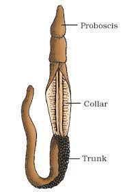
Hemichordates, on the other hand, have a primitive structure comparable to the notochord in the collar region. This phylum is made up of a tiny group of worm-like marine organisms that are organized at the organ-system level.This phylum is made up of marine deuterostome creatures. They are sometimes referred to as the Echinodermata's sister group. Hemichordatesare bilaterally symmetrical and triploblastic animals.The body is divisible into proboscis, collar, and trunk.Simple, open, and well-developed circulatory system is present. The alimentary canal is a straight, complete, or U-shaped tube.Excretion by a single glomerulus situated in the proboscis.Reproduction is mainly sexual. Sexes are usually separate.External fertilization takes place in seawater.Development can be direct or indirect.An important example of a hemichordate is Balanoglossus or acorn worm. Other species are Saccoglossus, Rhabdopleura, Atubaria, and Dendrograptus.
phylum chordata
- Books Name
- ACME SMART COACHING Biology Book
- Publication
- ACME SMART PUBLICATION
- Course
- CBSE Class 11
- Subject
- Biology
PHYLUM: CHORDATA
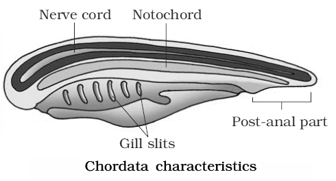
The most important characteristics of phylum chordata are:
1. They possess a notochord either throughout life or during early embryonic development.
2. Dorsal hollow nerve cord.
3. Paired gill slits on the lateral sides of pharynx.
4. Post anal tail.
5. Closed circulatory system.
Phylum chordata can also be divided as :
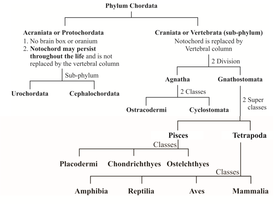
CLASSIFICATION OF PROTOCHORDATA OR ACRANIATES
Sub-Phylum 1. Urochordata
They are exclusively marine.
(i) This sub-phylum is also called Tunicata because the adult body is enclosed within a leathery test or tunic formed of a cellulose-like organic substance termed tunicin.
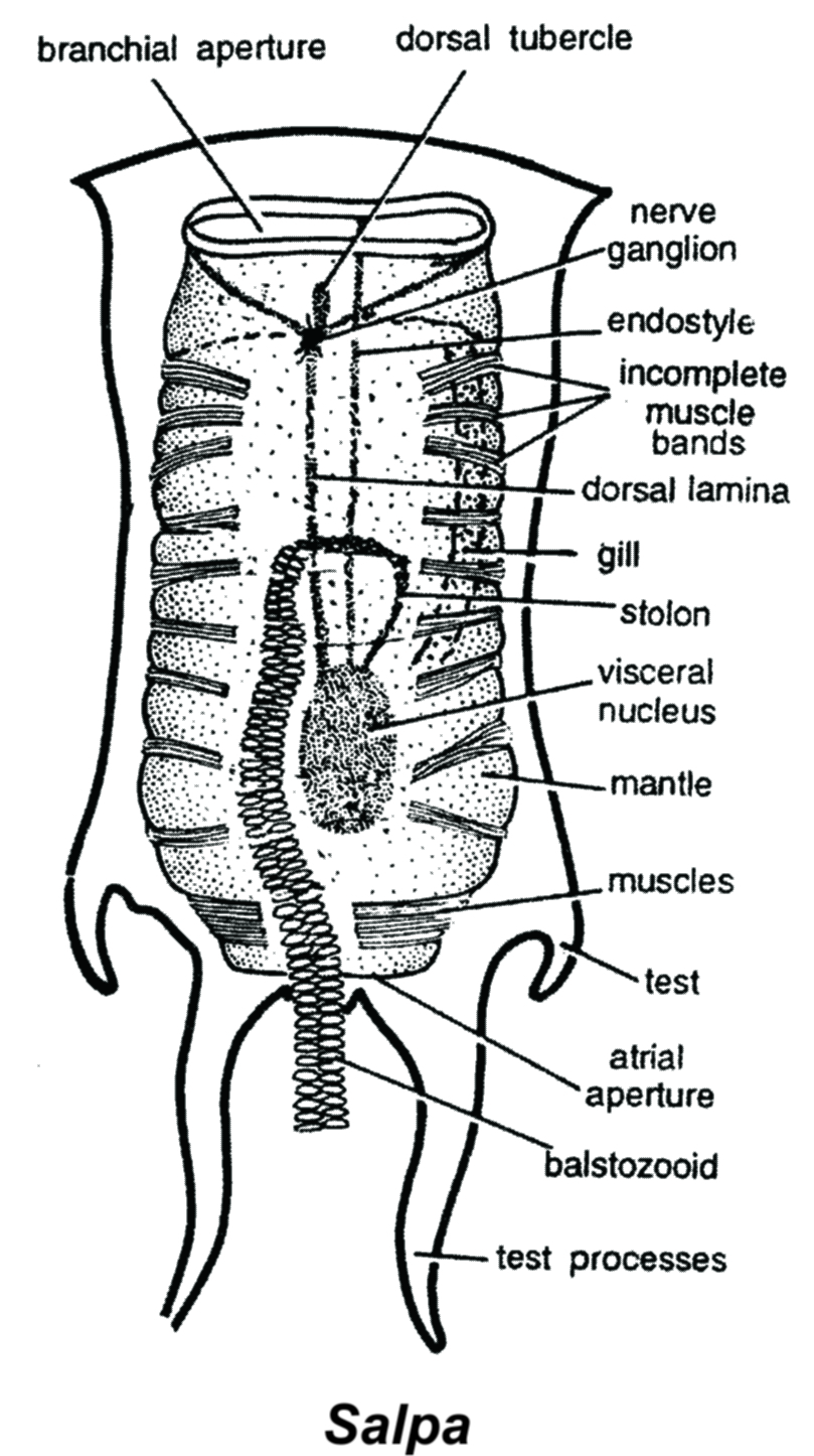
(ii) The larva (Ascidian tadpole) undergoes retrogressive metamorphosis, i.e., change from better developed larva to less developed adult, e.g., Herdmania (Sea Squirt).
(iii) Vanadocytes are present except in Herdmania. Vanadium gives green colour to blood. Excretion by neural gland, hence excretion is glandular.
Examples: Salpa, Doliolum, Ascidia.
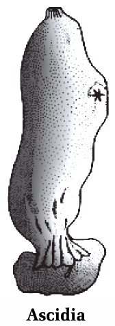
Sub-phylum 2. Cephalochordata
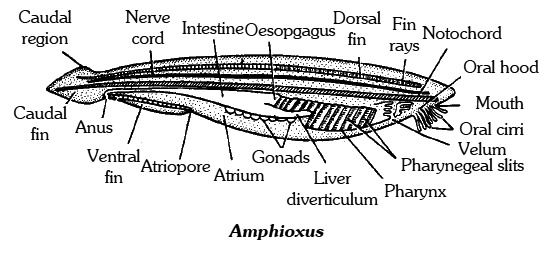
(i) The notochord extends upto anterior end of the body, hence this sub-phylum is named so.
(ii) The tail is present throughout life,e.g., Branchiostoma (Amphioxus).
Amphioxus / Branchiostoma has both ends pointed like lance. Hence it is commonly called lancelet.
VERTEBRATA OR CRANIATA
(i) These are advanced chordates that have cranium (cartilage or bony brain box) around brain.
(ii) Notochord is present only in the embryonic stages, it is replaced by cartilaginous or bony vertebral column (back bone) in the adult forms. They have paired appendages as fins and limbs.
(iii) Closed circulatory system, muscular and ventral heart 2, 3 or 4 chambered, lymphatic system present, erythrocytes and haemoglobin are present.
(iv) A pair of kidneys are present for excretion and osmoregulation; endocrine glands are well developed; they are generally unisexual; have single pair of gonads.
Division 1 : Agnatha (The Jawless Vertebrates)
They are the most primitive of all craniates.
The mouth does not possess jaws hence named Agnatha.
Vertebral column is represented only by small imperfect neural arches over the notochord.
They do not have exoskeleton and paired appendages.
They have single nostril. Internal ear has one or two semi-circular canals.
They are cold blooded.
It includes 2 classes :
1. Ostracodermi 2. Cyclostomata
1. Ostracodermi:
Ostracodermi are all extinct now and are called fossil agnatha.
These were shell skinned with exoskeleton of bony plates. e.g. Cephalaspis.
They were the first jawless fishes (originated in Ordovician period in Palaeozoic era).
2. Cyclostomata: Its general characters are :
(i) All living members of the class Cyclostomata are ectoparasites on some fishes. The body is devoid of scales and paired fins. Head and brain are poorly developed. They are also called jawless fishes.
(ii) Cyclostomes have an elongated body bearing 6 -15 pairs of gill slits in their gill pouch for respiration and have a sucking and circular mouth .
(iii) Single sex organ discharges the gametes in the well developed coelom.
(iv) Cranium and vertebral column are cartilaginous with persistent notochord.
(v) Stomach is absent.
(vi) A single dorsal nostril leads into closed nasal sac. A functional pineal eye is present just behind it.
(vii) Circulation is of closed type.
(viii) Kidneys are mesonephric.
Differences between Lamprey and Hag fish
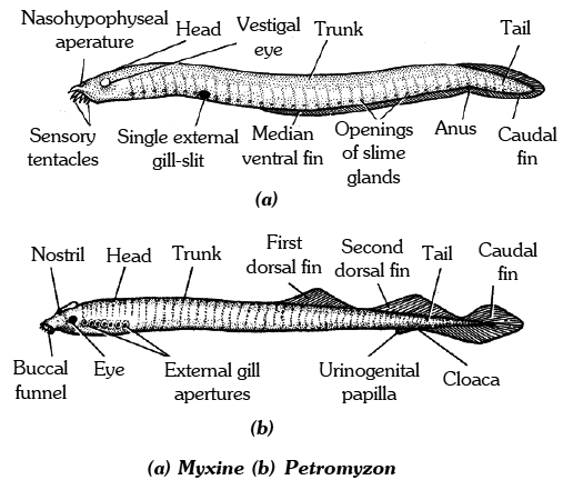
Concept Builder
1. Petromyzon , though marine, goes to fresh water for spawning i.e., anadromous. After spawning within a few days, they die.
2. Larva Ammocoete hatches out of eggs in about 14-21 days. Larval period may long from 3 to 7 years.
3. Ammocoete is connecting link between Amphioxus and the cyclostomes.
4. Lingual Cartilage is also a part of skeleton, that lies in the tongue region and supports it (Lingual = related to tongue).
5. Typhlosole is a fold of epithelium in the intestine. It prevents the rapid flow of food in the intestine and increases the absorptive surface area.
COMPERISION AMONG VARIOUS CLASSES OF VERTEBRATA

Division 2 : Gnathostomata (The Jawed Vertebrates)
It includes advanced vertebrates.
Embryonic notochord is usually replaced in adults by a vertebral column.
Paired fins or limbs are present.
Mouth has jaws hence it is named gnathostomata.
Paired nostrils are present.
Intemal ear has three semicircular canals. Girdles present.
Gnathostomata is divided into two super classes: Pisces and Tetrapoda.
CLASS 1 : PISCES
It includes true fishes. All are aquatic. The body bears fins. They are cold blooded.
(i) Monocondylic skull, slimy glands present on skin.
(ii) Vertebrae are Amphicoelous type
(iii) Neck absent, no upper or lower eyelids, no tympanum (only internal ear is present). Each eye has a well developed nictitating membrane.
(iv) Heart-two-chambered (S-shaped), venous heart with sinus venosus and conus arteriosus.
(v) Both renal portal and hepatic portal system are found. Hypophyseal portal system is also present.
(vi) Respiration occurs typically by gills.
(vii) Scales mesodermal or dermal, embedded in skin.
(viii) Kidneys -Mesonephric, ammonotelic (sharks-Ureotelic).
(ix) Lateral line sense organs are present.
Super class Pisces is divided into three classes :
Class I. Placodermi. (Extinct)
It includes the earliest fossil fishes which lived in fresh water. Body had an external protective armour of bony scales or plates. Primitive jaws with teeth were present. Skeleton was bony. Fins were mostly formed of large spines, e.g., Climatius (Spiny Shark).
(i) They appeared in Silurian period, flourished in Devonian and Carboniferous and became extinct in Permian .
(ii) The name placodermi means "armoured fish" or "Plate skinned".
(iii) Both paired and unpaired fins were present. Caudal fin heterocercal.
(iv) Autostylic jaw suspension; gill slits are covered by operculum.
Class II. Chondrichthyes.
This class includes cartilaginous fish, e.g., Scoliodon (dog fish), Torpedo (electric ray) etc. Sharks are fast swimming predators, whereas, the rays and skates are stout, bottom living scavengers and mollusc feeders.
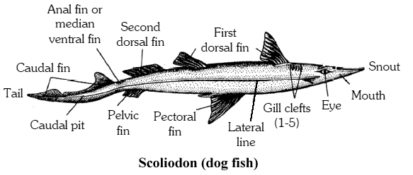
Class III. Osteichthyes.
It includes bony fish, e.g., Labeo, Hippocampus, Exocoetus, Catla, Clarias, Betta, Pterophyllum etc.
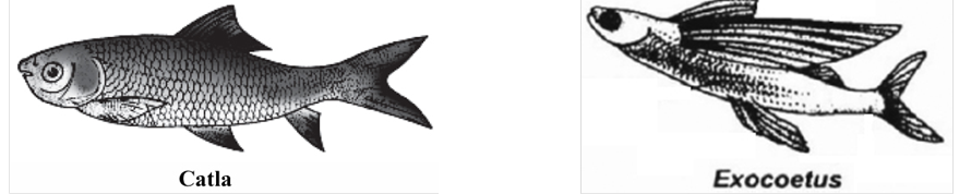
Differences between cartilaginous and bony fishes
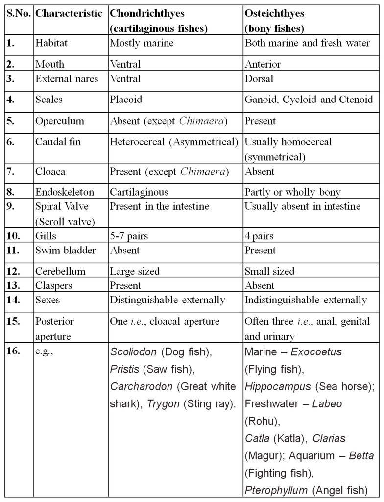
Some Important Fishes
Torpedo (Electric Ray) :
Torpedo is bottom dwelling, carnivorous fish. It stuns or kills the prey and enemies with electric shock from its electric organs. The latter are modified muscles. The skin is scale less.
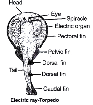
Pristis: (Saw fish)
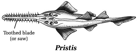
Head bears a series of strong tooth like denticles along the margin.
(a) Freshwater Species (b) Marine Species
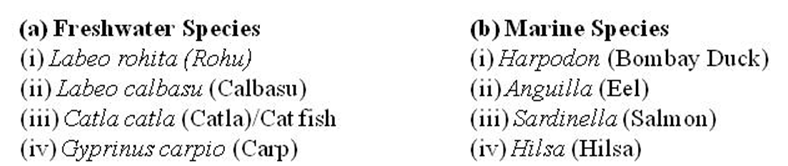
Some air breathing fishes, use their paired fins to move on land and gave rise to the first land vertebrates. A living fossil of this group is Latimeria, a lobe-finned fish.
Exocoetus (Flying Fish) :
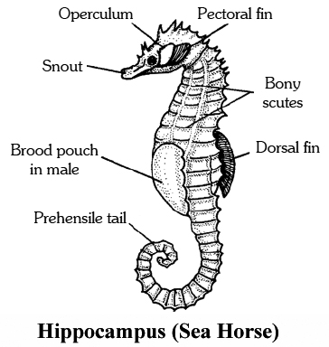
It does not fly but often leaps into the air upto about six metres high. The pectoral fins are modified into wing-like structures, with the help of which the fish glides.
Hippocampus (Sea Horse) :
The neck and the head of the fish are horse like and the tail is prehensile. The male bears a brood pouch in which the female lays eggs and the latter remain there till they hatch.
Labeo (Rohu) :
Labeo rohita and Labeo calbasu are the common fresh water edible carps. It is covered by large overlapping scales.
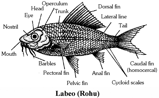
Clarias (Cat fish/Magur) :
Indian Cat Fish (Clarias batrachus) lives in ponds. It is carnivorous. The head bears sensory threads called barbels. The body is smooth and without scales. Other fresh water cat fishes of India are Mystus seengala (slinghara), Rita lita and Wallago attu (Malli).
Latimeria chalumnae (The Coelacanth) :
It was first caught in 1938 off the east coast of South Africa. It swims by curious rotating movements of its pectoral fins. All its fins except the anterior dorsal are lobed. It is the oldest living fish; and has survived till today without undergoing any change in it. It is therefore described as a living fossil.
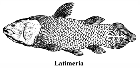
Lung Fishes: Sub-Class Dipnoi
There are three genera of living lung fishes: Neoceratodus, Lepidosiren and Protopterus. All have three chambered heart (two auricles and one ventricle).
4. Fins:
(i) Paired pectoral and pelvic fins: Act as balancers and brakes; provide lift and counteract pitching and rolling.
(ii) Anterior dorsal fin : Counteracts rolling and yawing.
(iii) Posterior dorsal and anal fin : Counteract yawing and rolling.
(iv) Caudal fin : Propels the body and provides lift.
Concept Builder
Types of caudal fins :
(a) Diphycercal or Protocercal : Most primitive kind of tail fin. The tail is symmetrical, both externally and internally as in protocercal but it is secondarily symmetrical.
(b) Heterocercal/Hypocercal tail: Epicaudal and hypocaudal lobes are of unequal sizes.
(c) Homocercal: Identical lobes. Externally symmetrical and internally asymmetrical.
Endothermic fishes: Although most fishes are ectothermic, a few species, such as blue fish tuna and swordfish are able to maintain a body temperature higher than the temperature of the water that surrounds them. Genetic studies of these endotherms have revealed that the ability to maintain high body temperature gives these fishes an adaptive advantage by allowing them to hunt in much colder waters than their competitors.
Fish taken out of water dies of suffocation due to lack of oxygen. This is because the gill filaments stick together when taken out of water, thereby reducing the surface area.
Word Roots and Origin : Operculum from the Latin meaning "Gover".
5. Scales:
(i) Cosmoid : Absent in living fishes; consists of four distinct layers -outermost ganoine, followed by cosmine, spongy bone and innermost compact bone e.g. extinct fishes.
(ii) Placoid: Backwardly directed spine arising from a basal plate e.g. chondrichthyes only.
(iii) Ganoid or rhomboid scales are thick, diamond or rhomboid shaped plates e.g. primitive bony fishes like Acipencer etc.
(iv) Cycloid: Thin, flexible, circular in outline, marked with concentric lines of growth which can be used for determining age e.g. lung fishes, Amia etc.
(v) Ctenoid: Bear numerous small comb like spines e.g. higher bony fishes like perch etc.
6. Air bladder is associated with respiration, hydrostasis, sound production and audition.
7. Parental care :
(i) Nest building: e.g., Gasterosteus, Amia (bow fin), Protopterus, Lepidosiren (nest is built by males of these fishes).
(ii) Shelter in mouth: e.g., Males of cat fish, Arius.
(iii) Brood pouch: e.g., Male Hippocampus
Common Food Fishes Of India
False Fishes
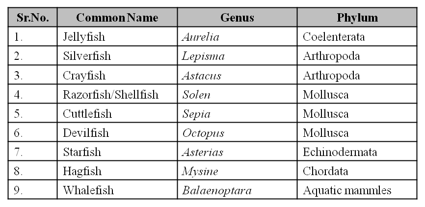
Concept Builder
1. Stone fish is the most poisonous fish.
2. Gambusia fish (Iarvivorous) was introduced into several tropical regions to control malaria.
3. Catadromous fishes live in fresh water and go down to sea for breeding e.g., Anguilla (european eel). When fishes migrate from the rivers to the sea, it is called catadromous migration.
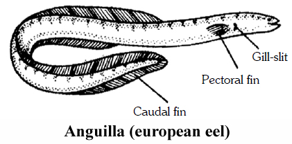
4. Anadromous fishes live in sea water and move to the rivers for breeding e.g., Hilsa, Salmon. When fishes migrate from sea to the rivers, it is called anadromous migration.
5. Hilsa is the only Indian fish that migrates from the seas to the river for breeding.
6. Most primitive fish (fossil): Climatius (Placodermi).
7. Anabas (climbing perch). It has accessory respiratory organs for breathing atmospheric air which enables the fish to take long overland excursions in search of earthworms.
8. Pomfret is the most widely eaten fish in India.
9. Dipnoi have incompletely divided three chambered heart.
10. Urinary bladder is absent in fishes.
11. Some elasmobranchs retain urea in blood to maintian hypertonicity.
12. Ampulla of Lorenzini are thermo receptors.
13. Electric organs of electric ray are modified muscles.
14. Bioluminescent fishes : Anamalops, Porichthyes etc.; Sound producing fishes: Mola, Batistes.
15. Poisonous glands are found in sting ray, eagle ray, Chimaera, Diodon and Tetrodon.
16. Fish byproducts are -Fish meals and fertilizers; fish flour, fish proteins, fish oil, steaming. Floatations, shagreen, fish glue, isinglass, leather, artificial pearls etc.
17. Weberian Ossicles were discovered by Waber (1820). These connect air bladder with internal ear.
18. Commercial name of integument of Scoliodon is "shagreen".
19. Typical trunk vertebrae of fish are amphicoelous, centrum peculiar as is supported by four wedge shaped calcified fibrocartilages forming a "Maltese Cross" and leaving uncalcified area.
20. The study of fish is called Ichthyology.
21. The study of skates, rays and sharks is called Torpedology.
22. Crossopterygians (Rhipidistia -Osteolepis genus) gave rise to amphibia.
23. Parental care is well developed in Hippocampus.
24. Isinglass is a gelatinous product obtained from the air bladders of certain fishes such as carps, salmons, cat fishes etc. It is used for making cements, jellies and for clarification of wines and beers.
25. Types of vertebrae :
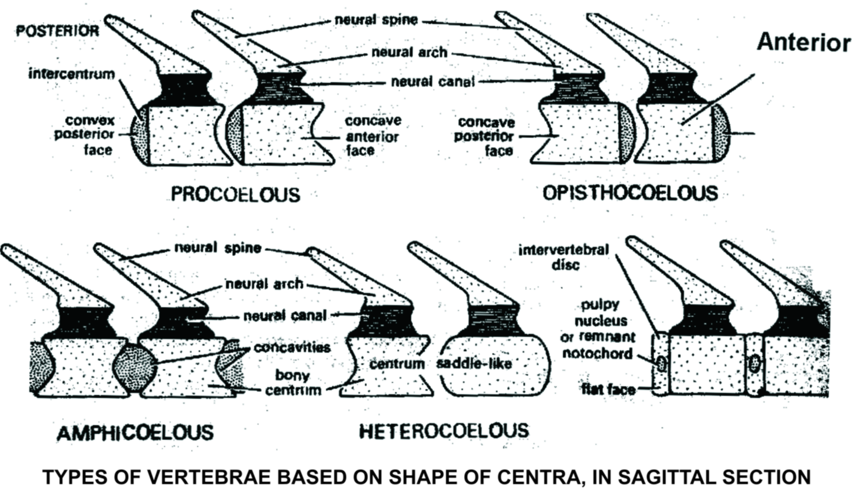
SUPER CLASS 2 (TETRAPODA) :
All gnathostomes excluding fish are included in this super-class.
Typically all tetrapods (Gk. Tetra = four + podos = foot) possess two pairs of limbs. Skin is adapted to withstand exposure to air.
They have lungs for aerial respiration. Their sensory organs are adapted for reception in air, such as vision, hearing, smelling etc.
Tetrapoda is divided into four classes: Amphibia, Reptilia, Aves and Mammalia.
CLASS : AMPHIBIA - The Vertebrates with Dual-life (Gk. Amphi = two or both; bios = life)
General Characters:
There are about 3,000 species of amphibians out of which 2,600 species are in order Anura.
They are present in the warmer regions of the world except Newts and Salamander which are present in cooler regions.
They occur only in fresh water and moist land. Amphibians are not found in sea water.
The other characters are :
1. They are poikilothermal animals. They are amphibious in nature, i.e., they can live on land as well as in water. They are mostly found in warm countries. They are ectothermic (cold blooded). Body is divisible into head and trunk. Tail may be present in some forms.
2. The skin is smooth or rough, having cutaneous glands which keep it moist. They are usually without scales, but if scales are present, they are hidden beneath the skin (e.g., Caecilians).
3. Two pairs of limbs are used for locomotion.
4. The gills are present at least in the larval stage; some adult forms also carry them in addition to lungs (e.g., Necturus).
5. Alimentary canal, urinary and reproductive tracts open into a common chamber called cloaca which opens to the exterior.
6. Skull is dicondylic, i.e., with two occipital condyles for articulation with vertebral column.
7. The respiratory organs are lungs, buccopharyngeal cavity, skin and gills.
8. The heart is three chambered, having two auricles and one ventricle. In the heart, there are present sinus venosus and truncus arteriosus. Both hepatic portal and renal portal systems are well developed.
9. Kidneys are mesonephric. Urinary bladder is present in frog. Larvae and tailed amphibians (e.g., Salamanders) are ammonotelic. Frogs and toads are ureotelic.
10. Ear consists of internal and middle ear. Tympanum (outer membrane) covers the middle ear. The middle ear has a single ear ossicle called Columella Auris.
11. Ten pairs of cranial nerves are present. Sexes are seperate, fertilization is external, oviparous and development indirect.
12. They return towaterfor breeding. Male lacks copulatory organs. Indirect development occurs. A fish like stage, the tadpole is present.
13. Male frog can croak louder than females because of the presence of vocal sacs.
Example : Bufo (Toad), Rana (Frog), Hyla (Tree frog), Salamandra (Salamander), Ichthyophis (Limbless amphibia).
Living Amphibians are divided into three orders:

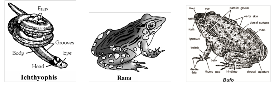
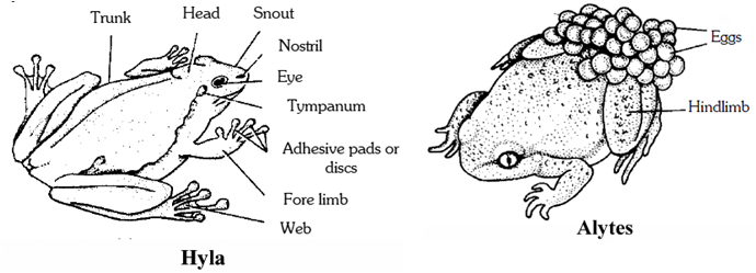
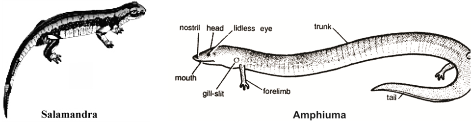
Concept Builder
1. Some frogs have developmen in amazing ways to prevent their eggs from drying out: The female surinam toad of South America carries her eggs in pits/pockets of skin on her back. As many as 60 young pass through the tadpole stage while embedded in her back and then emerge as small frogs.
2. In Frogs, external ears are absent, only Tympanum is seen externally: Frogs have a tympanic membrane on the surface of both sides of their head. The tympanic membrane of a human are called eardrums and each is protected inside ear canal.
3. Word Roots and Origins
(i) Caudata: from latin Caudata meaning "tail"
(ii) Nictitating: from the latin, nictare meaning to "wink"
Concept Builder
1. Seymouria is a connecting link between amphibia and reptiles.
2. Paedogenesis: Development of gonads and/or production of young ones by immature or larval forms e.g. Salamander (Ambyostoma).
3. Total neoteny is shown by Necturus, Siren and Proteus.
4. Toads are used in chinese medicines. Skin of toad secretes a substance that increases B.P when injected into humans.
5. Skin of tadpole secretes an enzyme, diastase.
6. Upper jaw of frog is derived from quadrate cartilage whereas lower jaw is derived from Meckel's cartilage.
7. First toe is called hallux.
8. Jaw suspension is autostylic, urostyle is last component of vertebral column.
9. Shapes of 2-7th vertebrae-Procoelous (typical); 8th vertebra-Amphicoelous; 9th vertebra - Acoelous.
10. Amphiuma-has the largest RBCs among vertebrates.
11. At the time of metamorphosis, the tadpole does not feed.
12. Blind worms (limbless amphibians) are unusual amphibians as only one species lives in water. All the other burrow underground and are rarely seen on the surface. Many do not even need water to breed.
13. Carboniferous Period-UAge of Amphibians".
14. The Arrow poison frogs secretes a powerful poison from the skin which can cause instant death.
15. Most poisonous frog-Golden dart poison frog from South America is the most poisonous frog. One adult frog contains enough poison to kill 200 people.
CLASS : REPTILIA
Their name refers to their creeping or crawling mode of locomotion.
Reptiles are cold blooded or poikilothermic animals.
There are only about 6,000 species of reptiles now living.
But fossils show that 200 million years ago, the reptiles were the most dominant animals on earth.
Herpetology is the study of reptiles.
The largest reptiles today are the 'komodo dragon' Varanus komodoensis of indonesia (a lizard) 3m in length, crocodiles (upto 7m), pythons (upto 10m) and giant tortoises weighing upto 600kg.
Important characters of this class are:
1. They are covered by dry and cornified skin having epidermal scales or scutes. Snakes and lizards shed their scales as 'skin cast'. Scales prevent loss of body water and so reptiles do not require a moist environment like amphibians.
2. Respiration is only through lungs, which is improved by the development of ribs.
3. Limbs -when present are two pairs, pentadactyl, digits have horny claws.
4. Mouth is terminal with conical teeth which are pleurodont in lizards and snakes and thecodont in crocodiles. Teeth are absent and replaced by horny beaks in turtles.
5. Endoskeleton is bony, skull is monocondylic (with one occipital condyle).
6. Heart is usually 3-chambered or partially 4-chambered. Interventricular septum is incomplete. Heart is completely 4-chambered in crocodiles. Two systemic arches are present. Erythrocytes are oval and nucleated.
7. Kidneys are metanephric. Excretion is uricotelic. 'Urinary bladder is absent in snakes and crocodiles.
8. Cranial nerves are 12 pairs. Jacobson's organ (vomeronasal organ) present in the roof of buccal cavity, concerned with smell, is well developed in snakes and lizards. They do not have external ear openings. Tympanum represent ears.
9. Sexes are separate. Males usually have a muscular copulatory organ. Fertilization is internal. Mostly are oviparous and development is direct. Some are viviparous. No metamorphosis.
Example : Chelone (Turtle), Testudo (Tortoise), Chameleon (Tree lizard), Calotes (Garden lizard), Crocodilus (Crocodile), Alligator (Alligator). Hemidactylus (Wall lizard), Poisonous snakes – Naja (Cobra), Bangarus (Krait), Vipera (Viper) etc.
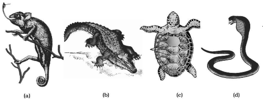
CLASSIFICATION
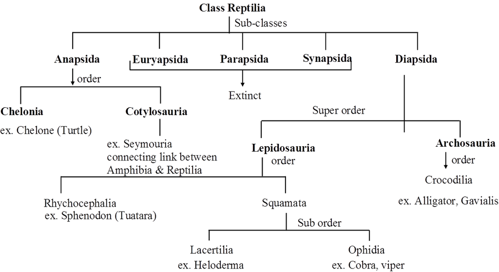
Classification of reptiles is based on temporal fossa on skull. The class reptilia is first divided into 5 sub-classes:
I. Anapsida, II. Euryapsida, III. Parapsida, IV-Synapsida and
V. Diapsida
Out of 5 sub-classes, the living reptiles are found only in two sub-classes,
[A] Anapsida and [B] Diapsida.
Concept Builder
1. Snake Charming :
Contrary to general belief, the snake charmer's cobra does not dance the tune of the flute.
Rather it follows the movement of the tip of the flute and the swaying body of its master, who really moves with the rhythm of the music.
Raising its head and spreading its hood, the cobra gets ready to strike the moving flute tip.
The charmer knows the striking distance and adjusts himself so that the snake orients towards the waving flute but keeps beyond the strike distance.
2. Rattle snake muscles :
Rattle snakes have the quickest moving skeletal muscles in the animal world.
Their tail muscle allows them to shake their rattle 90 times each second.
Most muscles would quickly tire from this kind of energetic activity, but the rattle snake can shake its tail for long periods of time.
The muscle's ability to use ATP in an efficient manner allows it to sustain a high level of activity.
Identification of poisonous and non-poisonous snake

Order: Crocodilia
Differences between Crocodile, Alligator and Gavialis

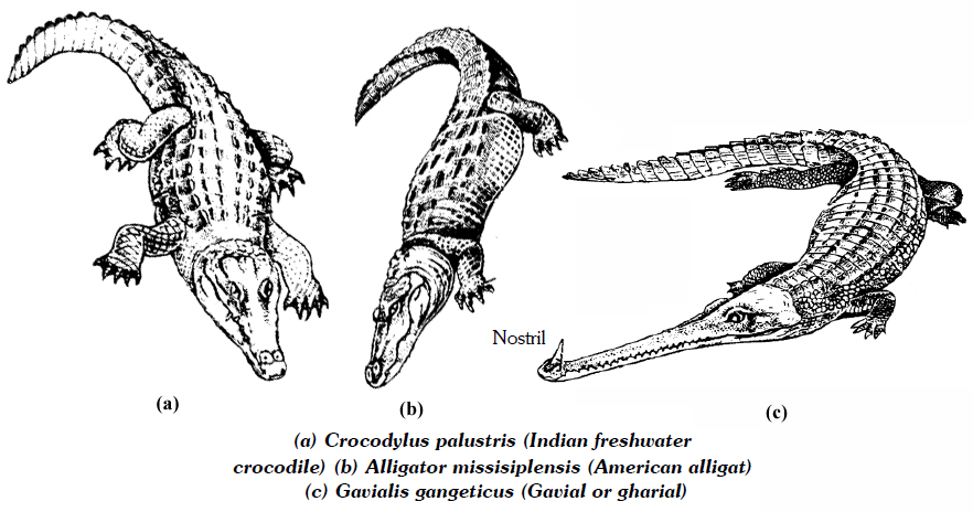
Concept Builder
1. Benadryl is used as an antidote to counteract the effect of haemotoxins.
2. Seymouria is considered as connecting link between amphibians and reptiles.
3. Mesozoic era is known as 'age of reptiles.'
4. Loreal pit of pit viper is thermoreceptor organ.
5. Reptiles without urinary bladder-snakes, crocodiles, alligators.
6. Many sea snakes are viviparous. Pit vipers are also mostly viviparous.
7. The oldest living animal in any Indian Zoo is the Aldabra giant tortoise.
8. Rattle snakes's tail emits a frightening sound which scares away the enemy.
9. The king cobra of India is the only snake in the world that builds a nest.
10. Largest snake-python; Smallest snake-Thread snake.
11. Largest lizard-Komodo dragon; Smallest lizard-Two species of dwarf gecko.
12. Long dinosaur-Diplodocus; Small dinosaur-Compsognafhus.
13. Largest living reptiles (i) Python, (ii) Crocodile (iii) Komodo dragon.
14. Hemidacfylus or wall lizard (Gecko) is a swift runneron smooth vertical surface due to the presence of vaccum pads.
15. Draco is flying lizard but it actually does not fly.
16. Heloderma is a poisonous lizard. (Gila monster)
17. Ophiosaurus known as 'glass snake', 'blind worm', or 'slow worm', limbless lizard.
18. Anguis is also blind worm and limbless.
19. Most Poisonous snake of world-Peninsular Tiger Snake.
20. Poisonous sea snake-Hydrophis
21. Heaviestdinosaurs-Brontosaurus
22. Biggestflesh eater dinosaur-Tyrannosaurus
23. Horned dinosaur-Stegosaurus
24. Varanus-Monitorlizard (largest living lizard).
25. Carapace is hard shield like structure present on the dorsal surface of tortoise and turtle.
26. Reptiles arose in the lower carboniferous time, from Labyrinthodon Amphibia.
27. Varanus is commonly called "Goye" or "monitor"; semi-aquatic; slightly laterally compressed tail.
28. Phrynosoma is found in deserts; called "Horned toad", skin hygroscopic; takes water in the form of dew; exudes red blood-like stream from eyes, whenever, terrified.
29. Iguana is 5-6 feet long; body and tail laterally compressed; arboreal in habit; herbivorous found in tropical America.
30. Sphenodon is the only living genus of Rhynchocephalia. It is commonly called Tuatara. Found only in New Zealand. It is called a living fossil as it resembles with Hanaesaurus of Jurassic age.
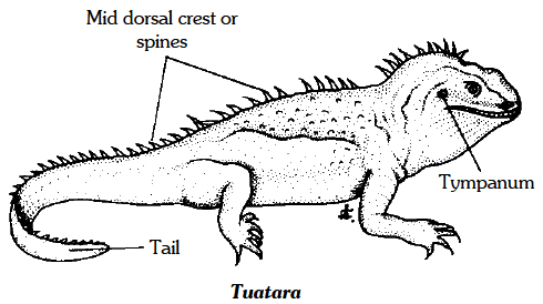
31. Largest living carnivorous reptile is Crocodilus.
32. Thermomorpha is a group of reptiles which are considered to be the ancestors of mammals.
33. Hedonic glands present in males secrete a sticky substance which hardens like spine and helps to hold the mate firmly.
34. Hemipenes-In Uromastix, cloaca possesses a pair of copulatory organs called hemipenes.
35. Russel's viper is the largest Indian viper.
36. Hafkin's Institute of Bombay is the main centre where antivenom is produced.
37. Limbs of Chameleon show syndactyly (fusion of digits) as an adaptation to arboreal life to form opposable finger to hold the twigs.
CLASS : AVES (Birds)
Study of birds is ornithology.
General Characters :
1. Birds are feathered bipeds, air breathing, truly flying veliebrates (other being bats from Class Mammalia).
2. Jaw bones are prolonged into a toothless beak or bill.
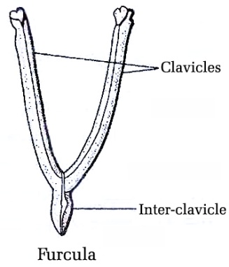
3. Limbs are two pairs. Forelimbs are modified as wings for flying. Hindlimbs or legs are large, each foot usually bears four, clawed toes. Hind limbs generally have scales.
4. Exoskeleton consists of feathers, scales and claws, which are epidermal derivatives. Skin without glands, the only cutaneous gland is uropygial gland or preen gland/oil gland at the base of tail.
5. Oesophagus is dilated into a crop for quick feeding and storage. Crop secretes 'pigeon millt' during breeding season. Stomach is divided into a glandular proventriculus and a muscular gizzard. The junction of small intestine and rectum is marked by a pair of rectal caecae. Cloaca of birds is three-chambered: antenior coprodaeum, middle urodaeum and a large posterior proctodaeum. There is no gall bladder in birds like pigeon.
6. Respiration with compact, spongy, nondistensible lungs continuous with thin walled air sacs. Air sacs reduce body weight, helps in double respiration and internal cooling. Larynx without vocal cords. A sound box or syrinx producing voice, lies at or near the junction Furcula of trachea and bronchi.
7. Heart is completely four-chambered as in mammals. Sinus venosus absent Only right aortic (systemic) arch persists in the adults. Renal portal system is vestigial. Erythiocytes are minute, oval and nucleated. The blood of the birds may be called the richest blood in the animal kingdom. It has more RBCs per cubic mm of blood than in any other animal.
8. Kidneys metanephric and three-lobed. Ureters open into cloaca. Urinary bladder is absent. Excretion is uricotelic.
9. Cranial nerves are 12 pairs.
10. Olfactory organs are poor. Middle ear contains a single ossicle. Eyes possess nictitating membrane, Pecten is a comb-like structure found in the eyes near blindspot. Pecten helps in the nutrition of eyeball. Pecten is found in all birds except Kiwi.
11. Sexes separate. Some birds show sexual dimorphism (e.g. , Parrot and Peacock). Male bird has a pair of abdominal testes and a pair of sperm ducts. A copulatory organ is absent in all birds except in ratitae, geese, ducks etc. Females are oviparous with a single (left) functional ovary and oviduct (Mullerian duct). Development is direct.
Different parts of oviduct secrete the following :
Ostium – Dense albumin
Magnum – White albumin
Uterus – Calcareous shell
Eggs of birds are cleidoic (with calcareous shell), macrolecithal (amount of yolk), meiolecithal (distribution of yolk). Extraembryonic membranes are present (Amniota).
12. Feathers are found only in birds. Feattlers are made up of a protein keratin and are water proof due to the oily secretion of preen glands. The arrangement of feathers on the body of birds is called Pterylosis. The featherless areas are called as Apteria. Four types of feathers are: quill feathers, contour feathers, filoplumes and down feathers. In absence of down feathers the birds will not be able to keep the body warm. Quill feathers in the wings are called 'remiges'. Quill feathers in the tail are called 'rectrices'. Filoplumes and Down feathers help in the insulation of the body.
13. Birds are the first vertebrates to have warm blood. Body temperature is regulated (homoiothermous).
14. Alaspuria -bastardwing -Feathers on 1st digit (thumb).
Example : Corvus (Crow), Columba (Pigeon), Psittacula (Parrot), Struthio (Ostrich), Pavo (Peacock), Aptenodytes (Penguin), Neophron (Vulture).
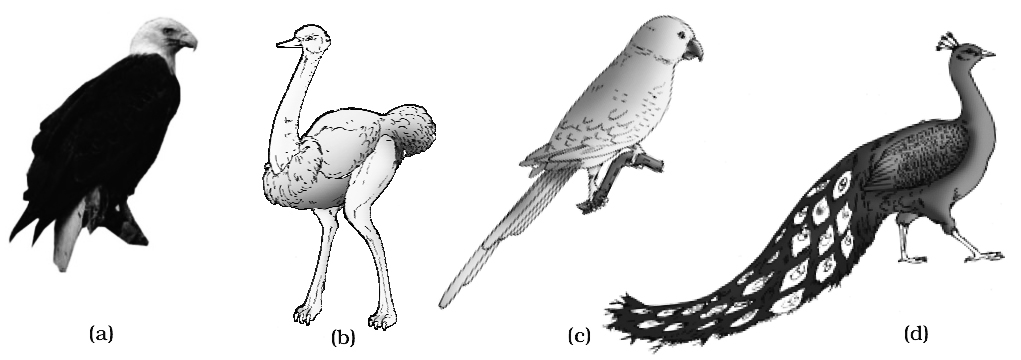
Concept Builder
Types of Feathers
1. Quills:
(a) They are large feathers found in wings and tail.
(b) It has a central axis called shaft.
(c) Small proximal part of the shaft is hollow, translucent and cylindrical and is termed as calamus.
(d) The long-distal, solid and opaque part of the stem is known as rachis.
(e) An umbilical groove extends all along the ventral side of rachis.
(t) Small hole on the proximal end of calamus is known as inferior umbilicus, the hole on the distal end of calamus is known as superior umbilicus.
(g) Each vane is composed of parallel filaments, the barbs. Each bears barbules.
(h) The distal barbules of each barb bear small hooklets.
(i) Emu bird have an after shaft as long as the main shaft.
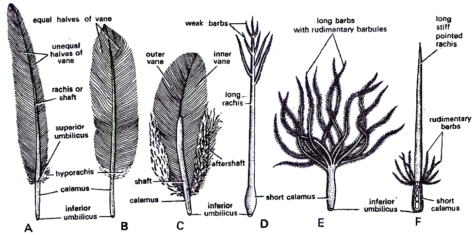
2. Coverts are small feathers found in the wings and tail. They fill up the gaps left between the bases of the quills. They have short calamus in comparison to quills.
3. Contours: Small feathers that cover the body and give it its shape. They resemble the quills but their barbs are not so strongly joined and can be separated easily.
4. Filoplumes: Occur beneath contour feathers, very small in size, consists of long rachis bearing at the tip a few weak free barbs with barbules.
5. Down feathers/Nestling downs: They cover the newly hatched bird. They consist of short calamus, reduced rachis bearing flexible barbs with short barbules. They are found beneath contours.
6. Bristles: Short calamus, a long rachis bearing a few vestigeal barbs at its base. Bristles occur nearthe mouth in fly-catchers.
7. The first digit of the hand (thumb or pollex), bears a tuft of small feathers known as bustard wing or alaspuria or false wing.
CLASSIFICATION
Class Aves is divided into two sub-classes: Archaeornithes and Neornithes.
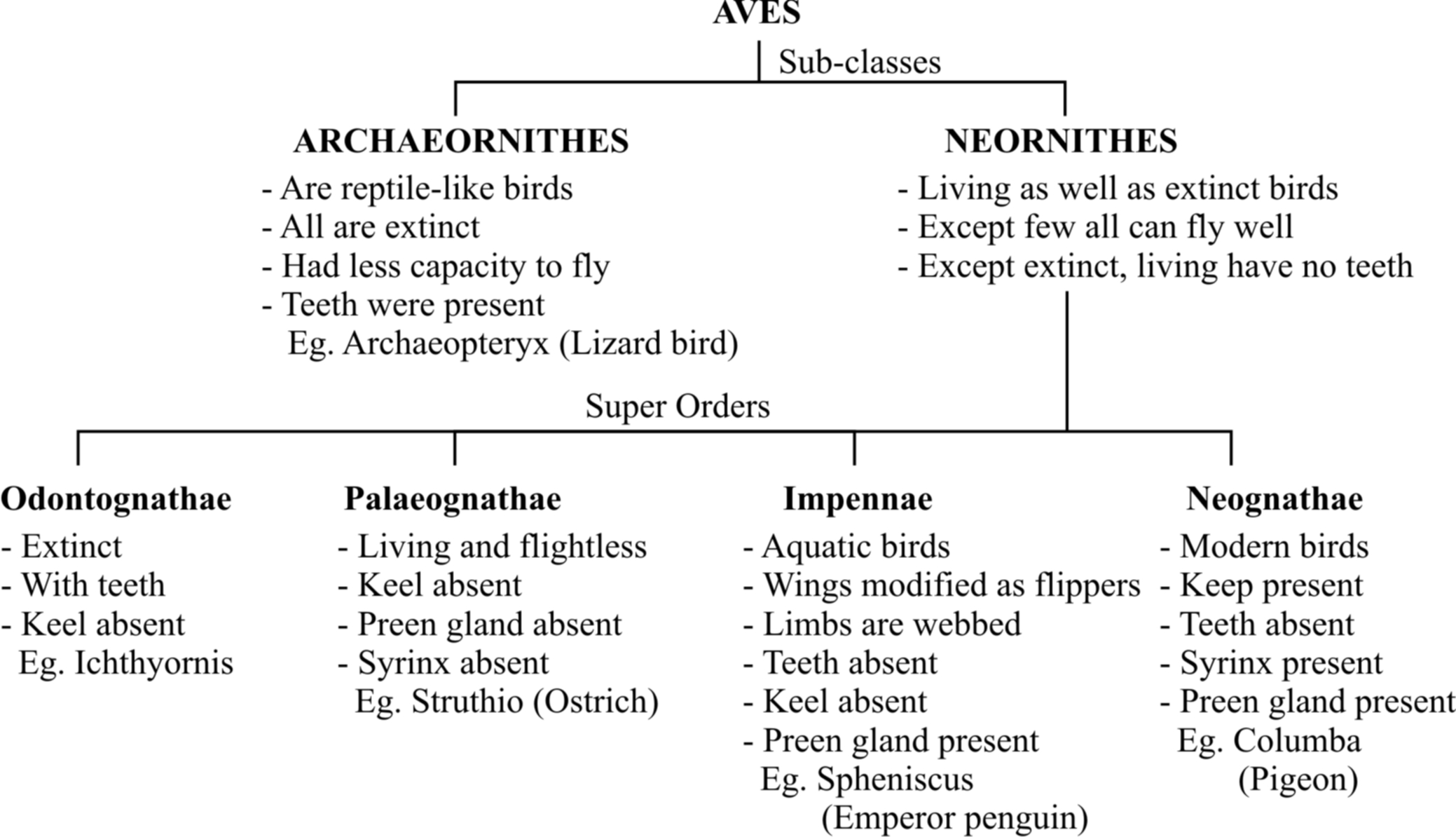
 Sub-class I : Archaeornithes
1. All extinct Jurassic period birds of Mesozoic era.
2. Wings primitive with little power of flight.
3. Tail long, lizard-like with two lateral rows of rectrices.
4. Hand with clawed fingers.
5. Skull with teeth in both jaws e.g., Archaeopteryx.
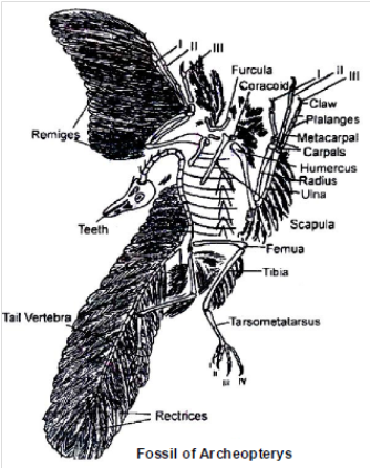
Sub-class II : Neornithes
1. Modern as well as extinct, post-Jurassic period birds.
2. Wings well developed and adapted for flight (with few exceptions).
3. Tail short and reduced.
4. Teeth absent except in some fossil birds.
5. Sternum with keel or carina.
There are four super orders in this subclass :
Super-order I : Odontognathae
e.g., Hesperornis (with teeth) extinct.
Super-order II : Palaeognathae (Ratitae)
e.g., Flightless running birds
Super-order III : Impennae
e.g., Penguins (Aptenodytes)
Penguins are found in Antarctica (South pole). They have paddle-like wings and cannot fly. Penguins are marine and lay eggs in ice.
Super-order IV : Neognathae (Carinatae)
e.g., Modern flying birds as Columba (pigeon), Psittacula (parrot), Neophron (vulture), Pavo (peacock).
Conceptual Questions
Q.1 Why might it be a disadvantage for an eagle to have a urinary bladder?
Q.2 Why might teeth be a disadvantage to a bird?
Ans. 1. Urine contains a greater amount of water, which is heavy. The extra weight would be a disadvantage in flight.
2. Teeth are heavy and require heavy jaws.
Concept Builder
1. Brood Parasite : Eudynamis (koel, kokila). The male sounds like kuoo-kuoo-kuoo. The female is generally heard during breeding season and has short and sharp call kik-kik-kik, otherwise she is mostly silent. It does not make any nest but lays eggs in the crow's nest. In this way koel is nest parasite. Male is black. Female is brown and profusely spotted barred with white Koel is known as the Indian nightingale because of its pleasant sound.
2. Eagle-Eyes: Eagles have much better vision than humans do. The birds keen eyesight allows it to see prey at great distances. Its visual acuity is 3-4 times higher than ours.

 Differences between Ratitae and Carinatae
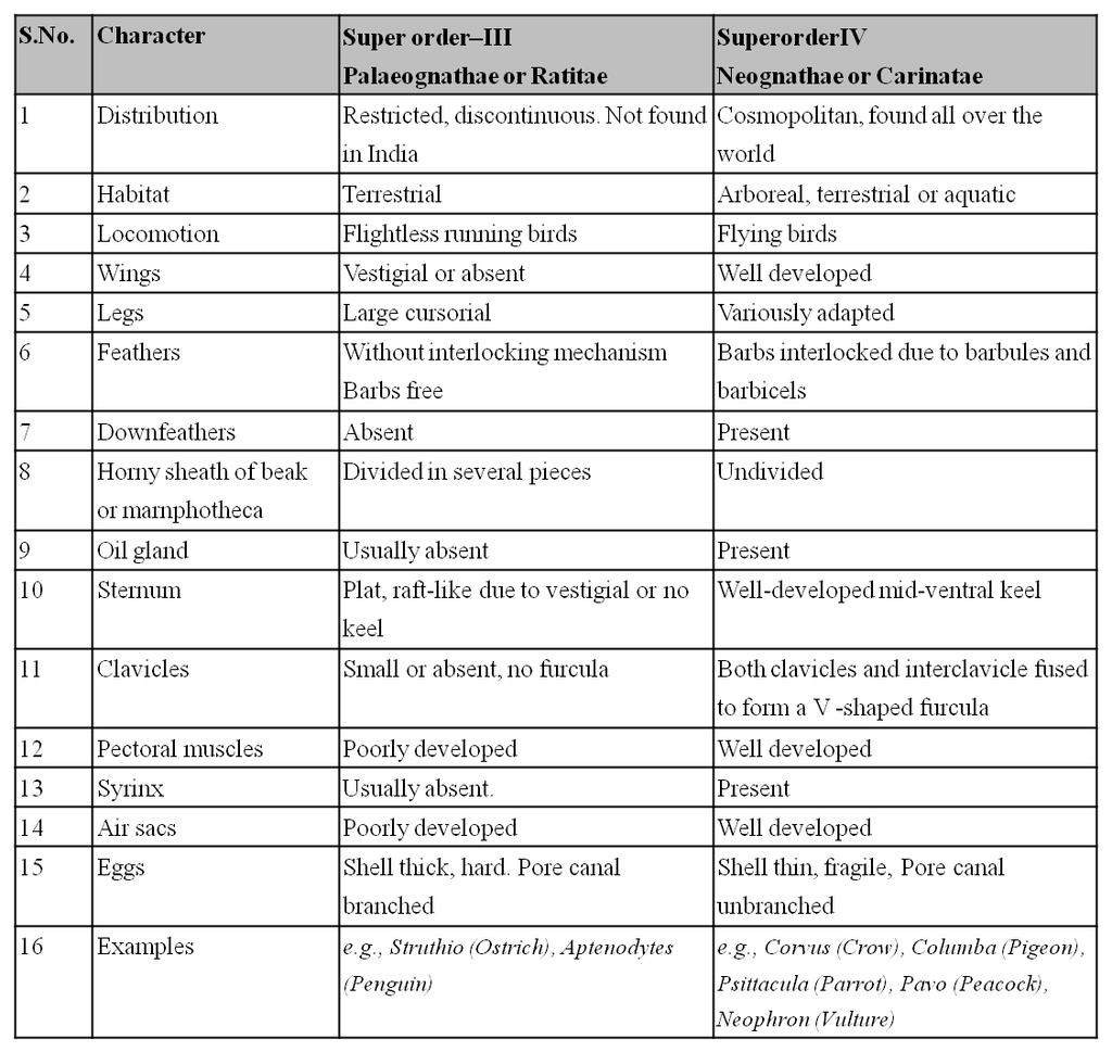
Concept Builder
1. Largest bird-Struthio camelus -8 feet height, 300 Lbs weight.
2. Smallest bird -Mellisuga helenae-5.5 cm long.
3. Smallest egg of bird-Mel/isuga (Humming bird).
4. The humming bird is the only bird which can fly backward as well as forward.
5. Largest egg of bird-Struthio-1.5 Litre capacity.
6. Largest sea bird-Diomedea epomorphora (Royal albatross) wing stretch 200-375 cms.
7. Deepest Diver bird-Aptenodytes torsteri (Emperor Penguin)
8. Fastest Swimmer bird-Pygoscelis papua (Penguin)
9. Fastest Runner bird-Struthio-60 kms/hr. speed.
10. Heaviest bird of prey-Vulture gryphus.
11. Fastest Flying bird-Swift-rarely touch the earth.
12. Second largest bird is Emu (Dromaeus) as is most primitive living bird.
13. Recently extinct bird is Dodo (Passenger birds) of Mauritius.
14. Incubation period of Hen's egg at 102°F (38.8°C) is 21 days.
15. Though polygamy is common in birds, Emu is strictly monogamous.
16. Penguins are flightless birds found in Antarctica, wings are paddle-like, act as flippers during swimming and are covered by scale-like feathers.
17. T.H. Huxley said "birds are glorified reptiles". The feathers are highly modified reptilian scales. Birds have scales on their legs. Their eggs resemble reptilian eggs in general but have a calcareous shell.
18. Among the Indian birds the sun birds are probably the smallest.
19. The Bursa Fabricii is a blind sac with much lymphoid tissue in the cloaca of some young birds. It produces lymphocytes (a type of white blood corpuscles). It is also called "cloacal thymus".
20. Columbia has the world's richest diversity of birds. '.
21. Uropygium (tail)-the projecting terminal portion of a bird's body from which the tail feathers arise.
22. Swifts use saliva for binding nest materials.
23. Kiwi lays the largest egg in proportion to its own size.
24. Himalayan Bearded Vulture is the largest Indian bird. Previously the Sarus was considered the largest Indian bird.
25. Ostrich has the largest eyes of any land animal.
26. Kiwi is the only bird known to use the sense of smell for finding food on the ground.
27. Indonesia has the most bird species facing extinction.
28. Red Billed Quelea (Quelea quelea) of Africa are the most abundant birds. Previously the house sparrow was considered the most abundant species of birds.
29. Jatinga is a village in Assam where a mass suicide of birds occurs.
30. The owl rotates its head through an angle of 2700 as its eyes do not rotate in their sockets. Each eye is fixed like a car headlight.
31. Copulatory organ (true penis) is present in ostrich, duck, swan and goose.
32. Famous Indian Ornithologist-Dr. Salim Ali.
33. National bird-Pavo cristatus (Peacock).
CLASS : MAMMALIA
They are terrestrial, warm-blooded (endothermous) or homeothermal animals so that their body temperature remains constant inspite of the changes in the temperature of the environment.
General characters of this class are :
1. The skin is covered with epidermal hair which acts as an insulating layer and allows high body temperature to be maintained. The hair may be partly lost as a secondary adaptation. In some aquatic mammals where hair is negligible, there is a subcutaneous layer of heat which provides insulation of heat and makes the warmblooded condition possible.
2. The skin has two kinds of glands, sudorific glands which produce sweat -evaporation of sweat controls the body temperature; second kind of glands are sebaceous glands which produce an oily secretion which makes the hair water-resisting. Modified sweat glands (sudoriferous) form mammary glands which produce milk in the females for nourishment of the young for some time after birth. Because they possess mammae (breasts) and mammary glands, they are called mammals. It is the most unique character of mammals.
3. Them is an external ear or pinna with an external auditory meatus. This is in addition to middle and internal ear.
4. In higher mammals, the anus is separated from the urinogenital aperture; consequently urinary and genital ducts have no connection with the digestive tube.
5. In males, testes have come to lie outside the body cavity in scrotal sacs (except in elephants and aquatic mammals and prototherian mammals).
6. Teeth are embedded in sockets of the jaw bone and are said to be thecodont. There are only two sets of teeth in life time, one deciduous or milk set and another permanent set, this condition is spoken of as diphyodont. There are four different types of teeth, hence mammals are heterodont.
7. A muscular diaphragm divides the coelom into a thoracic and an abdominal cavity; the thoracic cavity has a pericardial cavity containing the heart and two pleural cavities containing the lungs, the remaining viscera lies in the abdominal or peritoneal cavity.
8. The heart is 4-chambered with two auricles and two ventricles so that oxygenated blood lies in the left half and deoxygenated blood in the right half. This condition is also found in birds. Only the left aortic arch is present. Erythrocytes are round, biconcave and non-nucleated (except in camel where they are oval and non-nucleated).
9. Cerebral hemispheres and cerebellum are large-sized and highly developed with great increase in the cortex. The two cerebral hemispheres are joined by a transverse band of nerve fibres called corpus callosum. There are four solid optic lobes called corpora quadrigemina in the mid-brain. 12 pairs of cranial nerves.
10. The middle ear has three ear ossicles called malleus, incus and stapes; the internal ear has a spirally coiled cochlea as an efficient organ of hearing.
11. The neck generally has seven cervical vertebrae.
Example : Oviparous-Ornithorhynchus (Platypus); Viviparous -Macropus (Kangaroo), Pteropus (Flying fox), Camelus (Camel), Macaca (Monkey), Rattus (Rat), Canis (Dog), Felis (Cat), Elephas (Elephant), Equus (Horse), Delphinus (Common dolphin), Balaenoptera (Blue whale), Panthera tigris (Tiger), Panthera leo (Lion). etc.
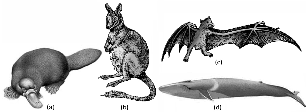
DIFFERENCE BETWEEN THREE GROUPS OF MAMMALS

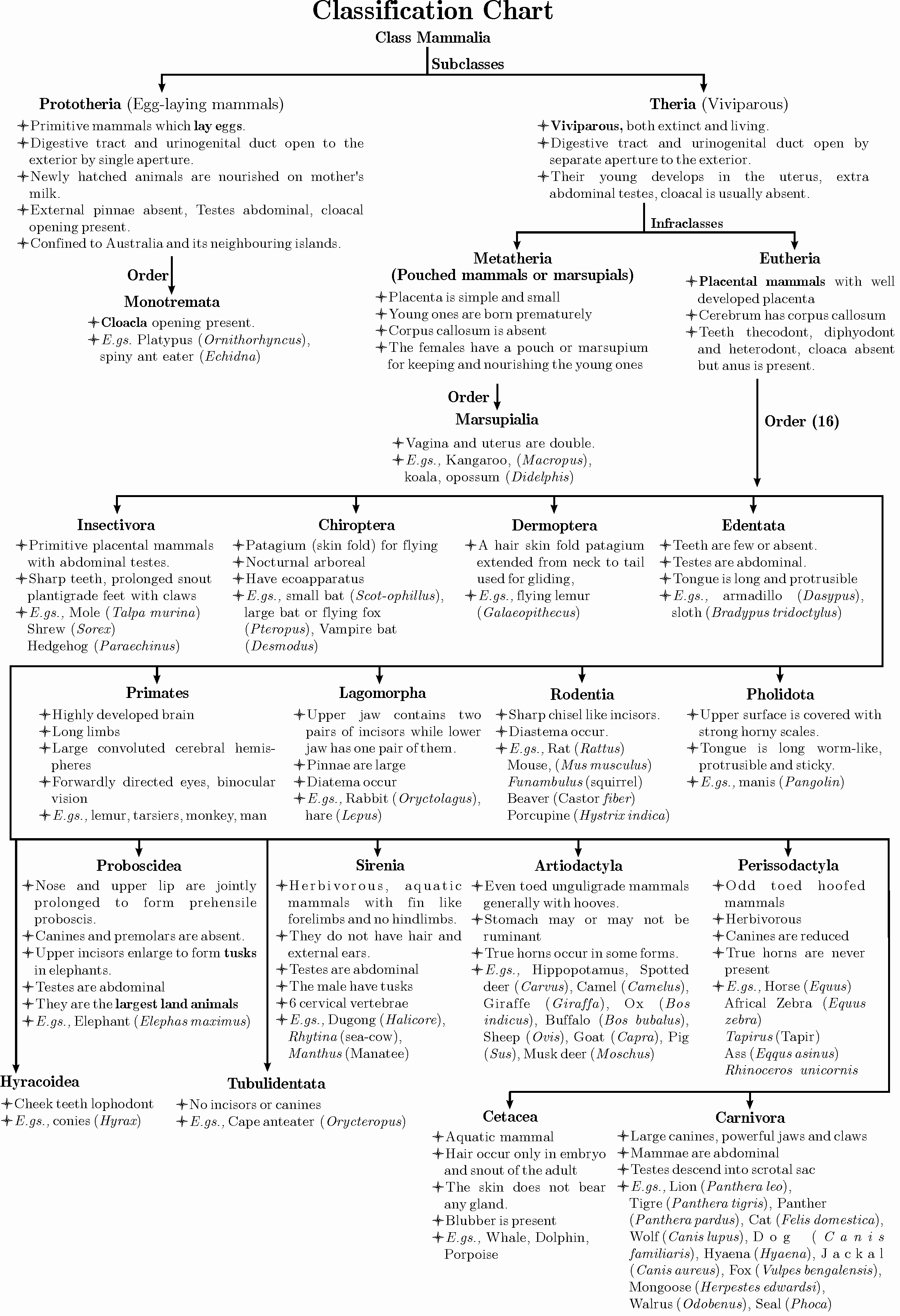
Concept Builder
1. How many vertebrae do giraffes have in their neck?
Most of the vertebrates, including giraffes and camel, have seven cervical (neck) vertebrae. At almost 6 m (20 ft) tall, the giraffe is the tallest of all vertebrates and its seven cervical vertebrae are greatly elongated
Exception : 3 toed sloth (Bradypus) with 9 cervical vertebrae and Manatee is with 6 neck vertebrae.
2. Armadillos : The nine-banded armadillo (Dasypus) is one of the few mammalian species whose range is expanding. In late 1800s, they lived only as far north as central Texas. Leprosy bacteria is cultured in the armadillos (Dasypus).
Concept Builder
1. Man remains plantigrade at rest or during locomotion. Uses only the toes for running, thus is called sub plantigrade.
2. Colour of human skin is yellow to orange due to carotene pigment in cells of stratum corneum and subdermal fat cells. Melanin is found in melanocytes.
3. In man sweat glands are absent at lip borders, clitoris, glans penis, nail beds, under surface of prepuce.
4. Sweat or sudoriferous glands secrete sweat. Sweat comprises of 95% water and 5% metabolic waste.
5. Spiny ant eater, scaly ant eaters, moles, cetaceans, sirenians and some edentates do not possess sweat glands.
6. Tusks of elephants are two incisors of upper jaw which constantly grow throughout life.
7. Man ![]()

8. Dog ![]()

9. Cat ![]()

10. Rat ![]()

11. Horse ![]()

12. Elephant ![]()

13. Primates such as monkeys, man, apes, etc., acquired 3 dimensional vision due to their most evolved motor understanding of visual sensation.
14. Deer, amongst vertebrates, proportionately to their body size, possess largest eyes.
15. Carnassial Teeth. Special shearing teeth in carnivores for cracking bones-4th premolar of upper jaw and first molar of lower jaw.
16. Milk Producing Male Mammal. Spiny Ant Eater/Echidna.
17. Monotreme. With a single aperture of cloaca for urinogenital and digestive tracts.
18. Largest Land Animal. Loxodonta africana/African Elephant. Height 3.5 m and weight 7 tonnes.
19. Tallest Land Animal/Mammal. Giraffe, upto 6 m.
20. Mouse and Rat. Mouse and Rat has first molar bigger than combined 2nd and 3rd molars.
21. Zebra Stripes. Stripes of no two zebras are alike.
22. Fastest Mammal. Acinonyx jubatus/(Cheetah) (extinct from India but present in Africa). Speed upto 100 km/hr.
23. Slowest Terrestrial Mammal. 3-toed Sloth/Bradypus, Tridactylus, 100-150 m/hr.
24. Slowest Aquatic Mammal. Sea Otter/ 10 km/hr.
25. Longest Gestation Period in Mammal. 609 days/20 months in Elephas maximus.
26. Shortest Gestation Period in Mammal. Opossum. 12-13 days.
27. Hom. Matted hair in Rhino. Antlers are solid, shed and regrown every year.
28. Tiger. Panthera tigris is National animal of India (declared in 1973).
29. Today Marsupials are found mainly in Australia except a few marsupials such as the American opossum are found outside that continent.
30. Rabbit is digitigrade because it moves on digits.
31. Plantigrade mammals are those whose palm and sole touch the ground when moving from one place to another e.g., bears and certain insectivores.
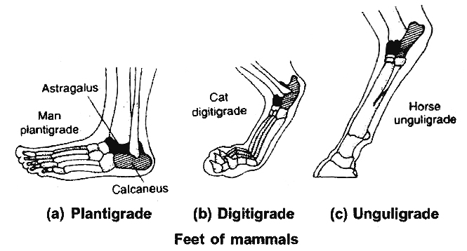
32. Unguligrade mammals are those whose only the tips of one or two fingers and toes remain in contact with the ground both at rest and locomotion. These are fastest running terrestrial mammals e.g., horses, deers, cows, donkeys etc.
33. Hides are prepared from dermis of animal skin.
34. Seals and whales have scanty hairs because heat insulation is done by blubber.
35. Horns of rhinoceros, scales of scaly ant eater and spines of porcupine are derivatives of hairs.
36. Retina of owls contains only rods (suitable for nocturnal habit). Also, the same occurs in shrews, hedge hog, rodents, bats etc.
37. Retina of fowls contains only cones (suitable for diurnal habit). Squirrels also have only cones.
38. Whales, mice, shrews and also some other mammals, but not rabbit and man, posses Harderian gland like that of frog.
39. Scrotum of mammals contains testes, in most of the mammals; this is a thermostatic chamber for testes and lies outside the abdominal cavity because sperms cannot develop in body temperature. Thus, these become close, if the temperature falls more than 2°C or come apart from the body, if temperature rises more than 2°C.
40. In insectivores, chiroptera and rodentia scrotum is formed only during breeding season, later move to abdominal cavity e.g., bat, otter.
41. Insectivores, edentates, proboscidians (elephants), cetaceans (whales) always have their testes inside the body cavity.
SUMMARY
1. Phylum Chordata includes animals which possess a notochord either throughout or during early embryonic life. Other common features observed in the chordates are the dorsal, hollow nerve cord and paired pharyngeal gill slits.
2. Some of the vertebrates do not possess jaws (Agnatha) wheares most of them possess jaws (Gnathostomata). Agnatha is represented by the class Cyclostomata. They are the most primitive chordates and are ectoparasites on fishes.
3. Gnathostomata has two super classes. Pisces and Tetrapoda. Classes chondrichthyes and osteichthyes bear fins for locomotion and are grouped under pisces. The chondrichthyes are fishes with cartilaginous endoskeleton and are marine and Osteichthyes class includes bony fishes.
4. Classes Amphibia, Reptilia, Aves and Mammalia have two pairs of limbs and are thus grouped under Tetrapoda. The amphibians have adapted to live both on land and water. Reptiles are characterised by the presence of dry and cornified skin. Limbs are absent in snakes. Fishes, amphibians and reptiles are poikilothermous (cold-blooded). Aves are warm-blooded animals with feathers on their bodies and forelimbs modified into wings for flying. Hind limbs are adapted for walking, swimming, perching or clasping. The unique features of mammals are the presence of mammary glands and hairs on the skin. They commonly exhibit viviparity.
phylum chordata
Phylum Chordata
This is the third most populous phylum in the animal kingdom. The existence of a notochord, a dorsal hollow nerve cord, a post-anal tail, and paired pharyngeal gill slits distinguishes animals belonging to the phylum Chordata. These are bilaterally symmetrical, triploblastic, coelomates that are organized at the organ-system level. Their circulatory system is closed. The Chordata phylum is subdivided into three subphyla:
The Chordata phylum is subdivided into three subphyla:
(a) Urochordata or Tunicata: Examples: Ascidia, Salpa, and Doliolum;
(b) Cephalochordata: Example: Branchiostoma; Amphioxus or Lancelet
(c)Vertebrata: Example: Horse, Humans
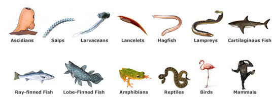
Subphyla Urochordata and Cephalochordata are collectively referred to as protochordate andare only found in the ocean. The notochord is only present in the larval tail of Urochordata, but it extends from the head to the tail area of Cephalochordata and is present throughout their lives.During the embryonic period, members of the Vertebrata subphylum have a notochord. In adulthood, the notochord is replaced by a cartilaginous or bony vertebral column. All chordates are vertebrates, however, not all chordates are vertebrates. Vertebrates have a ventral muscular heart with two, three, or four chambers, kidneys for excretion and osmoregulation, and paired appendages that can be fins or limbs.
The subphylum Vertebrata is further divided as shown below:
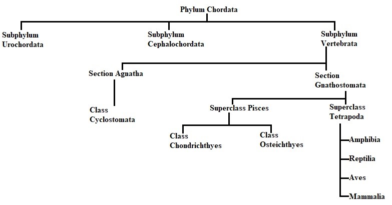
Agnatha comprises jawless organisms which consist of only one subdivision, Cyclostomata. Gnathostomata comprises animals having jaws and can be further divided into fin-bearing Pisces and limb-bearing Tetrapods.
A. Class Cyclostomata: Cyclostomata comprises loving members of the class who are ectoparasiteson some fishes. For respiration, they have an extended body with 6-15 pairs of gill slits. Cyclostomes have a circular sucking mouth with no jaws. Scales are missing from their bodies, and they have paired fins. The cranium and spinal column are made of cartilage. The circulation is closed. Cyclostomes are marine creatures that travel to freshwater to procreate. They die within a few days of spawning. After metamorphosis, their larvae return to the sea. Petromyzon (Lamprey) and Myxine (Hagfish) are two examples.
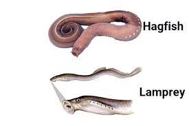
B. Class Chondrichthyes: They are streamlined aquatic organisms with a cartilaginous endoskeleton. The mouth is situated ventrally. The notochord remains throughout life. Gill slits are distinct and lack an operculum (gill cover). The skin is robust and covered in little placoid scales. Teeth are modified placoid scales that point backward. Their jaws are extremely strong. These creatures are predators. They must swim frequently to prevent sinking due to the lack of an air bladder. It has two chambers (one auricle and one ventricle). Some have electric organs (such as Torpedo) and others have poison stings (e.g., Trygon). They are cold-blooded (poikilothermous) animals, meaning they can't control their body temperature. Sexes are distinct. Males have claspers on their pelvic fins.Internal fertilization is present, and many of them are viviparous. Scoliodon(dogfish), Pristis (sawfish), Carcharodon (great white shark), and Trygon(Stingray)are among examples.
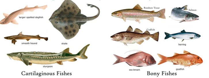
C.Class Osteichthyes: With a bony endoskeleton, it contains both marine and freshwater fish. Their physique is slim. The mouth is primarily terminal. They have four pairs of gills on each side, which are covered by an operculum. Cycloid/ctenoid scales cover the skin. The presence of an air bladder influences buoyancy. A two-chambered heart (one auricle and one ventricle). They're cold-blooded creatures. Sexes are distinct. External fertilization is common. They are generally oviparous and have a straight development. Examples includeFreshwater – Labeo (Rohu), Catla (Katla), Clarias (Magur); Marine – Exocoetus (Flying fish), Hippocampus (Sea horse); Aquarium – Betta (Fighting fish), Pterophyllum(Angelfish).
D. Class Amphibia: Amphibians can survive in both aquatic and terrestrial environments. They all have two sets of limbs. The body is divided into two parts: head and trunk. Some individuals may have a tail. The skin of amphibians is wet (without scales). Eyelids cover the eyes. The ear is represented by a tympanum. The alimentary canal, urinary system, and reproductive tract all open into a single chamber called the cloaca, which is visible from the outside. Gills, lungs, and skin are all used to breathe. There are three chambers in the heart (two auricles and one ventricle). They are cold-blooded creatures. Sexes are distinct. External fertilization is used. They are oviparous and have indirect development. Bufo (toad), Rana (frog), Hyla (tree frog), Salamandra(salamander), and Ichthyophis (Limbless amphibian) are a few examples.
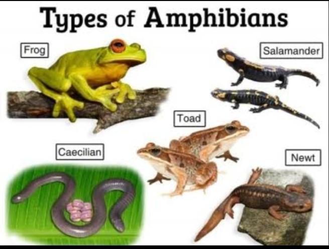
E. Class Reptilia: This class includes organisms having to creep or crawla mode of locomotion. They are generally terrestrial animals with dry, cornified skin, epidermal scales, or scutes covering their bodies. External ear holes are not present. Tympanum stands for the ear. Limbs are two pairs when present. Crocodiles have four chambers in their hearts, which are usually three. Reptiles are Poikilotherms. As skin cast, snakes and lizards shed their scales. Sexes are distinct. Internal fertilisation. They are oviparous and have straight development. Chelone (turtle), Testudo (tortoise), Chameleon (tree lizard), Calotes (garden lizard), Crocodilus (crocodile), and Alligator are just a few examples. Poisonous s.nakes — Naja (Cobra), Bangarus (Krait), Vipera, (Viper) etc. are also known as reptiles.
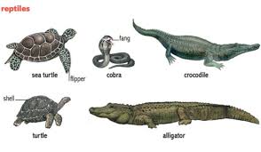
F.Class Aves: The presence of feathers distinguishes Aves (birds), and most of them can fly, except flightless birds (e.g., Ostrich and Emu). They have a beak. The forelimbs have been transformed into wings. Scales cover the hind limbs, which are modified for walking, swimming, and grasping tree branches. Except for the oil gland at the base of the tail, the skin is dry and lacks glands. The long bones are hollow with air spaces, and the endoskeleton is entirely ossified (bony) (pneumatic). The crop and gizzard are extra chambers in the digestive tract of birds. The heart has four chambers in total. They are warm-blooded (homoeothermic) creatures, which means they can keep their body temperature constant. Lungs are used to breathe. Respiration is aided by air sacs attached to the lungs. Sexes are distinct.Internal fertilization is present. They are oviparous and have direct development. Corvus (Crow), Columba (Pigeon), Psittacula (Parrot), Struthio (Ostrich), Pavo (Peacock), Aptenodytes (Penguin), and Neophron (Vulture) are some examples.
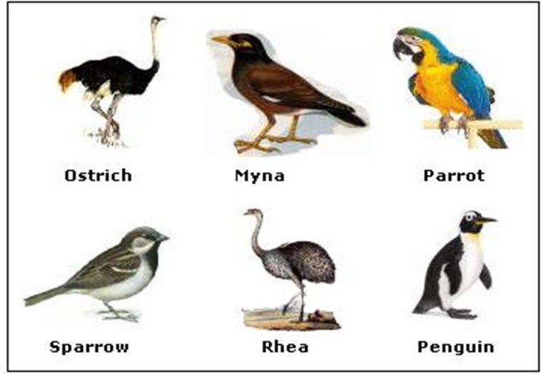
G. Class Mammalia: In the phylum Chordata, Mammalia is the most developed class. Polar ice caps, deserts, mountains, woods, grasslands, and gloomy tunnels are all places where mammals can be found. Some have developed the ability to fly or live in water. The existence of milk-producing glands (mammary glands), which nourish the young, is the most distinguishing mammalian trait. They have two pairs of limbs, one for walking, the other for running, climbing, digging, swimming, and flying. Mammalian skin is remarkable in that it has hair. External pinnae or ears are present. The jaw contains a variety of tooth kinds. The human heart has four chambers. They're the same temperature. Lungs are used to breathe. Sexes are distinct, and fertilization takes place internally.
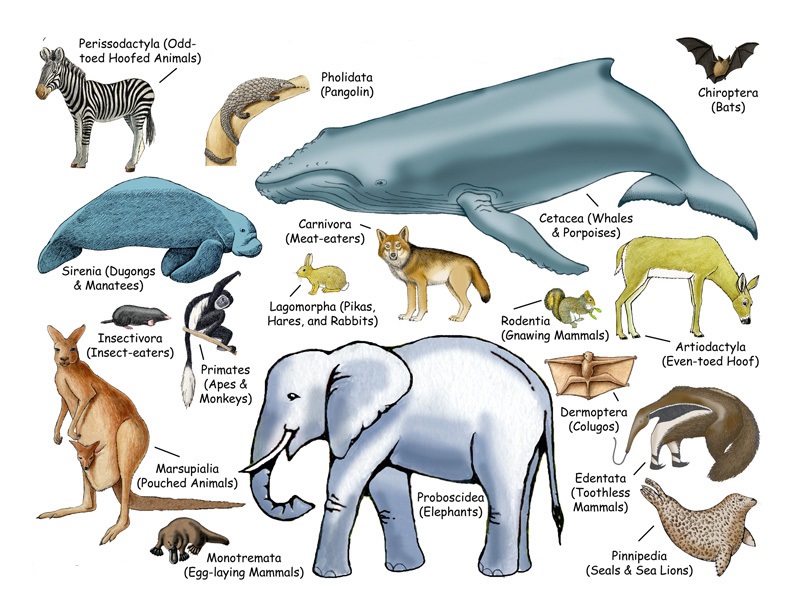
With a few exceptions, they are viviparous and develop directly. Macropus (Kangaroo), Pteropus (Flying fox), Camelus (Camel), Macaca (Monkey), Rattus (Rat), Canis (Dog), Felis (Cat), Elephas (Elephant), Equus (Horse), Delphinus (Common dolphin), Balaenoptera (Blue whale), Panthera tigris (Tiger), Panthera leo (Lion).
