 ACME SMART PUBLICATION
- Books Name
- ACME SMART COACHING Biology Book
- Publication
- ACME SMART PUBLICATION
- Course
- CBSE Class 11
- Subject
- Biology
MUSCLE TISSUE
Muscles cause movements of limbs and internal organs and also locomotion of the organism.
Cells of muscle tissue can shorten forcefully and again return to the relaxed state.
This specialised property is called Contractility.
It is based on the organised arrangement of some protein filaments in the cytoplasm of a muscle cell.
The cell shortens or relaxes according to the relative positions of different intracellular filaments.
Whenever adequately stimulated, muscle cells respond by contracting.
This property of the muscle tissue is responsible for various movements in an animal.
Muscle cells are usually called Muscle Fibres because they are thin and elongated.
In higher animals, some muscles remain associated with the skeleton, but many others form walls of visceral organs, blood vessels and heart.
Muscle tissue may be classified into striated, non-striated and cardiac muscles, according to their structure, location and functions.
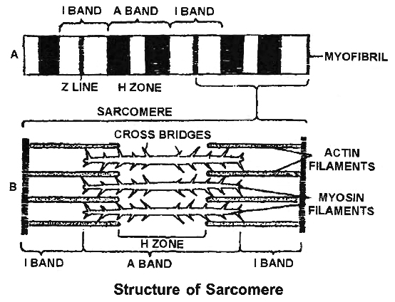
(i) Striated / Skeletal / Voluntary muscles are attached to bones by tendons. A voluntary muscle is composed of long bundles of striated muscle fibres. Each fibre is a long, unbranched, cylindrical cell. It shows transverse striations in the form of regular alternate dark (A) and light (l) bands.
At the centre of the I band is a fine, dense Z band or Z-line (Krause's membrane). The plasma membrane covering the fibre is called Sarcolemma. The cytoplasm inside the fibre is called Sarcoplasm.
The sarcoplasm contains many long, thin, unbranched, cross-striated cylindrical structures called Myofibrils. They are arranged along the long axis ofthe fibre. Dark A bands of neighbouring myofibrils are located side by side, so also are their light I bands. This gives crossstriated appearance to the entire muscle fibre also.
A-band has both actin and myosin filaments. The portion of A-band, where actin filaments are absent is called H-zone. Z-line or Krauze membrane is a dark membrane which bisects I band or isotropic band.
Muscle is rich in proteins. Most of these proteins occur as two types of filaments arranged longitudinally in myofibrils. The thick filaments are made up of the protein Myosin. Myosin filaments are located inside A bands.
Thin filaments are more numerous. They are composed of the protein Actin. From a fine, dense, dark Z band at the centre of each I band, actin filaments extend through the I band and encroach between myosin filaments upto a considerable distance into the A band.
Each segment of the myofibril from one Z band to the next, functions as a contractile unit and is called a Sarcomere.
Various parts of a sarcomere have a specific arrangement of actin and myosin filaments as given below.
I band – Has only actin filaments
A band – Has both actin and myosin filaments
H band – Has only myosin filaments
Z line – A membrane to which actin filaments are attached on both the sides.
(ii) Non-Striated or Smooth muscle fibres do not show cross-striations, instead, they look smooth. Smooth muscles cannot be moved voluntarily. So they are also called Involuntary Muscles. Functionally, smooth muscles are of two types. Single-Unit Smooth Muscles are composed of muscle fibres closely joined together.
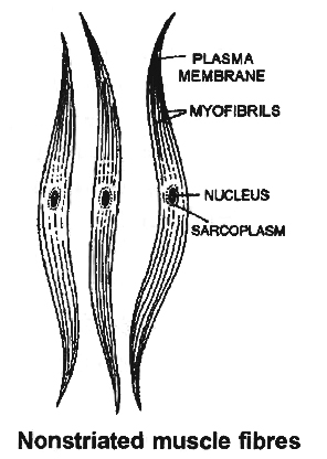
All its fibres contract together as a single unit. They may contract automatically and rhythmically. Such smooth muscles occur on the walls of hollow visceral organs such as the urinary bladder and the gastrointestinal tract. Multi-Unit Smooth Muscles are composed of more independent muscle fibres, not so closely joined together. Individual fibres of such smooth muscles contract as separate unit. These occur at hair roots and in the walls of large blood vessels. e.g., Erector pili muscles.
Smooth muscle fibres are elongated spindle-shaped cells. They are packed parallel to each other in branching bundles. Each fibre contains a single, spindle shaped nucleus at its thick central part. The smooth muscle fibre is generally shorter than a striated muscle fibre. Mitochondria and other organelles are less extensive and protein filaments are not regularly arranged to give rise to striations.
Table : Differences between Single-unit and Multi-unit Smooth Muscles

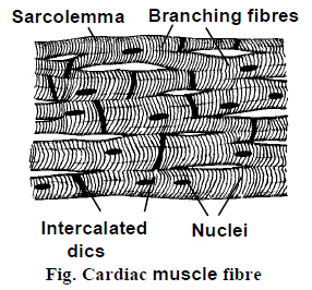
(iii) Cardiac muscle occurs in the heart. It possesses considerable automatic rhythmicity and generates its own wave of excitation. The excitation can also pass directly from one fibre to another in the cardiac muscles. It is not under voluntary control. It shows cross-striations, but striations are much fainter than those of striated muscle.
Between the cardiac muscle fibres, intercalated discs are present. They are specialised regions of cell membrane of two adjacent fibres. The intercalated discs function as boosters of contraction wave and permit the wave of contraction to be transmitted from one cardiac fibre to another.
Cardiac muscle cells are short cylindrical cells joined end to end to form rows. They possess abundant cytoplasm with myofibrils (sarcoplasm) and numerous mitochondria and glycogen granules.
This is because they need a large amount of energy. Faint but regular, alternate dark and light bands give rise to cross-striations in the cardiac muscle fibres and indicate regular and alternate arrangements of thin and thick filaments in the fibre.
Sarcomeres are also present. Cardiac muscle cells frequently branch to form junctions with neighbouring cells. Where two cardiac muscle cells meet end to end, dense zig-zig junction is formed between them. It is called an Intercalated Disc. Longest refractory period is present in cardiac muscles.
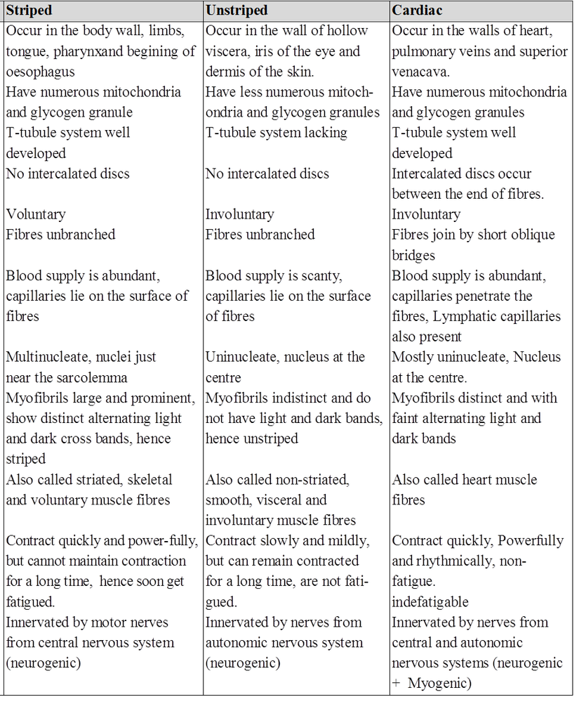
Differences between striated, non-striated and cardiac muscles.
Muscular Tissue
Many long, cylindrical fibers are organized in parallel arrays in each muscle. Myofibrils are a type of tiny fibril that makes up these fibers. Muscle fibers contract (shorten) in response to stimulus, then relax (lengthen) and uncontract in a coordinated manner. Their actions cause the body to move in order tochange the environment and maintain the postures of the various body parts. The sarcolemma is the muscle cell membrane, and the sarcoplasm is the muscle cell cytoplasm. The excitability of the sarcolemma allows it to conduct electrical impulses that occur during depolarization. Muscles are involved in all of the body's movements in general. Skeletal, smooth, and cardiac muscles are the three types of muscles. Skeletal muscle tissue is tightly bound to the bones. Striated (striped) skeletal muscle fibers are bundled together in a parallel pattern in a typical muscle like the biceps.Several bundles of muscle fibers are encased in a strong connective tissue sheath.Tendons connect skeletal muscles to bones, and they are responsible for all motions of bodily components in relation to one another. Skeletal muscle, unlike smooth and cardiac muscle, is controlled by the user. Shoulder muscles, hamstring muscles, and abdominal muscles are all examples of skeletal muscles. Smooth muscle fibers have no striations and taper at both ends (fusiform). They are held together by cell connections and bundled in a connective tissue sheath. This type of muscle tissue can be found in the walls of internal organs such as the blood vessels, stomach, and intestine. Smooth muscles are referred to as "involuntary" since they cannot be controlled directly. Cardiac muscle tissue is only found in the heart, where it performs coordinated contractions that allowthe heart to pump blood through the circulatory system.The only contractile tissue found in the heart is cardiac muscle tissue. The plasma membranes of heart muscle cells are fused.At some fusion points, communication junctions (intercalated discs) allow cells to contract as a group, meaning that when one cell receives a signal to contract, its neighbors are also stimulated to contract.
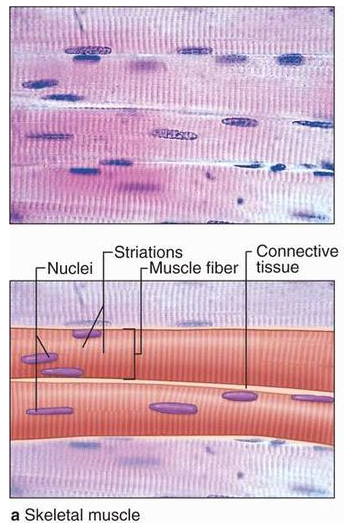
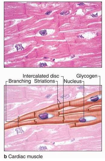
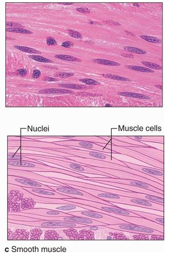
Figure 8: Muscular Tissue
