ACME SMART PUBLICATION
- Books Name
- ACME SMART COACHING Biology Book
- Publication
- ACME SMART PUBLICATION
- Course
- CBSE Class 11
- Subject
- Biology
THE TISSUE SYSTEM
In response to division of labour, tissues are classified into three systems:
A. Epidermal tissue system
It consists of epidermis and its associated structures.
The epidermal cells are living, parenchymatous and compactly arranged (without intercellular spaces).
In aerial parts, epidermis is covered by cuticle.
The epidermal cells secrete a waxy substance called cutin, which forms a layer of variable thickness (the cuticle) within and on the outer surface of its all walls.
It helps in reducing the loss of water by evaporation.
Cuticle is absent in roots.
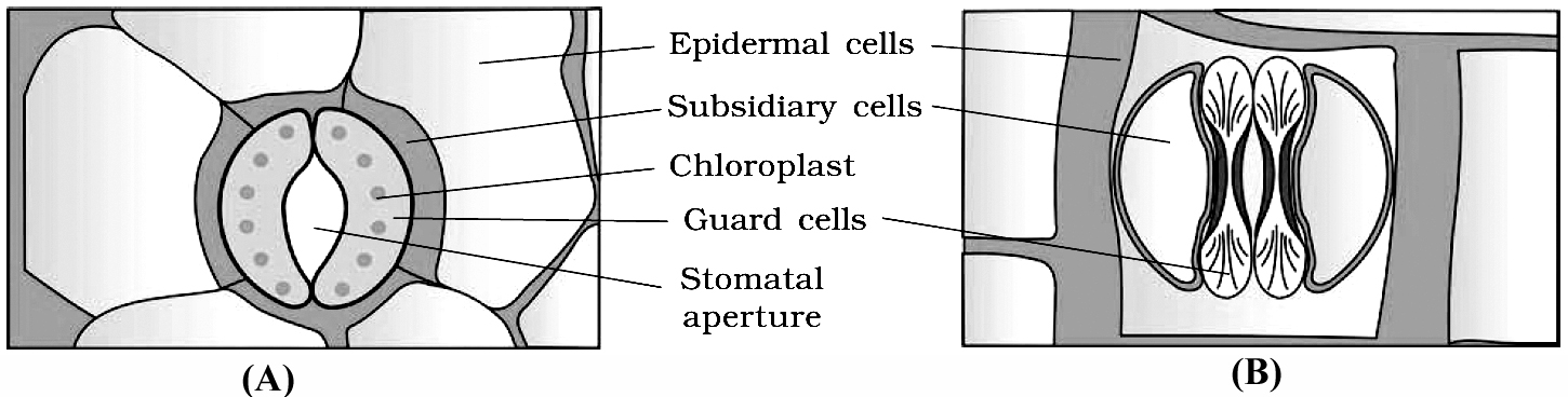
Stomata are structures present in the epidermis of leaves.
Stomata regulate the process of transpiration and gaseous exchange.
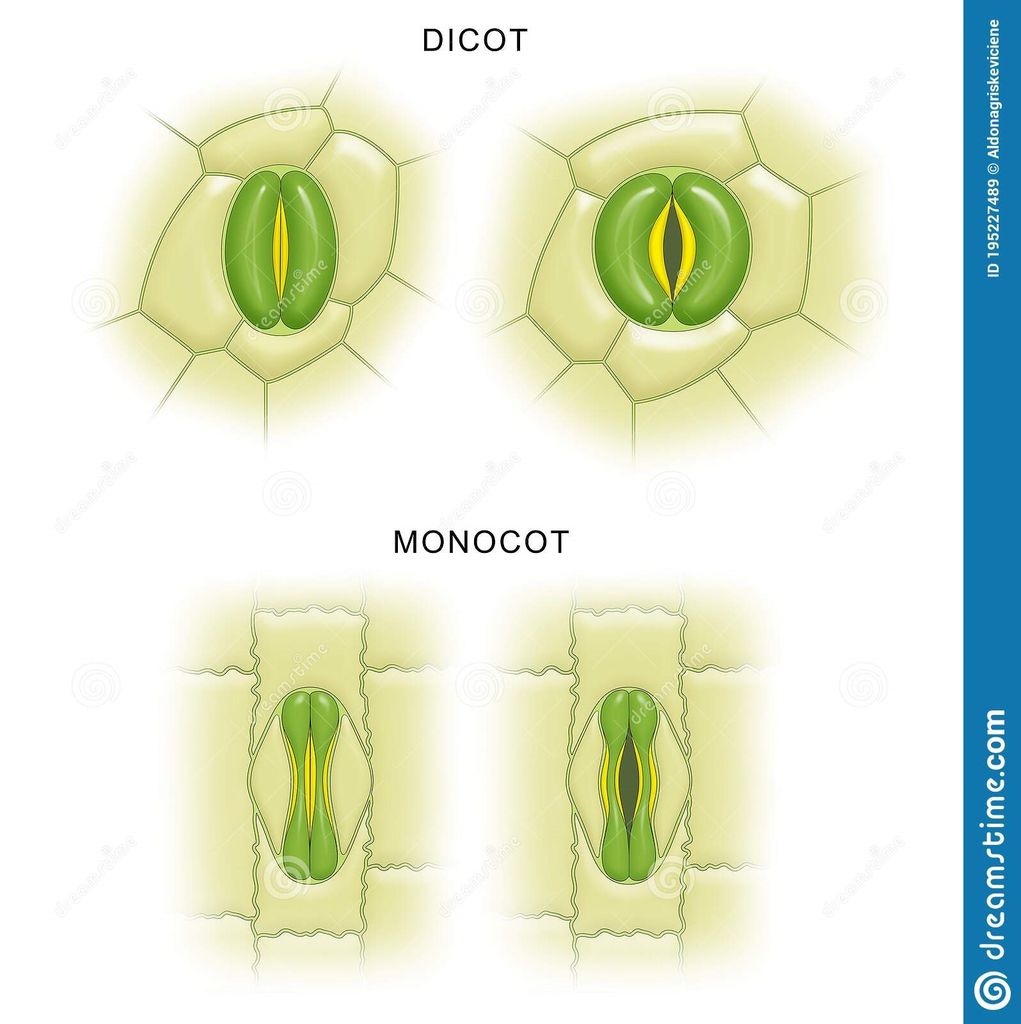
Each stoma is composed of two bean shaped cells known as guard cells.
In grasses, the guard cells are dumb-bell shaped.
The outer walls of guard cells (away from the stomatal pore) are thin and the inner walls (towards the stomatal pore) are highly thickened.
The guard cells possess chloroplasts and regulate the opening and closing of stomata.
Sometimes, a few epidermal cells, in the vicinity of the guard cells become specialised in their shape and size and are known as subsidiary cells.
The stomatal aperture, guard cells and the surrounding subsidiary cells are together called stomatal apparatus.
Mostly epidermis is single layered parenchymatous, but is multilayered in leaf of Ficus and Nerium.
Epidermis is mainly protective in nature (external protective tissue).
In grass leaves, motor or bulliform cells are present in upper epidermis.
On stem, the epidermal hairs are called trichomes, which are usually multicellular.
They may be branched or unbranched and soft or stiff. They may even be secretory. These help in preventing water loss due to transpiration.
Concept Builder
In grasses and Equisetum, silica is present in the epidermal cells.
The epidermal cells containing cystoliths are called lithocysts, these are found in Ficus leaves.
B. Ground or fundamental tissue system
It extends from epidermis upto the centre of axis (excluding vascular tissue).
The ground tissue constitutes the following parts :
(a) Cortex. It lies between epidermis and the pericycle. It is further differentiated into
(i) Hypodermis. It is collenchymatous in dicot stem and sclerenchymatous in monocot stem. It provides strength.
(ii) General cortex. It consists of parenchymatous cells. Its main function is storage of food.
(iii) Endodermis (called starch sheath in dicot stem). It is mostly single layered and is made up of parenchymatous, barrel shaped, compactly arranged cells. The inner and radial wall of root endodermis cells have casparian strips. These thick walled endodermal cells are interrupted by thin walled cells just outside the protoxylem patches. These thin walled endodermal cells are called passage cells.
Endodermis behaves as water and air tight dam to check the loss of water and entry of air in xylem elements.
(b) Pericycle. It lies between endodermis and vascular tissue. It is mostly single layered and parenchymatous in roots and sclerenchymatous (mixed with parenchyma) in stem. The pericycle cells just opposite the protoxylem are the seat for the origin of lateral roots. In dicot roots, pericycle form a part of cambium and whole of cork cambium.
(c) Pith. It occupies the central part in dicot stem and monocot root. It is mostly made up of parenchymatous cells. In dicot root, pith is completely crushed by the metaxylem elements. In dicot stem the pith cells between the vascular bundles become radially elongated and are known as primary medullary rays or pith rays. They help in lateral translocation.
C. Vascular tissue system
Vascular bundles found in stelar part constitute vascular tissue system.
Xylem, phloem and cambium forms the major part of the vascular bundle.
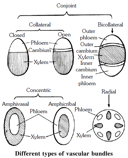
Vascular bundles may be of following types -
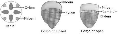
(a) Radial. When the xylem and phloem are arranged on different radii, alternating with each other, e.g., roots.
(b) Conjoint. When xylem and phloem combine in the same bundles and are present on the same radius, e.g., stem. Conjoint vascular bundles may be:
(i) Collateral. Xylem is towards inner side and phloem towards outside.
(ii) Bicollateral. When xylem is surrounded on its both sides by the phloem and cambium e.g., members of Cucurbitaceae and Solanaceae.
• Open. Cambium is present between xylem and phloem, e.g., dicot stem.
• Closed. Cambium is absent between xylem and phloem, e.g., monocot stem.
Concept Builder
Concentric. When one vascular tissue surrounds the other. They are of two types:
(i) Amphicribal or Hadrocentric. The xylem is surrounded on all sides by phloem e.g., ferns.
(ii) Amphivasal or Leptocentric. The phloem is surrounded on all sides by xylem e.g., Yucca, Dracaena.
The Tissue System
There are three types of tissue systems based on their structure and location.
A. The epidermal tissue system
Epidermal cells, stomata, and epidermal appendages like trichomes and hairs make up the epidermal tissue system, which covers the entire plant body. The epidermis is the major plant body's outermost layer. It is made up of a continuous layer of elongated, compactly packed cells. The epidermis is normally one layer thick. Epidermal cells are parenchymatous, with a big vacuole and a little quantity of cytoplasm along the cell wall. The cuticle, a waxy thick covering on the outside of the epidermis that resists water loss, is commonly present. The roots have no cuticle. The epidermis of leaves has features called stomata. The process of transpiration and gas exchange is regulated by stomata.Guard cells enclose the stomatal pore in each stoma, which are two bean-shaped cells. The guard cells in grasses are formed like a dumbbell. Guard cells have thin outer walls that face away from the stomatal pore and robust interior walls that face the stomatal pore. Guard cells have chloroplasts and control stomata opening and closing. A few epidermal cells near the guard cells can become specialized in shape and size, and these cells are known as subsidiary cells. The Stomatal apparatus is made up of the stomatal orifice, guard cells, and surrounding subsidiary cells. Hairs are found on the epidermis cells.Root hairs are unicellular epidermal elongations that assist absorb water and minerals from the soil. Trichomes are epidermal hairs that grow on the stem. In the shoot system, trichomes are frequently multicellular. They might be soft or stiff, branching or unbranched. They could even be hidden. The trichomes aid in the prevention of water loss by transpiration.
B. The ground tissue system
It is also called the foundational tissue system.The ground tissue is made up of all tissues save the epidermis and vascular bundles. Simple tissues such as parenchyma, collenchyma, and sclerenchyma make up this layer. In the cortex, pericycle, pith, and medullary rays, as well as the primary stems and roots, parenchymatous cells are commonly seen. The mesophyll is the ground tissue in leaves that is made up of thin-walled chloroplast-containing cells. Ground tissue includes parenchyma (photosynthesis in the leaves and storage in the roots), collenchyma (shoot support in areas of active growth), and sclerenchyma (shoot support in areas where growth has ceased) and is the site of photosynthesis, provides a supporting matrix for the vascular tissue, provides structural support for the stem, and helps to store water and sugars, depending on the cell type and location in the plant.
C. The vascular tissue system:
Also called conducting tissue system. The vascular system is made up of two complicated tissues: phloem and xylem. Vascular bundles are made up of the xylem and phloem. Between the phloem and xylem in dicotyledonous stems, there is cambium. Because of the existence of cambium, such vascular bundles have the ability to produce secondary xylem and phloem tissues, and are hence referred to as open vascular bundles. The cambium is absent from the vascular bundles of monocotyledons. As a result, they are referred to as closed since they do not generate additional tissues. Radial arrangement occurs when xylem and phloem within a vascular bundle are placed in an alternative fashion along distinct radii, as in roots.The xylem and phloem are located along the same radius of vascular bundles in conjoint type vascular bundles. In stems and leaves, vascular bundles are common. Phloem is usually exclusively seen on the outer side of the xylem in conjoint vascular bundles.