 ACME SMART PUBLICATION
- Books Name
- ACME SMART COACHING Biology Book
- Publication
- ACME SMART PUBLICATION
- Course
- CBSE Class 11
- Subject
- Biology
MITOSIS
It was first observed by Strasburger in plant cells and by Flemming in animal cells.
It may be defined as, "The exact replication of a parent cell into two identical daughter cells having the identical number, and kind of chromosomes, the identical DNA and identical hereditary instructions as found in the parental cell".
It mainly occurs in somatic cells of animals and in meristematic tissue cells of plants for multiplication of undifferentiated cells.
In mitosis the cell cycle involves series of changes which occur in a newly formed cell to become fully grown and to be ready for cell division.
It has two phases :
(1) Interphase (as described earlier)
(2) Mitotic phase or M-phase.
It consist of the following four sub-stages :
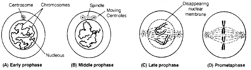
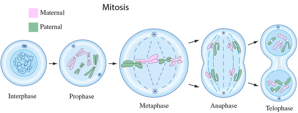
(a) Prophase: It is the first phase of mitosis which is longest of the M-phase.
Following changes occur during this stage:
(i) The chromatin fibres become shorter and thicker due to the process of coiling and folding and get condensed into distinct thread like chromosomes in late prophase.
(ii) Each chromosome appears double and consists of two coiled sister chromatids joined by a centromere. Their ends are not visible in early prophase. Therefore, the chromosomes appear like a ball of wool. It is also called spireme stage.
(iii) Cells at the end of prophase, when viewed under the microscope, do not show golgi complex, endoplasmic reticulum, nucleolus and the nuclear envelope.
(iv) The centriole now begins to move towards opposite poles.
(v) Fine radiating microtubules appear around each pair of centrioles to form the astral rays. A pair of centrioles and astral rays constitute a star like aster body.
(b) Metaphase
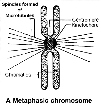
 (i) The complete disintegration of the nuclear envelope marks the start of the second phase of mitosis. By this stage condensation of chromosomes is completed. At this stage, metaphase chromosome appears to be made up of two sister chromatids, which are held together by the centromere.
(ii) Small disc shaped structures called kinetochores, serve as the sites of attachment of spindle fibres to the chromosomes that are moved into position at the centre of the cell.
Hence, the metaphase is characterised by all the chromosomes coming to lie at the equator and getting aligned along metaphase plate or equatorial plate through spindle fibres to both the poles.This chromosomal movement to equator is called as congression.
(iii) Spindle fibres which connect the pole to the chromosomes at the kinetochores are called chromosomal fibres (Tractile fibres) and those which extend without interruption from one pole to other are the continuous fibres.
(iv) The morphology of chromosomes can be easily studied at this stage as they are in a highly condensed stage and are distinctly visible. Chromosomes can be counted at this stage.
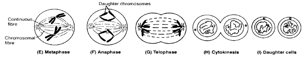
(c) Anaphase
(i) It is of shortest duration.
(ii) The centromere of each chromosome splits into two, resulting into daughter chromatids (chromosome of future nuclei).
(iii) These begin to migrate towards respective poles. The centromeres always lead the daughter chromosomes (towards the poles) and the arms trail behind.
(iv) As the daughter chromosomes move apart, fine microtubules in the form of interzonal fibres appear in between them.
(v) Expansion of interzonal fibres and dissolution of microtubules of chromosomal fibres is supposed to be the possible reason for anaphasic movement.
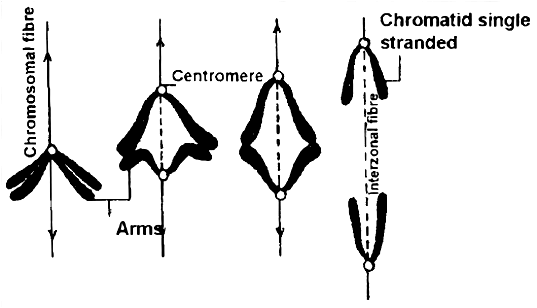
(vi) The daughter chromosomes now assume V, J and I shapes depending upon the position of centromere on them. Thus shape of chromosomes can be observed during this phase.
(vii) Finally, the polar migration of daughter chromosomes after reaching the opposite poles end, which mark the end of anaphase.
(d) Telophase :
(i) Daughter chromosomes undergo decondensation and uncoiling at each pole, so they lose their individuality.
(ii) The chromatin gets surrounded by discontinuous segments of nuclear membrane from elements of ER, which become fully developed at the end of telophase.
(iii) New nucleoli. ER and golgi complex reform.
(iv) The spindle fibres and astral rays gradually disintegrate and disappear. These become absorbed in the cytoplasm.
(v) Each daughter nucleus now enters into the interphase of cell cycle. The end of telophase marks the end of karyokinesis.
Cytokinesis : Karyokinesis results in formation of two nuclei inside a cell and now it is followed by division of cytoplasm (Cytokinesis), thus forming two cells (daughter cells).
(a) Cytokinesis occurs by two methods :
(i) Cell furrow method : This is characteristic of animal cells. Due to absence of rigid cell wall here, the more flexible plasma membrane forms the outer layer of cell. A furrow or invagination appears in plasma membrane at centre of equator, which deepens gradually and finally two daughter cells are separated.
(ii) Cell plate method : This is characteristic of plant cells. Here, vesicles provided by Golgi apparatus unite to form phragmoplasts, which join to form cell plate. Cell plate is first laid down in centre and then proceeds towards periphery (i.e., centrifugal plate formation). Cell wall materials are now laid down on both sides of cell plate, resulting in two daughter cells.
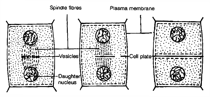
Cytokinesis by cell plate method in plant cells
Concept Builder
Mitotic Poisons
All those substances or chemicals which affect the cells during mitosis or prevent them from entering into normal mitotic divisions are called mitotic poisons. The various mitotic poisons are:
(i) The enzyme ribonuclease acts as poison at prophase. Azide and cyanide also inhibit prophase.
(ii) Mustard gas causes agglutination of chromosomes.
(iii) Chalones also inhibit mitosis. They are small peptides or glycoproteins in the extracellular fluid.
(iv) The alkaloid colchicine inhibits the formation of mitotic spindle (inhibits polymerisation of microtubules) and holds the cells in metaphase. The chromosomes and DNA undergo replication but remain within the same cell. The nucleus does not divide. This increases the number of chromosome sets per cell. This process leads to endopolyploidy or endomitosis in which nucleus contains multiple sets of chromosomes instead of the normal two sets as found in a diploid cell. Such cells are called polyploid cells.
(v) X-rays cause uncontrolled mitosis and induces breakage of chromosomes.
Abnormal Mitosis
(i) Intranuclear mitosis (Premitosis) : In Amoeba, Yeast, other fungi and a number of algae, the nuclear envelope does not degenerate during mitosis. Spindle is intranuclear.
(ii) Dinomitosis : Dinoflagellates possess condensed chromosomes even in non-dividing nuclei. Nuclear envelope does not degenerate. Division of chromosomes occurs with the help of special channels that develop in the nucleus.
(iii) Free Nuclear Division : Sometimes, repeated mitosis occur without subsequent cytokinesis. It produces multinucleated condition, e.g.:-Rhizopus, Vaucheria, Slime moulds, etc.
Significance of Mitosis
(i) It is an equational division since the number of chromosomes and the genetic constitution of the daughter cells remain the same as found in parent cells. Thus, mitosis helps in the survival of a species and continuation of its race. It provides a complete set of genetic information to each cell.
(ii) Mitosis occurs in somatic cells (n or 2n) and gonad cells for the multiplication of cell number. Thus, mitosis is related to the growth of an individual from zygote to adult stage. It also provides an opportunity for the growth and development of organs and body of organisms.
(iii) Mitosis produces new cells to replace the old worn out and injured cells. Thus, it helps in repairing of cells, healing of wounds and regeneration of body cells. Blood cells, intestinal cells and skin cells are regularly regenerated and replaced by mitosis.
(iv) It is a method of restoring nucleocytoplasmic ratio.
(v) Mitosis helps the organisms in both sexual and asexual reproduction.
Mitosis and its significance
A cell prepares for cell division by replicating its chromosomes, segregating them, and creating two identical nuclei during the mitotic phase. The cell's contents are often evenly divided into two daughter cells with identical genomes after mitosis.Mitosis is divided into the following four stages:
1. Prophase:
Interphase's S and G2 phases are followed by prophase, the initial step of mitosis. The newly synthesised DNA molecules are not distinct but rather entangled in the S and G2 phases. The beginning of chromosomal material condensing characterises prophase. During the process of chromatin condensation, the chromosomal material is untangled. The centriole, which underwent duplication during interphase's S phase, now starts to migrate in the direction of the cell's opposing poles.Thus, the following distinctive occurrences can indicate the end of prophase:
- Compact mitotic chromosomes are created when chromosomal material condenses.Two chromatids are observed to be joined together at the centromere to form chromosomes.
- The beginning of the mitotic spindle's construction, the microtubules, and the proteinaceous elements of the cell cytoplasm aid in the process.
- The golgi complex, endoplasmic reticulum, nucleolus, and nuclear envelope are absent from cells near the end of prophase when they are observed under a microscope.
2. Metaphase:
The second phase of mitosis begins when the nuclear envelope completely disintegrates, and as a result, the chromosomes are dispersed throughout the cell's cytoplasm. The chromosome condensation process is now complete, and the chromosomes may be seen clearly under a microscope. Therefore, this is the period at which it is easiest to study the shape of chromosomes. The two sister chromatids that make up the metaphase chromosome at this point are joined by the centromere. Kinetochore refers to a little disc-shaped structure at the centromere surface. These structures act as the points of attachment for the spindle fibres, which are created by the spindle fibres, to the chromosomes that are placed at the cell's centre.Thus, all of the chromosomes align at the equator during the metaphase, with one chromatid of each chromosome attached by its kinetochore to spindle fibres from one pole and its sister chromatid connected by its kinetochore to spindle fibres from the opposite pole. The term "metaphase plate" refers to the chromosomes' alignment plane during metaphase. Metaphase's primary characteristics are:
- Spindle fibres adhere to chromosomal kinetochores.
- Chromosomes are transferred to the spindle equator, where they are positioned along the metaphase plate and along the spindle fibres to both poles.
3. Anaphase:
Each chromosome on the metaphase plate splits simultaneously at the start of anaphase, and the two daughter chromatids, which are now known as the chromosomes of the future daughter nuclei, start to move in opposite directions. The centromere of each chromosome is at the pole and, as a result, at the leading edge, with the arms of the chromosome trailing behind as they advance away from the equatorial plate. Events that define the anaphase stage are:
- Centromeres divide, and chromatids dissociate.
- Chromatids shift to the polar opposites
4. Telophase:
The chromosomes that have reached their respective poles decondense and lose their identity at the beginning of the last stage of mitosis, known as telophase. The individual chromosomes are no longer visible, and the two poles tend to accumulate a mass of chromatin material. The essential events at this stage include:
- Chromosomes cluster at opposing spindle poles, losing their identity as distinct elements.
- The nucleolus, Golgi complex, and ER remodel themselves
- The nuclear envelope forms around the chromosomal clusters.
Cytokinesis:
In addition to segregating duplicated chromosomes into daughter nuclei (karyokinesis), mitosis also divides the cell into two daughter cells by a separate process known as cytokinesis, marking the completion of cell division. This occurs when a furrow forms in the plasma membrane of an animal cell. The cytoplasm of the cell is split in half by the furrow, which eventually merges in the centre. Plant cells, on the other hand, are surrounded by a cell wall that is rather inextensible; as a result, they go through cytokinesis using a different process. In plant cells, wall construction begins in the cell's middle and extends outward to meet the lateral walls that already exist.The basic precursor known as the cell-plate, which symbolises the middle lamella between the walls of two neighbouring cells, is formed before the new cell wall can be fully formed. Organelles like mitochondria and plastids are dispersed between the two daughter cells during cytoplasmic division. In some organisms, cytokinesis does not follow karyokinesis, which results in a multinucleate state and the development of syncytium.
Significance of Mitosis :
Only diploid cells often undergo mitosis, also known as equational division. However, haploid cells can also divide through mitosis in some lower plants and social insects. Understanding the importance of this division in an organism's life is crucial. Typically, mitosis produces daughter cells that are diploid and have the same genetic makeup. Mitosis is responsible for multicellular organisms' growth. The ratio of the nucleus to the cytoplasm is disturbed as a result of cell expansion. Therefore, cell division is required to re-establish the nucleo-cytoplasmic ratio. Cell repair is one of mitosis' most important functions.Blood cells, stomach lining cells, and the outermost layer of the epidermis all undergo continuous replacement. Plants grow continuously throughout their lives as a result of mitotic divisions in the meristematic tissues known as the apical and lateral cambium.
