 ACME SMART PUBLICATION
- Books Name
- ACME SMART COACHING Biology Book
- Publication
- ACME SMART PUBLICATION
- Course
- CBSE Class 11
- Subject
- Biology
FROG (Rana tigrina)
Phylum : Chordata
Class : Amphibia
Order : Anura
Genus : Rana
Species : tigrina
The most common frog found in India is the Indian bullfrog.
It is the largest frog and is named as bullfrog because of its large size and loud call.
Indian bullfrog is found in fresh water marshes, ditches, ponds, and shallow lakes.
They undergo aestivation (summer sleep) in summer and hibernation (winter sleep) in winter.
They are carnivorous (feeding upon other animals, insects, etc.), poikilothermic i.e. the body temperature changes with environment.
They develop protective coloration to camouflage, i.e., to hide in surroundings.
Frogs belong to the Phylum Chordata, Subphylum Vertebrata or Craniata, Superclass Gnathostomata, Class Amphibia and Genus Rana.
The most common species is known as Rana tigrina (Indian bull frog).
The scientific name of common toad is Bufo melanostictus. Frogs exhibit sexual dimorphism.
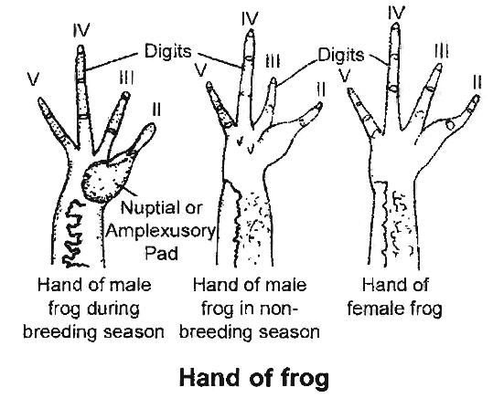
Male and female are distinguishable externally only during breeding season when the males develop nuptial pad in the first digit of forelimbs.
Vocal sacs are well developed in males so they produce louder sound as compared to the females which are devoid of vocal sacs.
The vocal sacs help to produce mating calls.
Total number of bones is 153.
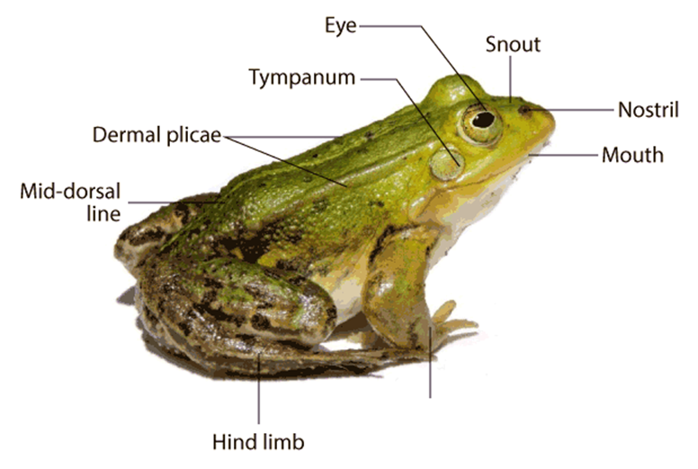
External Morphology
Skin is made up epidermis and dermis. Mucous glands are present in the dermis and their ducts open at the surface.
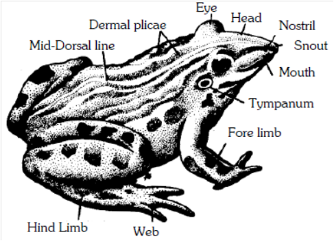
Blood capillaries and pigment cells (chromatophores) are present in the dermis.
Skin is without scales or any other cover or exoskeleton.
Body is divisible into head and trunk, neck is absent. The trunk is provided with a pair of fore and hind-limbs. The hind-limbs are much larger and muscular than the fore-limbs. Fore-limbs end in four digits and the hind limbs end in five digits. The digital formula of fore-limbs is 02233. The digital formula of hind limbs is 22343.
Shank or crus is associated with hind limbs.
Sexual dimorphism :
The male () and female () frogs exhibit certain differences in their external features, which become more pronounced during breeding season.
Generally, male frogs are larger than females.
During breeding season, however, the females become bloated with large ovaries and numerous ova, and appear considerably larger.
Only the males possess a pair of ventro-Iateral, wrinkled pouch-like vocal sacs located a little behind the mouth.
These sacs become specially large and distensible, in breeding season.
By inflating these repeatedly with air from the lungs, the males produce a loud croaking sound meant to call the females for copulation (amplexus).
The sound is actually produced by a pair of vocal cords in the larynx; the sacs only increase its pitch, like resonators.
The females produce a low pitch sound by their vocal cords alone.
The forelimbs in both male and female frogs bear small articular pads dorsally at the joints of digits, but the males also possess a special nuptial, copulatory or amplexusary pad on ventral side of the first finger of each forelimb.
Normally, these pads appear merely as rough patches but during breeding season, these become thick and sticky.
In amplexus, the male strongly grips a female under her armpits by means of these pads.
Internal Morphology
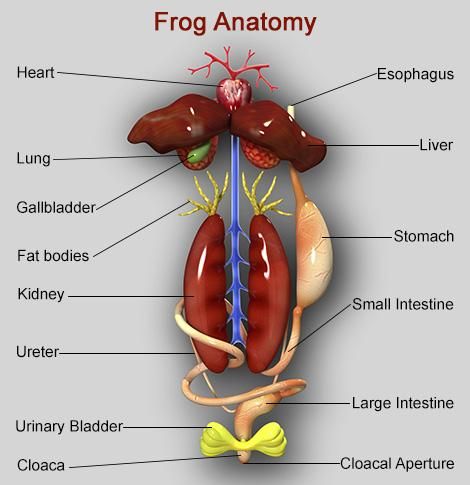
Digestive System
Since the frogs are carnivorous, their alimentary canal is short in length.
Tadpole larva is herbivorous so alimentary canal is very long and coiled in the form of spring.
The mouth is present as a terminal, wide opening.
It opens into bucco-pharyngeal cavity, which contains numerous maxillary teeth arranged along the margin of the upper jaw and vomerine teeth are present in the roof of the buccopharyngeal cavity.
The lower jaw is toothless. Salivary glands are absent.
Opening of eustachian tube, vocal sacs (only in male) gullet and glottis can be seen clearly in the bucco-pharyngeal cavity.
The muscular tongue is bilobed at the tip and free from behind. It is used for capturing the prey.
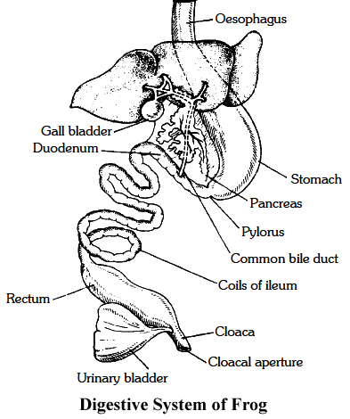
The gullet opens into a narrow and short tube-like oesophagus, which continues in large and distended stomach.
It contains a thick muscular layer, which helps in converting food into chyme.
It secretes gastric juice containing HCl and proteolytic enzymes. Stomach is followed by a coiled small intestine.
Intestinal wall has numerous finger-like folds called villi and microvilli, projecting into its lumen to enhance surface area for absorption of digested food.
The first part of the small intestine lying parallel to stomach is called duodenum. Intestine continues into a wider rectum, opening into cloaca.
The urinary bladder opens into cloacal chamber through the ureter.
The gastric and intestinal glands occur in the walls of stomach and intestine respectively.
The other important digestive glands associated with the alimentary canal are liver and pancreas.
Liver secretes bile which is temporarily stored in gall bladder before being released into the duodenum.
Bile helps in digestion of food by changing its pH from acidic to alkaline and by emulsifying the fats.
Liver does not secrete any digestive enzymes. Pancreas is an irregular, elongated gland, situated in a thin mesentery and lies parallel to the stomach.
It produces pancreatic juice containing digestive enzymes like trypsin, amylopsin, etc.
Respiratory System
Three types of respiration: cutaneous, buccopharyngeal and pulmonary occur.
Cutaneous respiration on land is through the body surface. During hibernation and aestivation, frog respires only through this method.
Buccopharyngeal respiration occurs through the lining of buccal cavity. It occurs only when frog is out of water. The mucus membrane of the buccal cavity is moist which dissolves oxygen whose diffusion occurs into the blood capillaries.
Pulmonary respiration: Lungs in frog are not efficient respiratory organs because only mixed air enters into them and they mainly function as hydrostatic organs.
Lungs are a pair of thin walled, translucent sacs with inner surface divided into alveoli by septa. Pulmonary respiration has a maximum frequency of 20/minute.
It occurs when more energy is required. Mouth and gullet are kept closed during pulmonary respiration.
Respiratory movements in pulmonary respiration are because of buccopharyngeal cavity which acts as a force pump.
These movements are carried out by set of paired muscles -sternohyal and pterohyal muscles.
Sternohyal muscles are attached with hyoid and coracoid processes, clavicles of the pectoral girdle and on contraction, depress the buccal floor enlarging the buccopharyngeal cavity.
Pterohyals are attached in between hyoid and pro-otics of the skull and on contraction lift the floor of buccal cavity.
With the depression of buccal floor, air enters buccal cavity through the nares.
External nares are then closed by pushing tuberculum prelinguale and the movable premaxillae.
It is followed by raiSing of the buccal floor by pterohyal muscles which reduces the volume and the air is pushed into the lungs where exchange of gases takes place.
Buccal floor is again lowered enlarging its volume which draws air into the buccal cavity.
External nares are opened fol'lowed by raising the buccal floor, pushing the air out through external nares.
Sound producing organ of frog is laryngo-tracheal chamber.
It is supported by one cricoid, two arytenoids and two prearytenoid cartilages.
It has a pair of muscle strands (vocal cords) which actually prod uce sound. Male frog has vocal sacs which act as resonating chambers.
Circulatory System
Circulatory system is closed type.
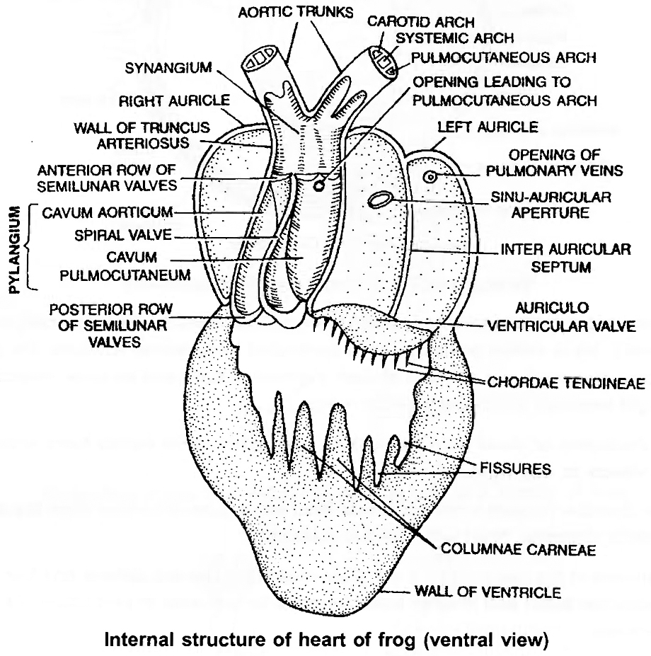
The heart lies enclosed by a thin, transparent, two layered sac, pericardium.
Frog's heart is a three chambered structure made of two upper auricles and a single lower ventricle.
The two additional chambers connected to the heart of frog are sinus venosus and truncus arteriosus.
Frogs also possess two well developed portal systems: Renal portal system and Hepatic portal system. Frog also has two pairs of lymph hearts.
Nervous System
The nervous system is organised into a central nervous system (brain and spinal cord), a peripheral nervous system (cranial and spinal nerves) and an autonomic nervous system (sympathetic and parasympathetic chains ofganglia).
There are ten pairs ofcranial nerves.
Brain is enclosed in a bony structure or brain box (cranium) which has two occipital condyles for attachment with the first vertebra (atlas).
The brain is divided into fore-brain, mid-brain and hind-brain.
Forebrain includes olfactory lobes, paired cerebral hemispheres and unpaired diencephalon.
The mid-brain is characterised by a pair of optic lobes.
Hind-brain consists of cerebellum and medulla oblongata.
The medulla oblongata passes out through the foramen magnum and continues into spinal cord which is contained in the vertebral column.
Jacobson's organ, also called Vomero-nasalorgan opens into nasal chamber and acts as additional olfactory organ.
Eye
Eye is guarded by immovable upper eyelid, movable lower eyelid and transparent nictitating membrane.
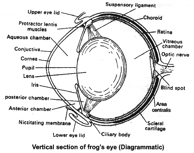
Vertical section of frog's eye (Diagrammatic)
Outersclerotic ring is cartilaginous and cornea is the part exposed out. Middle, highly vascular, pigmented layer is choroid.
Iris is yellow pigmented and perforated by a central aperture, the pupil.
Retina is the innermost coat of eyeball and consists of inner pigmented layer and an outer receptor layer.
Rods and cones are light sensitive structures found in retina.
Rods have rhodopsin or visual purple meant for night vision while cones have iodopsin responsible for colour vision in day light.
The anterior chamber present in front of lens is filled with aqueous humour while the chamber behind the lens is posterior chamber, filled with vitreous humour.
Eye ball is moved in the eye orbit by a set of six muscles -two are oblique and four are recti.
Besides, there are retractor bulbi and levator bulbi muscles for intrusion or protrusion of eye ball into the eye orbit, respectively.
Ear
Ear of frog has only middle and internal ear.
Tympanic membrane is present at the body surface.
Middle ear has single bone called columella auris.
Its outer end is attached to the ear drum while inner end to the stapedial plate.
Pressure of air in middle ear is controlled by eustachian tubes.
Membranous labyrinth or internal ear consists of utriculus, sacculus and semicircular canals. Endolymph fills the membranous labyrinth .
Excretory System
The main organ of excretion is a pair of kidneys.
These are compact, dark red and bean like structures situated little posteriorly in the body cavity on both sides of vertebral column.
The frog excretes urea, thus is a ureotelic animal.
Urea is carried by blood into the kidney where it is separated and excreted.
Each kidney is composed of several structural and functional units called uriniferous tubules or nephrons.
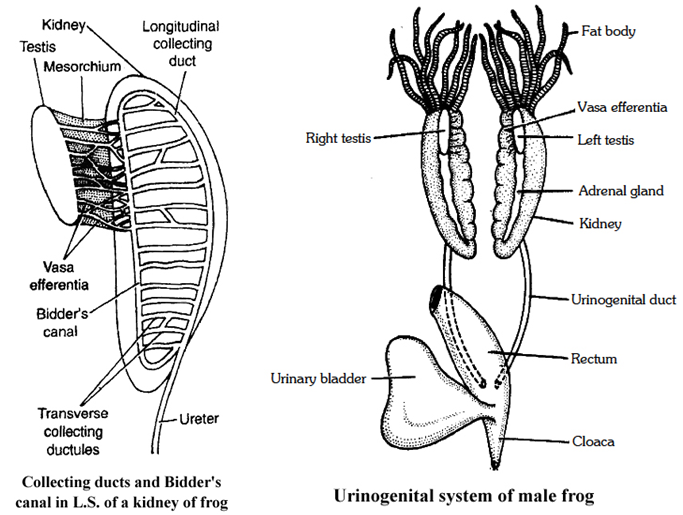
Ureter emerges from the kidney as urinogenital ducts in the males.
A common ureter opens into the cloaca.
A thin-walled urinary bladder is present ventral to rectum which also opens in the cloaca.
Reproductive System
Male reproductive organs consist of a pair of yellowish ovoid testes, which are found adhered to the upper part of kidneys by a double fold of peritoneum called mesorchium.
Vasa efferentia are 10-12 in number and after arising from testes, run through the mesorchium and enter the kidneys of their side.
In kidneys, these open into Bidder's canal which finally communicates with the urinogenital duct.
This duct emerges from the kidneys and finally opens into the cloaca.
The cloaca is a small, median chamber that is used to pass faecal matter, urine and sperms to the exterior.
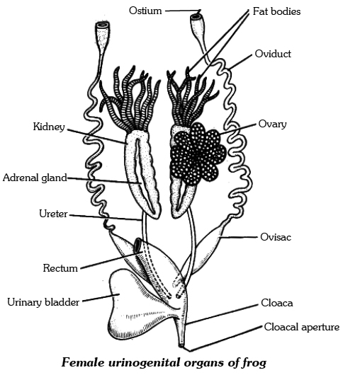
A pair of ovaries, situated near kidneys, comprises the female reproductive organs.
However, these have no functional connection with kidneys.
A pair of oviduct opens into the cloaca, separately.
The release of ovum in female is termed as spawning.
A mature female can lay 2,500 to 3,000 ova at a time.
Fertilisation is external and takes place in water.
Development involves a larval stage called tadpole.
Tadpole undergoes metamorphosis to form the adult.
SUMMARY
Earthworm, Cockroach and Frog show characteristic features in body organisation.
In Pheretima posthuma (earthworm), the body is covered by cuticle.
All segments of its body are alike except the 14th, 15th & 16th segment, which are thick and dark and glandular, forming clitellum.
A ring of S-shaped chitinous setae is found in each segment.
These setae help in locomotion.
On the ventral side spermathecal openings are present in between the grooves of 5 & 6, 6 &7, 7 & 8 and 8 & 9 segments.
Female genital pores are present on 14th segment and male genital pores on 18th segment.
The alimentary canal is a narrow tube made of mouth, buccal cavity, pharynx, gizzard, stomach, intestine and anus.
The blood vascular system is of closed type with heart and valves.
Nervous system is represented by ventral nerve cord.
Earthworm is hermaphrodite.
Two pairs of testes occur In the 10th and 11th segment, respectively.
A pair of ovaries are present on 12th and 13th intersegmental septum.
It is a protandrous animal with crossfertilisation.
Fertilisation and development takes place in cocoon secreted by the glands of clitellum.
The body of Cockroach (Periplaneta americana) is covered by chitinous exoskeleton.
It is divided into head, thorax and abdomen.
Segments bear jointed appendages.
There are three segments of thorax, each bearing a pair of walking legs.
Two pairs of wings are present, one pair each on 2nd and 3rd segment.
There are ten segments in abdomen.
Alimentary canal is well developed with a mouth surrounded by mouth parts, a pharynx, oesophagus, crop, gizzard, midgut, hindgut and anus.
Hepatic caecae are present at the junction of foregut and midgut.
Malpighian tubules are present at the junction of midgut and hindgut and help in excretion.
A pair of salivary gland is present near crop.
The blood vascular system is of open type.
Respiration takes place by network of tracheae.
Trachea opens outside with spiracles.
Nervous system is represented by segmentally arranged ganglia and ventral nerve cord.
A pair of testes is present in 4th and 5th segments and ovaries in 4th, 5th and 6th segments.
Fertilisation is internal.
Female produces 10-40 ootheca bearing developing embryos.
After rupturing of single ootheca sixteen young ones, called nymphs come out.
The Indian bullfrog, Rana tigrina, is the common frog found in India.
Body is covered by skin.
Mucous glands are present in the skin which is highly vascularised and helps in respiration in, water and on land.
Body is divisible into head and trunk.
A muscular tongue is present, which is bilobed at the tip and is used in capturing the prey.
The alimentary canal consists of oesophagous, stomach, intestine and rectum, which open into the cloaca.
The main digestive glands are liver and pancreas.
It can respire in water through skin and through lungs on land.
Circulatory system is closed with single circulation.
RBCs are nucleated.
Nervous system is organised into central, peripheral and autonomic.
The organs of urinogenital system are kidneys and urinogenital ducts, which open into the cloaca.
The male reproductive organ is a pair of testes.
The female reproductive organ is a pair of ovaries.
A female lays 2500-3000 ova at a time.
The fertilisation and development are external.
The eggs hatch into tadpoles, which metamorphose into frogs.
Frog
Frogs are members of the phylum Chordata's class Amphibia, which can live on land and in freshwater. Rana tigrina is the most common frog species in India. They do not have a steady body temperature; rather, their body temperature changes depending on the ambient temperature. Cold-blooded animals are known as poikilotherms. You may have also noticed that the frogs' color changes while they are in grasses or on dry soil. They can change their color to hide from their adversaries (camouflage). The term for this protective coloring is mimicry. During the summer and winter, the frogs are not visible. They take refuge in underground tunnels during this time to avoid the excessive heat and cold.Summer sleep (aestivation) and winter sleep (hibernation) are the two types of sleep.
(i) Morphology: Due to the presence of mucus, the skin is smooth and slippery. Moisture is always retained on the skin. The dorsal side of the body is olive green in color with dark irregular markings. The skin is uniformly pale yellow on the ventral side. The frog does not drink water; instead, it absorbs it through its skin. A frog's body is divided into two parts: head and trunk. There is no neck or tail. A pair of nostrils can be found above the mouth. The eyes protrude and are protected by a nictitating membrane while in the water. A membranous tympanum (ear) receives sound signals on either side of the eyes. Swimming, walking, leaping, and digging are all assisted by the forelimbs and hindlimbs.The hind limbs have five fingers and are larger and more muscular than the forelimbs, which have four. Webbed digits on the feet aid with swimming. Sexual dimorphism is seen in frogs. Male frogs can be recognized from female frogs by the presence of sound-producing vocal sacs and a copulatory pad on the first digit of the forelimbs.
(ii) Anatomy: Frogs' bodily cavities house many organ systems with well-developed structures and functions, including the digestive, circulatory, respiratory, nervous, excretory, and reproductive systems. The alimentary canal and digestive glands make up the digestive system. Because frogs are carnivores, the alimentary canal is short, resulting in a shorter gut. The pharynx connects the mouth to the buccal cavity, which leads to the esophagus. The esophagus is a short tube that connects the mouth to the stomach, then to the intestine, rectum, and finally to the cloaca. Bile is produced by the liver and stored in the gall bladder. Pancreatic juice, which contains digestive enzymes, is produced by the pancreas, a digestive gland. The bilobed tongue is responsible for capturing food.
Food is broken down by the action of Hydrochloric acid and gastric secretions released from the stomach's walls. Chyme, or partially digested food, is transferred from the stomach to the duodenum, the first segment of the small intestine. Through a shared bile duct, the duodenum receives bile from the gall bladder and pancreatic secretions from the pancreas. Pancreatic fluids digest carbs and proteins while bile emulsifies fat. The intestine is where the final digesting takes place. The many finger-like folds in the inner wall of the intestine called villi and microvilli absorb digested food. The undigested solid waste goes through the cloaca and into the rectum. Frogs have two different strategies to breathe on land and in water.
Skin serves as an aquatic respiratory organ in water (cutaneous respiration). Diffusion exchanges dissolved oxygen in the water through the skin. The buccal cavity, skin, and lungs serve as respiratory organs on land. Pulmonary respiration is the process of breathing through the lungs. The lungs are a pair of elongated, pink-colored sac-like structures located near the top of the trunk (thorax). Air enters the lungs via the nostrils, buccal cavity, and lungs. Gaseous exchange occurs through the skin during aestivation and hibernation. Frogs have a well-developed closed circulatory system. Frogs, like humans, have a lymphatic system. The heart, blood arteries, and blood make up the blood vascular system. Lymph, lymph channels, and lymph nodes make up the lymphatic system.
The heart is a muscle structure located in the upper body cavity. It has three chambers, two atria, and one ventricle, and is surrounded by a pericardium membrane. The right atrium is connected to the sinus venosus, a triangle structure. It receives blood through the vena cava, which are large veins. On the ventral side of the heart, the ventricle opens into a sac-like conus arteriosus. The arteries transport blood from the heart to all regions of the body (arterial system). The venous system collects blood from various regions of the body and transports it to the heart.
Frogs have a unique venous connection between the liver and gut, as well as the kidney and lower body.The first is known as the hepatic portal system, whereas the second is known as the renal portal system. Plasma and cells are found in the blood. RBCs (red blood cells) or erythrocytes, WBCs (white blood cells) or leucocytes, and platelets are the blood cells. RBCs are nucleated and contain the pigment haemoglobin, which is red in color. Lymph is not the same as blood. It is deficient in proteins and RBCs. During circulation, the blood transports nutrients, gases, and water to their proper locations. The pumping activity of the muscular heart is responsible for blood circulation. A well-developed excretory system is responsible for the removal of nitrogenous wastes. A pair of kidneys, ureters, cloaca, and urine bladder make up the excretory system.
On both sides of the vertebral column, they are compact, dark red, bean-like structures located a bit posteriorly in the body cavity. Uriniferous tubules or nephrons are structural and functional units that make up each kidney. Male frogs have two ureters that arise from their kidneys. The ureters serve as a urinogenital channel, leading to the cloaca. The ureters and oviduct open separately in the cloaca in females. The rectum, which likewise opens in the cloaca, has a thin-walled urine bladder ventral to it. The frog is a ureotelic animal because it excretes urea. Blood transports excretory wastes to the kidney, where they are sorted and expelled. The frog's control and coordination system is highly developed.
It involves both the nervous and endocrine systems. Hormones released by the endocrine glands are responsible for the chemical coordination of the body's many organs. The pituitary, thyroid, parathyroid, thymus, pineal body, pancreatic islets, adrenals, and gonads are the most significant endocrine glands in frogs. The nervous system is arranged into a central nervous system (brain and spinal cord), a peripheral nervous system (cranial and spinal nerves), and an autonomic nervous system (sympathetic and parasympathetic) (sympathetic and parasympathetic). The brain gives rise to ten pairs of cranial nerves. The brain is protected by a bony structure known as the brain box (cranium). The forebrain, midbrain, and hindbrain are the three parts of the brain. The olfactory lobes, paired cerebral hemispheres, and unpaired diencephalon makes up the forebrain.
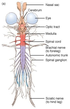
A pair of optic lobes distinguish the midbrain. The cerebellum and medulla oblongata make up the hindbrain. The medulla oblongata exits the foramen magnum and continues into the spinal cord, which is contained within the vertebral column. The sense organs of the frog are touch (sensory papillae), taste (taste buds), smell (nasal epithelium), vision (eyes), and hearing (ears) (tympanum with internal ears). Eyes and internal ears are well-organized structures, with the rest consisting of cellular aggregations around nerve endings. Frog eyes are a pair of spherical structures in the orbit of the skull. These are basic eyeballs (possessing only one unit). In frogs, the external ear is missing, leaving just the tympanum visible.
The ear is both a hearing and a balancing organ (equilibrium). Male and female reproductive systems in frogs are well-organized. A pair of yellowish ovoid testes are discovered adhering to the top section of the kidneys by a double fold of peritoneum called the mesorchium in male reproductive organs. The number of Vasa efferentia that emerge from the testes is 10-12. On their side, they enter the kidneys and open into Bidder's canal. Finally, it connects with the urinogenital duct, which runs from the kidneys to the cloaca. The cloaca is a small, midline chamber where faeces, urine, and sperms are passed to the outside. A pair of ovaries are among the female reproductive organs.
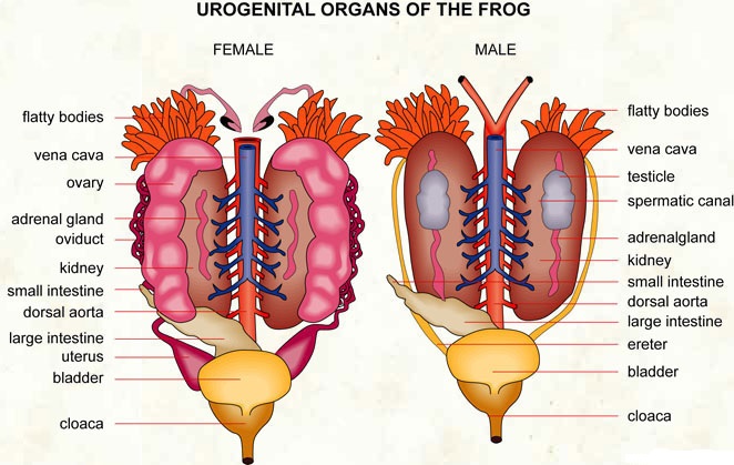
The ovaries are close to the kidneys but have no functional relationship with them. Separate oviducts emerge from the ovaries and open into the cloaca. At any given time, a mature female can lay between 2500 and 3000 ova. External fertilization takes place in water. Tadpoles are in the larval stage of development. Tadpoles go through metamorphosis to become adults. Frogs are advantageous to humans because they consume insects and protect crops. Frogs keep the ecology in balance since they are a vital link in the food chain and food web. Humansconsume the muscular legs of frogs in several regions.
