 ACME SMART PUBLICATION
- Books Name
- ACME SMART COACHING Biology Book
- Publication
- ACME SMART PUBLICATION
- Course
- CBSE Class 11
- Subject
- Biology
COCKROACH (Periplaneta americana)
Phylum : Arthropoda
Class : Insecta
Genus : Periplaneta
Species : americana
Morphology
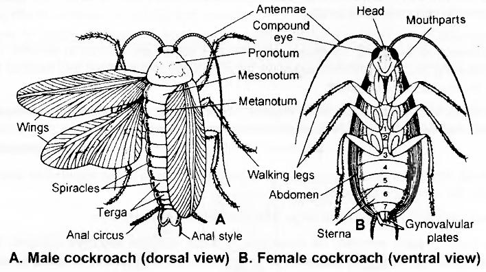
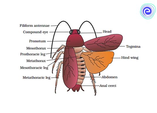
The body is divided into three distinct regions head, thorax, abdomen.
Theirsize ranges from 1/4 inches to 3 inches (0.6-7.6 cm).
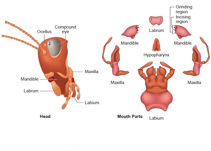
Head is hypognathus (facing downwards), and is formed by the fusion of six segments.
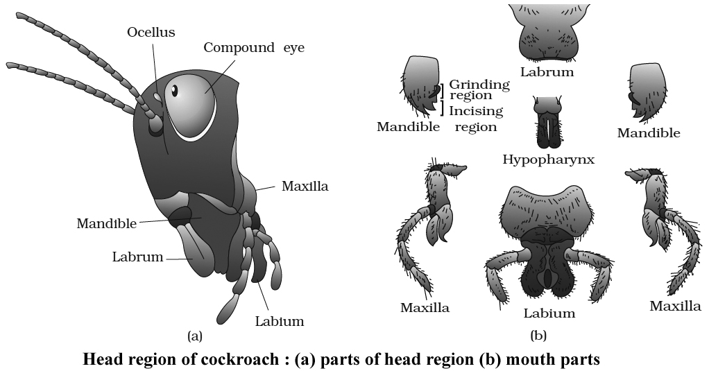
Anteriorly, the head bears mouth which is provided with appendages collectively called mouth parts which are used in chewing, cutting and swallowing.
The mouth parts consist of a pair of mandibles and maxillae, labium forming the lower lip and a labrum forming upper lip. Within the cavity enclosed by mouthparts, there is a median flexible lobe called hypopharynx which acts like a tongue.
Thorax consists of 3 segments - the prothorax, mesothorax and metathorax.
A pair ofwings arise from mesothorax which are thick and leathery and are called Elytra or Tegmina.
A pair of membranous wings, used in flying arise from metathorax.
In houseflies and mosquitoes the metathoracic wings are reduced to halteres for balancing.
Salient Features of Peripianata americana (Cockroach) Habit and Habitat
Cockroaches are world wide and found in such places where darkness, warmth, dampness and plenty of organic debris is available.
Cockroaches are nocturnal and omnivorous.
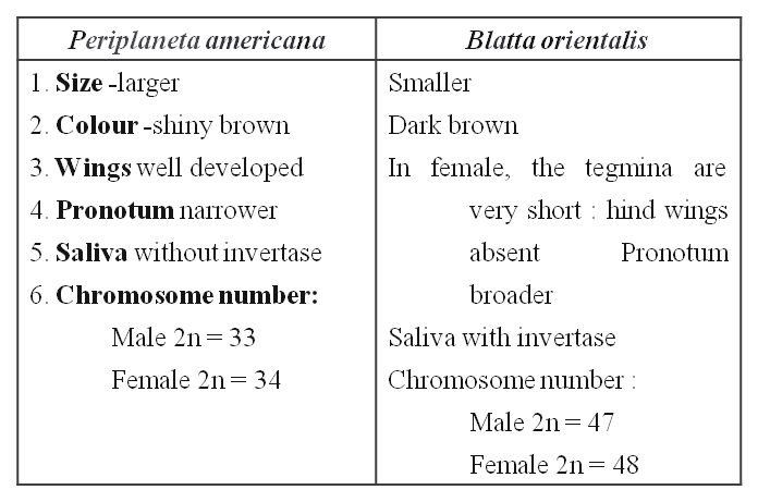
Three species of cockroaches are commonly found in India (a) Periplanata americana, (b) Blatta orientalis, and (c) Blatta germaniaca.
P. americana is the largest and most common . It is commonly called, American cockroach, Bombay canary or Ship cockroach. In both the sexes, wings are present which are larger than the body.
Blatta orientalis is a black or dark brown medium sized species. The female has rudimentary wings which are not helpful in flight. Male has wings which are short and are not present up to the end of the body.
Blatta germanica : It is the smallest of the domestic species of the cockroaches. It is pale yell:ow-brown in colour.
External features -(P. americana)
The body is narrow, elongated, bilaterally symmetrical and flattened, measuring about 3 to 4½ cms (34-53 mm) in length and 1½ to 2 cms in breadth.
The colour is reddish brown with a pale yellow area around the edge of pronotum and two dark patches over it.
Exoskeleton
The entire body of the cockroach is covered with a thick, hard, chitinous cuticle, which is secreted by epidermis, forming the exoskeleton.
The exoskeleton of each segment consists of four plate like pieces called sclerites. Dorsal sclerite is called tergum or tergite, ventral is called sternum or sternite and two lateral sclerites are called pleura or pleurites.
The sclerites of each segment are joined with each other and with those of the adjacent segments by means of soft and flexible articular or arthrodial membranes. This gives the sclerites, some freedom of movement upon each other at their edges.
The stiff, immovable bristles or spines covering the body and its appendages are in fact the outgrowths of the cuticle while the movable hair like setae occurring at some places are secreted by special trichogen cells of the hypodermis lying below the cuticle.
Segmentation
Embryologically, the body of cockroach is formed of 20 segments -6 in head, 3 in thorax and 11 in abdomen.
Due to complete or incomplete fusion between some segments during development, the number of distinct segments is reduced in the adult.
All segments of the head are fused. Thorax consists of 3 segments. Only 10 segments are retained in the abdomen of the adult. Of these, only the first seven are distinct in females and first nine in males. The remaining hinder abdominal segments become small and modified into extemal genitalia that are hidden under the last distinct segment.
When wings are removed, the three regions -head, thorax and abdomen become distinctly visible. A small, soft and mobile neck or cervicum connects the head with thorax.
Head
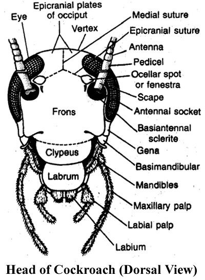
It is small, triangular and its narrow end is bent downwards in hypognathous position i.e., at an angle of 900 with the long axis of the body.
On each lateral side, it bears a large and blackish compound eye.
At the base of each antenna, on inner side, a small rounded and light coloured area called fenestra or ocellar spot representing simple eye is present. Endoskeleton of head is called tentorium.
All sclerites of the head are fused , forming a strong head capsule exhibiting only faint lines of fusion.
Cephalic appendages: The head appendages include 1 pair antennae, 1 pair mandibles, 1 pair first maxillae and 1 pair second maxillae (fused as single labium or lower lip), one labrum or upper lip and one hypopharynx.
Antennae : These are a pair of long, thread like appendages, extending forward from an antennal socket located dorsally upon head capsule near the eye. These are very mobile and act as tactile, thermal and olfactory receptor organs. Each is formed of several small segments called podomeres. The first basal podomere, called scape, is the largest. The second called pedicel, is narrow and elongated. The remaining long, slender and many jOinted part of each antenna is called flagellum.
The Mouth Parts
The remaining cephalic appendages are small and located around the mouth. Hence, together these appendages comprise the mouth parts of the cockroach. These help in 'biting and chewing' its food.
Labrum (upper lip) : It is the broad, flattened terminal sclerite of the dorsal side of head capsule, movably articulated to the clypeus acts as upper lip. It has epipharynx (chemoreceptors) on its inner side.
Mandibles: Thick, hard and triangular appendages beneath the labrum, one on each lateral side of the mouth, which bear pointed, teeth like processes called denticles.
First maxillae: Located on each side of the mouth next to mandibles. These serve to hold food particles in between the mandibles for cutting and chewing. They also bear olfactory receptors.
Labium (lower lip): The second maxillae are fused together forming a single large structure which covers the mouth from ventral side, hence called the 'lower lip' or labium. It bears tactile and gustatory sensory setae.
Hypopharynx: It is a small, cylindrical mouthpart, sandwiched between first maxillae and covered by labrum and labium on dorsal and ventral sides respectively. It bears several sensory setae on its free end, and the opening of common salivary duct upon its basal part.
Thorax
(i) It comprises of three segments -prothorax, mesothorax and metathorax.
(ii) The three thoracic segments are covered by relatively thicker and larger tergites called nota.
(iii) The notum of prothorax called pronotum is very large and covers the neck also. Each of the mesonotum and metanotum bears a pair of wings.
Thoracic Appendages
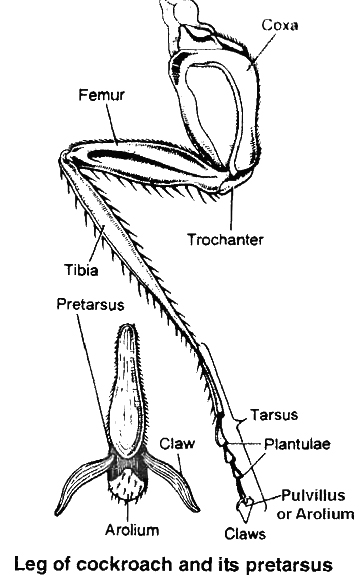
Each thoracic segment bears a pair of walking legs. Each walking leg consists of five segments:
(i) Coxa.
(ii) A triangular short rod like trochanter, articulated with coxa and femur.
(iii) A long, spiny femur.
(iv) A spring like tibia which represents the longest segment.
(v) A long tarsus, divided into five tarsomeres or podomeres. The last tarsomere is called pretarsus forming the claws and bearing an adhesive arolium or pulvillus. Similar but smaller adhesive pads called plantulae are located at each joint of tarsus.
Abdomen in both males and females consists of 10 segments. A typical abdominal segment has a dorsal tergum, ventral sternum and between them a narrow membranous pleuron on each side which bears spiracles. In females, the sclerites of 8th and 9th are overlapped by corresponding sclerites of the 7th segment.
The seventh sternum is boat shaped and together with eighth and ninth sterna forms a brood orgenital pouch. In males only 8th is overlapped by the 7th segment. The tenth segment bears a pair of 15 jointed filamentous structures called as anal cerci. Ventral to these in the males, the 9th segment bears a pair of short, thread like anal styles which are absent in females. Between one sclerite and the other, there is a flexible arthrodial membrane.
Abdomen

It is the largest and the broadest, relatively more flattened and softer part behind the thorax.
There are ten tergites. In both males and females, the 8th and 9th tergites are mostly covered by the 7th. The 10th tergum is somewhat bowl-shaped and posteriorly bifurcated into two lobes.
Ventrally, the abdomen has 9 sternites in male and 7 in females.
In females the last sternite (7th) is larger and boat shaped and together with indistinct 8th and 9th sternites it forms a chamber Anal styles like structure called gynatrium, posterior part of this chamber is MALE called oothecal chamber. Behind this chamber, 7th sternite Opening of oothecal chamber bifurcates into two prominent oval plates called apical lobes. Female gonopore is located between them.
In males, the 9th sternite bears a pair of spine like anal-styles.
In both male and female cockroaches, several small chitinous appendages are located around the gonopore. These help in reproduction and hence called gonapophyses.
At several places, certain processes of exoskeleton extend into the body and form endoskeletal elements which provide attachment to muscles and hence called apodemes.
Abdominal Appendages
Abdominal segments lack locomotory appendages. There are certain small structures associated with gonopore, which are different in male and female cockroaches.
The 10th tergum posteriorly bears a pair of many jointed anal cerci. They bear minute sensory hair sensitive to sound and other vibrations.
The 9th sternum of males bears, in addition, a pair of small and spine-like, unjointed anal styles.
Digestive System of Cockroach
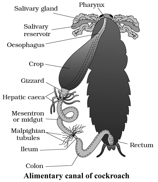
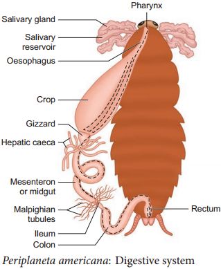
The alimentary canal is long and somewhat coiled, divisible into three main parts namely foregut, midgut and hindgut.
Foregut (stomodaeum) is lined by cuticle and is differentiated into five parts: buccal chamber, pharynx, oesophagus, crop and gizzard. Crop is used for storage of food.
Gizzard is muscular and internally provided with six cuticular teeth which crush the food.
A stomodaeal valve is present between gizzard and mesenteron.
Midgut (mesenteron or ventriculus) is short, tubular and lined with glandular endoderm.
At the anterior end of mesenteron, there are 6-8 blind glandular hepatic or gastric caecae which secrete digestive enzymes.
Internally mesenteron is not lined by cuticle but it is covered by a very thin and transparent peritrophic membrane formed of chitin and proteins.
Peritrophic membrane is secreted by gizzard, it serves to protect the wall of midgut from abrasion due to friction of food particles.
Peritrophic membrane is permeable to digested food and enzymes in the mesenteron.
Hindgut (proctodaeum) is broader than midgut and it comprises ileum, colon and rectum.
The wall of rectum is provided with six rectal papillae. They help in the absorption of water and salts.
Cockroach is omnivorous. It feeds on all sorts of organic debris.
The digestive enzymes of saliva are mainly zymase and amylase.
Most of the nutrients of food are digested in the crop.
Absorption of digested food takes place in mesenteron.
Respiratory System of Cockroach
The blood of cockroach is not responsible for the transportation of gases, it serves as a stationary medium for exchange of gases.
A complicated system of numerous, shiny, transparent and branched air tubes or tracheae are found for gaseous exchange in the haemocoel cavity. There are 6 longitudinal tracheal tubes -2 dorsal, 2 ventral and 2 lateral which are interconnected by transverse commissures. Chitinous rings prevent collapse of trachea.
Atmospheric air enters into and escapes out from this system through ten pairs of slit-like apertures called stigmata or spiracles located on lateral sides of the body. Two pairs of these are thoracic and eight pairs are abdominal. The openings of spiracles are regulated by the sphincters.
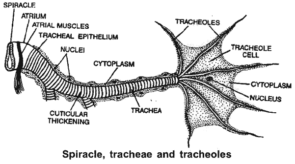
Thoracic spiracles are somewhat larger. One pairofthese is between prothoraxand mesothorax and the other between mesothoraxand metathorax, upon respective pleurites.
The first pair ofabdominal spiracles are dorsolateral upon tergite offirstabdominal segment, butthe remaining seven pairs are present upon the pleurites of second to eighth segments.
Each spiracle is surrounded by a ring-like sclerite called peritreme.
Mechanism
Several tergo-sternal muscles extend vertically between the tergites and sternites of all abdominal segments.
Harmonious contractions and relaxations ofthese at regular intervals cause rhythmic expansion and compression of abdomen leading to inspiration (with relaxation) and expiration (with contraction) of air.
At rest, the oxygen requirement is less, tracheolar ends get filled with tissue fluid.
The movement of O2 is along the pressure gradient as the tracheolar ends are losing oxygen to the cells for performing cellular respiration.
O2 requirement increases during activity.
Tracheolar fluid is withdrawn out of Tracheoles.
Alternate expansion and contraction of abdominal cavity occurs involving tergosternal muscles and abdominal muscles.
High level of CO2 in abdominal cavity make tergo-sternal muscles and abdominal muscles to contract pushing out the air from tracheal system to the outside through spiracles.
With relaxation abdomen expands i.e., tracheal trunks and tracheae expand and as a result, air rushes into tracheae and tracheoles via spiracles, it results in inspirations.
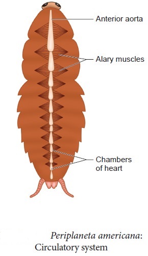
Circulatory System of Cockroach
Blood vascular system is open and lacunar type. Body cavity contains blood (haemolymph) which bathes viscera in it, therefore known as Haemocoel.
Blood vascular system consists of a tubular heart, a blood vessel called anterior aorta and a system of ill defined blood spaces or sinuses.
Heart
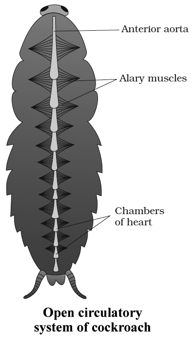
It is a long elongated tube situated in the mid dorsal line of thorax and the abdomen immediately beneath the terga.
Heart consists of thirteen chambers.
The last two posterior chambers are very small.
The chambers are separated from one another by deep constrictions.
The opening of each chamber into another is guarded by valves which allow blood from behind forward.
The Blood Sinuses
The large body cavity or haemocoel is divided by two membranous horizontal partitions, into three wide and flattened sinuses-the dorsal pericardial sinus containing the 'heart', the middle perivisceral sinus containing the gut, and the ventral perineural sinus or sternal sinus containing the nerve cord.
The partition between pericardial and perivisceral sinuses is called dorsal diaphragm and between perivisceral and perineural sinuses is called ventral diaphragm.
The sinuses intercommunicate by pores in the respective diaphragms.
A pair of fan like, triangular alary muscles in the floor of the pericardial sinus in each segment reinforce the dorsal diaphragm by their broad bases and also connect it, by their pointed tips with the tergite of the segment.
Circulation of Haemolymph
The pumping force that propels the haemolymph is provided by the pulsations of the 'heart'. The respiratory movements of abdomen and contraction of alary muscles increase this force.
From the pericardial sinus, the haemolymph enters into heart through ostia. When the heart is filled it contracts from behind forwards. This is its systole phase. Soon the heart becomes relaxed in its diastole phase. Then the next systole follows after a short time interval called diastasis. Thus heart pulsates about 50 times/minute.
During systole, the valve like ostia close, preventing back flow of haemolymph into the pericardial sinus. Therefore, some of its haemolymph is pumped into segmental vessels while most of it is poured into the head sinus through the terminally opening anterior aorta.
From the head sinus, the haemolymph flows backwards into the thorax and abdomen. While flowing backwards from head sinus, the haemolymph remains in the ventral part due to presence of Oesophagus in dorsal part. So, it fills into the perineural sinus.
From the perineural sinus, the haemolymph now flows into the perivisceral sinus through the pores of ventral diaphragm in abdominal region.
Then from perivisceral sinus, it flows into pericardial sinus through the pores of dorsal diaphragm. Then, during heart's diastole, it fills in the heart through the ostia.
Excretion in Cockroach
Cockroach is uricotelic. In cockroach, following structures help in excretion:
(i) Malpighian tubules
(ii) Fat bodies
(iii) Nephrocytes
(iv) Cuticle
(v) Uricose glands in some species.
Malpighian tubules:
Malpighian tubules are attached at the junction of midgut and hindgut. Excretory products, dissolved in haemolymph are absorbed by malpighian tubes are discharged into hindgut.
Fat bodies:
Some fat bodies are also present in haemocoel which have mycetocytes, urate cells, oenocytes and trophocytes.
Nephrocytes :
Nephrocytes present in lateral wall of heart and help in excretion and store nitrogenous waste.
Uricose glands:
In some species, in males, uricose glands are present on the periphery of mushroom glands. These glands synthesize uric acid . Malpighian tubules are analogous to mammalian kidneys. Fat bodies are analogous to vertebrate liver.
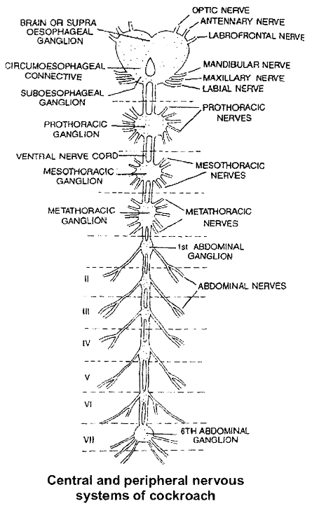
Nervous System of Cockroach
It consists of a series of fused, segmentally arranged ganglia joined by paired longitudinal connectives on the ventral side.
Central Nervous System :
It consists of brain or supra oesophageal ganglion. Brain gives off a pair of short, stout cords, the circumoesophageal connectives, that encircle the oesophagus and pass downwards and backwards over the suboesophageal ganglion situated below oesophagus.
From the suboesophageal ganglion passes backwards into the thorax, a double ventral nerve-cord, which bears three ganglia in the thorax and six in the abdomen.
Peripheral Nervous System :
It consists of nerves, which are given off from the ganglia so as to innervate all the parts of the body.
Sympathetic or Somatogastric Visceral Nervous System:
It consists of a frontal ganglion which is situated on the dorsal side of the oesophagus in the head.
From this ganglion, a median unpaired recurrent nerve reaches the visceral ganglion situated on the crop.
Various nerve branches are given off from the visceral ganglion.
The frontal ganglion is joined with the central nervous system by nerves which connect it to circumoesophageal commissures.
Sense Organs of Cockroach
Receptor cells are present on general body surface.
Proprioreceptors :
They are for hearing or receiving sound vibrations. Auditory receptors are present on antennae and anal cerci.
Thigmoreceptors :
They are receptors for touch. Present on antennae, maxillary palps and legs.
Olfactory receptors: They receive various smells. Present on antennae and palps.
Gustatory receptors: They are for sense of taste. Present on maxillae and labial palps.
Eyes
Cockroach has compound eyes. Each compound eye is formed of about 2000 hexagonal ommatidia.
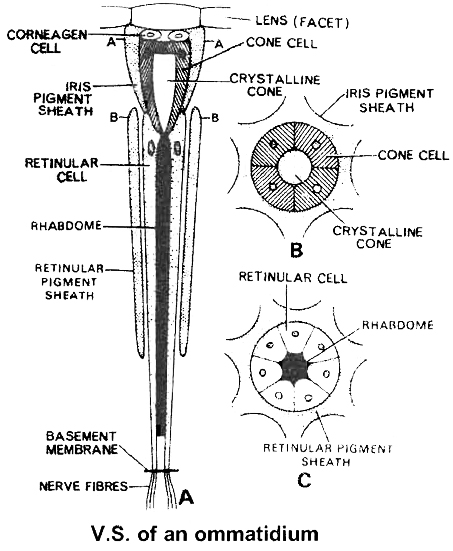
Each ommatidium has a biconvex lens or cornea. Below the lens there are corneagen cells which secrete the lens.
Below the corneagen cells is a transparent crystalline cone surrounded by four vitrellae or cone cell.
The vitrellae secrete the crystalline cone. All this forms the focusing or dioptrical region.
Below the cone there is refractive body the Rhabdome, surrounded by seven retinular cells.
Each ommatidia is isolated from the other by iris pigment sheath and retinal pigment sheath.
The image formed is apposition or mosaic vision, composed of as many separate but adjacent images as there are ommatidia.
In mosaic vision, images are sharp but separate and the eye can use only in bright light.
In cockroach vision is mosaic and apposition image is formed (although cockroach is nocturnal).
If pigmented iris sheath is removed from the compound eye of insects, only superposition image will be formed.
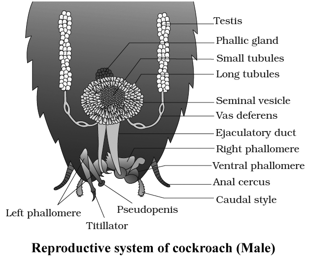
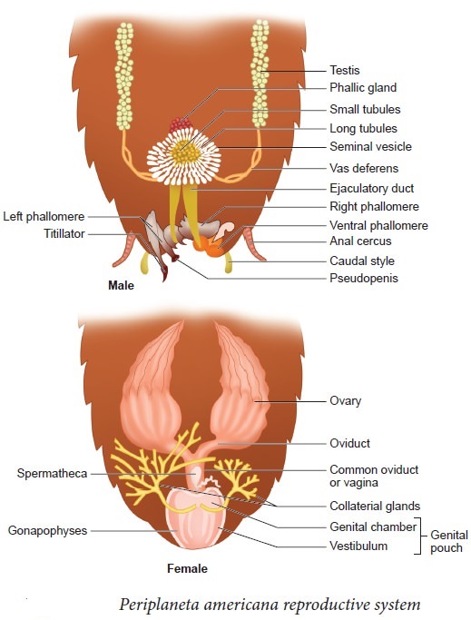
Reproductive System of Cockroach (Male)
In cockroach, sexes are separate, so it is dioecious.
Testes of cockroach are located in the abdominal segments 4, 5 and 6.
Mushroom gland consists of two types of tubules, (a) the long slender tubules the utriculi majores or peripheral tubules and (b) short tubules, the utriculi breviores, making up the major part of the gland. It is present in the 6-7th abdominal segments which functions as an accessory reproductive gland .
Small seminal vesicles are also found associated with mushroom glands.
All sperms of a seminal vesicle are glued together into a large bundle called spermatophore.
Spermatophore has three layered wall: inner layer secreted by utriculi majores; middle layer secreted by ejaculatory duct and outer layer secreted by phallic gland or conglobate gland
There are three asymmetrical chitinous structures called male gonapophyses or phallomeres. Right phallomere, left phaliomere (largest) and ventral phaliomere (smallest).
Reproductive System of Cockroach (Female)
Female organs consist of ovaries, oviducts, vagina, genital chamber, spermathecae, collaterial glands and female gonapophysis (ovipositor processes).
Ovaries of cockroach are located in the abdominal segments 2 to 6. Each ovary consists of eight ovarioles.
Two oviducts from each side open into a common oviduct or vagina which opens into genital chamber by female genital pore. A pair of spermathecae (left larger pyriform sac) are present near female genital pore.
A pair of collaterial glands also open in genital chamber.
Genital pouch or gynatrium is divisible into a genital chamber in front and oothecal chamber (vestibulum) behind.
Female genitalia consists of 3 pairs of chitinous processes hanging from the roof of oothecal chamber into its cavity.
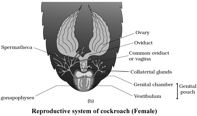
Ootheca of cockroach contains fourteen to sixteen fertilized eggs. Ootheca of cockroach is formed of a protein secreted by collaterial glands. On an average, females produce 9-10 oothecae.
Nymphs of cockroach emerge out from ootheca. A nymph resembles an adult in general structure but lacks wings and mature reproductive organs. The next to last nymphal stage has wing pads but only adult cockroach have wings.
Instar is a stage in the development of insect (larval instar, nymphal instar). Period between two successive moults in insects is termed stadium.
In Periplaneta americana, the nymph grows by moulting about 13 times to reach the adult form and Blatta orientalis, it moults 6 times.
Comparison of Periplaneta and Blatta
Cockroach
Cockroaches are dark or black-bodied animals belonging to the Phylum Arthropoda's class Insecta. Cockroaches with bright yellow, red, and green colorshave also been documented in tropical areas. Their antennae, legs, and a flat extension of the upper body wall that conceals the head vary in size from 14 inches to 3 inches (0.6-7.6 cm). They are nocturnal omnivores who live in moist environments all over the world. They have become occupants of human homes, making them major pests and disease vectors.
(i)Morphology: The common cockroach, Periplaneta americana, has wings that extend past the tip of the abdomen in males and is around 34-53 mm long inadults. The cockroach's body is divided into three separate regions: the head, the thorax, and the abdomen. A strong chitinous exoskeleton covers the entire body (brown). Hardened plates called sclerites (tergites dorsally and sternites ventrally) are linked to each other by a thin and flexible articular membrane in each segment of the exoskeleton (arthrodial membrane).
The triangular head is positioned anteriorly at right angles to the longitudinal axis of the body. It is made up of the fusion of six segments and has a flexible neck that allows it to move freely in all directions. A pair of compound eyes can be found on the head capsule. Membranous receptacles in front of the eyes sprout a pair of thread-like antennae. Sensory receptors in antennae aid in environmental monitoring. Appendages on the anterior end of the skull produce biting and chewing mouth parts. A labrum (upper lip), a pair of mandibles, a pair of maxillae, and a labium make up the mouthparts (lower lip). The tongue (hypopharynx) is a flexible lobe that resides within the space encompassed by the mouthparts.
Prothorax, mesothorax, and metathorax are the three sections of the thorax. The neck is a short extension of the prothorax that connects the head to the thorax. Walking legs are attached to each thoracic section. The mesothorax produces the first pair of wings, whereas the metathorax produces the second pair. Tegmina, or opaque dark and leathery forewings (mesothoracic), cover the hind wings when at rest. The rear wings are transparent and membranous, and they help the animal fly.
Both males and females have ten segments in their abdomen. The 7th sternum is boat-shaped in females, and it creates a brood or genital pouch with the 8th and 9th sterna, which contains the female gonopore, spermathecal pores, and collateral glands in the front section.The genital pouch or chamber is located at the back end of the abdomen, between the 9th and 10th terga and the 9th sternum. Dorsal anus, ventral male genital hole, and gonapophysis are all found here. Males have a pair of short, threadlike anal styles that females lack. Anal cerci are a pair of jointed filamentous structures found on the 10th segment of both sexes.
(ii) Anatomy: The alimentary canal is separated into three sections in the body cavity: foregut, midgut, and hindgut. The mouth opens into a short tubular pharynx, which leads to the oesophagus, a tiny tube conduit.
The esophagus expands into a sac-like structure called a crop, which is used to store food. Gizzard or proventriculus follows the crop. It possesses a thick outer layer of circular muscles and a thick inner cuticle that forms six teeth-like chitinous plates. The gizzard assists in the crushing of food particles. The cuticle covers the entire foregut. At the confluence of the foregut and midgut is a ring of 6-8 blind tubules called the hepatic or gastric caeca, which release digestive juice.Another ring of 100-150 yellow-colored thin filamentous Malpighian tubules exists at the midgut-hindgut junction. They aid in excretory product clearance from the hemolymph. The ileum, colon, and rectum make up the hindgut, which is larger than the midgut. Through the anus, the rectum opens up.
Cockroaches have an open blood circulatory system. The blood vessels are underdeveloped and open into the air (hemocoel). The blood bathes the visceral organs in the haemocoel (hemolymph). Colorless plasma and haemocytes make up the haemolymph. The cockroach's heart is an extended muscular tube that runs along the mid-dorsal line of the thorax and abdomen. It is divided into funnel-shaped chambers on both sides by ostia.Through the ostia, blood from the sinuses enters the heart and is pumped anteriorly to the sinuses. The respiratory system is made up of a network of trachea that open through ten pairs of small openings on the body's lateral side called spiracles. Air is carried to all parts by thin branching tubes (tracheal tubes separated into tracheoles). The sphincters control the opening of the spiracles.
Diffusion occurs at the tracheoles to exchange gases. Malpighian tubules are responsible for excretion. Glandular and ciliated cells line each tubule. They take in nitrogenous waste and convert it to uric acid, which is then expelled through the hindgut. As a result, It is known as uricotelic.
The fat body, nephrocytes, and urecose glands all contribute to excretion. On the ventral side, the cockroach's nervous system is made up of a series of fused, segmentally arranged ganglia connected by paired longitudinal connectives. The thorax has three ganglia and the abdomen has six. Cockroaches have a neurological system that runs throughout their bodies. A portion of the nervous system is located in the skull, while the rest is located on the ventral (belly) side of the animal. Because of this, even if a cockroach's head is removed, it can survive for up to one week. The brain is represented in the head area by the supra-oesophageal ganglion, which sends nerves to the antennae and compound eyes.
Antennae, eyes, maxillary palps, labial palps, anal cerci, and other sensory organs are found in cockroaches. The complex eyes are located on the head's dorsal surface. About 2000 hexagonal ommatidia make up each eye. A cockroach can obtain several photos of an object by using many ommatidia. Mosaic vision is a type of vision that has a higher sensitivity but lower resolution and is common at night (hence called nocturnal vision).
Cockroaches are dioecious, with well-developed reproductive organs in both sexes. A pair of testes, one on each lateral side in the 4th -6th abdominal segments, make up the male reproductive system.
A narrow vas deferens develops from each testis and opens into the ejaculatory duct via the seminal vesicle.The male gonopore is located ventral to the anus and opens into the ejaculatory duct. In the 6th and 7th abdominal segments, there is a mushroom-shaped gland that serves as an accessory reproductive gland. Male gonapophysis or phallomeres depict the external genitalia (chitinous asymmetrical structures, surrounding the male gonopore). The sperm are retained in the seminal vesicles and cemented together in spermatophores, which are expelled during copulation. Two enormous ovaries lie laterally in the 2nd – 6th abdominal segments, forming the female reproductive system. Each ovary is made up of eight ovarian tubules, also known as ovarioles, which contain a chain of growing ova. Each ovary's oviducts join to form a single median oviduct (also known as the vagina), which exits into the genital chamber.The 6th segment, which enters the vaginal chamber, contains a pair of spermatheca. Spermatophores are used to transport sperm. The fertilized eggs are enclosed in oothecae capsules. The ootheca is a 3/8" (8 mm) length dark reddish to a blackish-brown capsule. They're dropped or bonded to a suitable surface, usually near a food source in a crack or crevice with high relative humidity. Females produce 9-10 oothecae on average, each holding 14-16 eggs. P. americana has a paurometabolous development, which means it develops through the nymphal stage. The nymphs have the appearance of adults. To attain adulthood, the nymph must molt 13 times.Wing pads are present in the next to last nymphal stage, however, only adult cockroaches have wings.
Many cockroach species are found in the wild and have yet to be identified as economically important. In and around the human habitat, a few species flourish. They are pests because their stinky excreta spoils food and contaminates it. By polluting food, they can spread a range of bacterial infections.
