 Science Made Easy
 ACERISE INDIA
Cell as a basic unit of life;
- Books Name
- Science Made Easy Science Book
- Publication
- Science Made Easy
- Course
- CBSE Class 9
- Subject
- Science
Introduction -
• Cell is the structural and functional unit of life. It is the basic unit of life.
• It is discovered by Robert Hook in 1831 in cork slice with the help of primitive microscope.
• Leeuwenhoek (1674), discovered the free living cells in pond water with the improved microscope
• Robert Brown discovered the nucleus in the cell in 1831.
• Purkinje coined the term ‘protoplasm’ for the fluid substance of the cell in 1839.
The cell theory-
• The theory that all the plants and animals are composed of cells and the cell is the basic unit of life,
was presented by two biologists, Schleiden and Schwann.
• The cell theory was further expanded by Virchow by suggesting that all cells arise from preexisting
cells.
tructure and functions of animal and plant tissues (four types in animals; meristematic and permanent tissues in plants).
- Books Name
- Science Made Easy Science Book
- Publication
- Science Made Easy
- Course
- CBSE Class 9
- Subject
- Science
Introduction
→ A group of cells that are similar in structure and/or work together to achieve a particular function
forms a tissue.
→ Most of the tissues in plants are supportive, which provides them with structural strength.
→ These tissues are dead, since dead cells can provide mechanical strength as easily as live ones
and need less maintenance.
→ Plant Tissues are of two types Meristematic & Permanent tissues.
Meristematic Tissue
→ These are simple living tissues having thin walled compactly arranged immature cells which are
capable of division and formation of new cells.
Features of Meristematic tissues:
→ Thin primary cell wall (cellulosic).
→ Intercellular spaces are absent (compact tissue).
→ Generally vacuoles are absent, dense cytoplasm & prominent nuclei are present.
→ Large numbers of cell organelles are present.
→ Active metabolic state, stored food is absent.
→ Actively dividing cells are present in growing regions of plants, example: root & shoot tips.
Classification of Meristematic Tissues on the Basis of Origin
• Primary (Promeristem)
→ Derived directly from the meristems of embryo.
→ They consist of cells derived from primary meristem.
→ They add to primary growth of plants.
• Secondary Meristematic Tissues
→ Formed by permanent tissues.
→ These are having cells derived from primary permanent tissue.
→ They usually add to the diameter of plants.
Classification of Meristematic Tissues on the Basis of Location
• Apical Meristem
→ It is present at the growing tips of stems and roots.
→ Cell division in this tissue leads to the elongation of stem & root, thus it is involved in primary
growth of the plant.
• Intercalary Meristem
→ It is present behind the apex.
→ It is the part of apical meristem which is left behind during growth period.
→ These are present at the base of leaf & internode region.
→ These lead to the increase in the length of leaf (Primary), example: in grass stem, bamboo stem
mint stem etc.
• Lateral Meristem
→ It is also called as secondary meristem.
→ It occurs along the sides of longitudinal axis of the plant.
→ It gives rise to the vascular tissues.
→ Causes growth in girth of stem & root.
→ They are responsible for secondary growth.
Permanent Tissue
→ The permanent tissues are composed of those cells which have lost their capability to divide.
→ They have definite shape, size and thickness. The permanent tissue may be dead or living.
→ The division & differentiation of the cells of meristematic tissues give rise to permanent tissues.
→ In cell differentiation, developing tissue and organs change from simple to more complex form
to become specialized for specific functions.
→ The cells of permanent tissue loose the capacity to divide and attain a permanent shape, size
and function.
• Permanent tissues are classified into two types on the basis of Structure and Composition i.e.
Simple Permanent Tissues and Complex Permanent Tissues.
Simple Permanent Tissues
→ These are made up of same type of cells which are similar structurally and functionally.
→ They include two types of tissue Protective tissues and Supporting Tissues.
• Protective Tissues: These tissues are primarily protective in function.
→ They consist of Epidermis and Cork/Phellem.
(i) Epidermis
→ Epidermis forms one cell thick outermost layer of various body organs of plants such as leaves,
flowers, stems and roots.
→ Epidermis is covered outside by cuticle. Cuticle is a water-proof layer of waxy substance called
cutin which is secreted by the epidermal cells.
→ Cuticle is very thick in xerophytes.
→ Cells of epidermis of leaves are not continuous at some places due to the presence of small
pores called as stomata.
→ Each stomata is guarded by a pair of bean-shaped cells called as guard cells. These are the one
epidermal cells which possess chloroplasts, the rest being colourless.
Functions of Epidermis
→ The main function of epidermis is to protect the plant from desiccation and infection.
→ Cuticle of epidermis cuts the rate of transpiration and evaporation of water and prevents wilting.
→ Stomata in epidermis allow gaseous exchange to occur during photosynthesis respiration.
→ Stomata also helps in transpiration.
(ii) Cork or Phellem
→ In older roots and stems, tissues at the periphery become cork cells or phellem cells.
→ Cork is made up to dead cells with thick walls and do not have any intercellular spaces.
→ The cell walls in cork deposit waxy substance called as suberin.
→ The cells of cork become impermeable to water and gases due to the deposition of suberin.
→ The cork cells are without any protoplasm but are filled with resins or tannins.
Functions of Cork
→ Cork is protective in function. Cork cells prevent desiccation, infection and mechanical injury.
→ Imperviousness, lightness, toughness, compressibility and elasticity make the cork commercial
valuable.
→ Cork is used for insulation, as shock absorber in linoleum.
→ Cork is used in the making of a variety of sport goods such as cricket balls, table tennis, shuttle
cocks, wooden paddles etc.
• Supporting Tissues: These are supportive in function.
→ There are three types of Supporting tissues i.e. Parenchyma, Collenchyma and Sclerenchyma.
(i) Parenchyma
→ It is the fundamental tissue.
→ Tissue first time evolved in bryophyte.
→ Thin walled cells, oval or spherical in structure.
→ Cell wall mainly composed of cellulose & pectin.
→ Large central vacuole for food & water storage.
→ Primary function is food storage.
→ Some parenchyma involved in excretory substance storage are so called as idioblast, storing
such as resin, tannin, gums & oils.
→ In typical parenchyma chlorophyll is absent.
→ Chloroplast containing parenchyma tissue are chlorenchyma which perform
photosynthesis such as mesophyll of leaves.
→ In hydrophytic plants aerenchyma (a type of parenchyma containing air spaces) provides
buoyancy.
→ Parenchyma provides turgidity to cells.
(ii) Collenchyma
→ It is the living mechanical tissue.
→ Elongated cells with thick corners.
→ Localized cellulose & pectin thickening.
→ Provides flexibility to plant parts & easy bending of various parts of plant.
→ Present only in herbaceous dicot stem.
→ Present at thin margin of leaves.
→ Few chloroplasts may be present.
→ Gives mechanical strength & elasticity to the growing stems.
(iii) Sclerenchyma (Scleras–hard) Strengthening tissue.
→ Composed of extremely thick walled cells with little or no protoplasm.
→ Cells are dead & possess very thick lignified walls.
→ Lignin is water-proof material.
→ Intercellular spaces are absent.
• Cells of sclerenchyma are of two types Sclereids and Fibres.
• Sclereids
→ These are also called grit cells or stone cells.
→ These are small cells, where lumen is so small due to higher thickening of cell wall, as present in
drup fruit (mango, coconut, walnut) in legume seeds (Macrosclereid).
• Fibers
→ They are very long, narrow, thick, lignified cells. Lumen is large as compared to sclereids.
They are generally 1-3 mm long.
→ In the thick walls of both the fibres and sclereids are present thin areas called as pits.
→ Sclrenchyma Fibres are used in the manufacture of ropes, mats & certain textile fibres.
→ Jute and coir are obtained from the thick bundle of fibres.
Difference between Parenchyma, Collenchyma and Sclerenchyma

Complex Permanent Tissues
→ It consists of more than one type of cells which work together as a unit.
→ It helps in transportation of organic materials, water & minerals.
→ It is also known as conducting or vascular tissue.
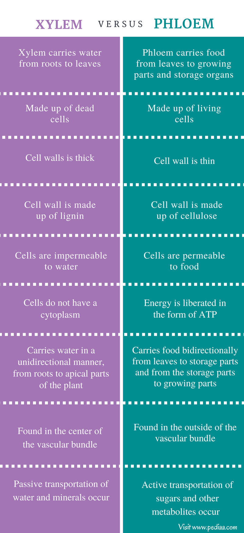
→ Xylem & phloem together form vascular bundles.
Xylem
→ It is also known as wood and is a vascular and mechanical tissue.
→ Thick walled cells are found in the form of tubular passages.
→ Xylem consists of four types of cells called as elements Tracheids, Vessels, xylem parenchyma
and xylem sclerenchyma.
(i) Tracheids
→ They are elongated angular dead cells (primitive elements) mainly involved in conduction of
water and minerals in gymnosperms.
(ii) Vessles
→ They are advance element (generally found in angiosperms).
→ Vessels are cylindrical tube like structures placed one above the other end to end which form a
continuous channel for efficient conduction of water.
(iii) Xylem parenchyma
→ They are small & thick walled parenchymatous cells subjected for storage of starch (food).
(iv) Xylem sclerenchyma
→ Thy are non-living fibres with thick walls and narrow cavities provide mechanical support.
→ Except xylem parenchyma all other xylem elements are dead.
→ The annual rings present in the trunk of a tree are xylem rings.
→ By counting the number of annual rings, we can determine the age of a tree.
Phloem
→ They also consist of both parenchymatous and schlerenchymatous cells.
→ Phloem consists of four types of element which are Sieve tubes, Companion cells, Phloem fibre
and Phloem parenchyma.
(i) Sieve tubes
→ Sieve tubes are slender tube like structures made up of elongated, thin walled cells placed end to
end.
→ The end walls of sieve tube cells are perforated by numerous pores, called as sieve plates.
→ Nucleus of sieve cell degenerates at maturity. However, cytoplasm persists, because of
protoplasmic continuation of sieve tube with companion cell through plasmodesmata.
→ Sieve cells possess slime protein or protein which is concerned with growth and repair of sieve
cells.
(ii) Companion cells
→ Companion cells have dense cytoplasm and prominent nuclei.
→ Sieve cells & companion cells are so called sister cells because they originate from single mother
cell.
(iii) Phloem fibre
→ They give mechanical support to sieve tubes.
(iv) Phloem parenchyma
→They store food and help in radial conduction of food.
(v) Leptome
→ Main part of phloem involved in conduction of food, which is sieve tube.
→ In xylem, only unidirectional movement is possible while in phloem bidirectional movement can
occur.
→ In phloem, except phloem sclerenchyma all elements are living.
Diversity of plants and animals
- Books Name
- Science Made Easy Science Book
- Publication
- Science Made Easy
- Course
- CBSE Class 9
- Subject
- Science
Introduction
→ All living organism are grouped on the basis of their similarities and increasing complexities into different complexities.

→ Biodiversity means the variety of living organisms present on a particular region.
→ There are about 20 lac organisms known on the earth which differ from one another in externaform, internal structure, mode of nutrition, habitat, etc.
• Taxonomy:
It is a branch of biology which deals with identification, nomenclature andclassification of organisms.
Carolus Lannaeus is called the father of taxonomy.
Health and its failure.
- Books Name
- Science Made Easy Science Book
- Publication
- Science Made Easy
- Course
- CBSE Class 9
- Subject
- Science
Introduction
→ ‘Health’ is a state of being well enough to function well physically, mentally and socially.
→ Disease: Any disturbance in the structure or function of any organ or part of body.
→ The various causes of diseases are pathogens (virus, bacteria), lack of nutritious diet/balanced
diet and lack of public health services.
→ Acute diseases occur suddenly and lasts for a short duration while chronic diseases develop
slowly and lasts for long period of time.
→ The diseases/infections can be prevented by life style (exercise, proper sleep, enough relaxatio
modification, taking balanced diet, good personal health and hygiene and also maintaining a cle
and healthy surrounding.
→ Treatment involves killing of the microbes/pathogens.
Health
→ Health is a state of physical, mental and social well-being.
• The conditions necessary for good health are:
(i) Good physical and social environment.
(ii) Good economic conditions.
→ Good physical and social environment includes clean surroundings, good sanitation, proper
garbage disposal and clean drinking water.
→ Good economic conditions includes job opportunities for earning to have nutritious food and to
lead a healthy life.
→ Personal and Community Issues Both Matter for Health
→ All those activities which people do both individually and in groups for the development of their
society, constitute the community health.
→ Personal and community health are supplementary to each other.
→ We protect ourselves by keeping our body clean.
→ For this, we also require a good and healthy environment in our surroundings.
→ We can have this only by the means of community health and development.
→ So, both personal and community health are inter-related.
Differences between Being Healthy and Disease-free
Being Healthy Being Disease-free
It is a state of being well enough to function well
physically, mentally and socially.
It is a state of absence from diseases.
It refers to the individual, physical and social environment.
It refers only to the individual.
The individual has good health. The individual may have good health or poor health.
Disease and Its Causes
What does disease look like?
→ When a person is affected by a disease either the functioning or the appearance of one or mor
systems of the body will change for the worse.
→ These changes give rise to symptoms and signs of disease.
→ On the basis of the symptoms the physicians look for the signs of a particular disease and
conduct tests to confirm the disease.
Types of Diseases
(i) Acute Diseases: Acute diseases which last for only very short period of time and affect body
suddenly and quickly. Example: Cold, cough, typhoid etc.
(ii) Chronic Diseases: The diseases which last for a long time, even as much as a life time, are cal
chronic diseases. Example: Diabetes, tuberculosis, elephantiasis etc.
Causes of Diseases
• Diseases are caused by:
→ Pathogens like virus, bacteria, fungi, protozoans or worms.
→ Poor health and under nourishment.
→ Hereditary and genetic disorder.
→ Lack of proper treatment of immunization.
→ Environmental pollution (air, water etc.)
prokaryotic and eukaryotic cells
- Books Name
- Science Made Easy Science Book
- Publication
- Science Made Easy
- Course
- CBSE Class 9
- Subject
- Science
→ Types of organisms
• On the basis of no. of cells, organisms are of two types:
(i) Unicellular Organism
(ii) Multicellular Organism
(i) Unicellular Organism: These organisms are single celled which perform all the functions.
Example: Amoeba, paramecium, bacteria.
(ii) Multicellular Organism: Many cells grouped together to perform different function in the body
and also form various body parts. Example: fungi, plants, animals.
• The shape and size of cell are different according to the kind of function they perform. There is
division of labour in cells.
• Each cell has certain kind of cell organelles to perform different type of function like mitochondria
for respiration.
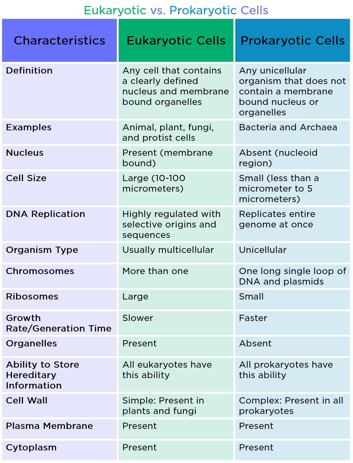
→ Types of cells
• There are two types of cells:
(i) Prokaryotes
(ii) Eukaryotes
multicellular organisms
- Books Name
- Science Made Easy Science Book
- Publication
- Science Made Easy
- Course
- CBSE Class 9
- Subject
- Science
Multicellular Organism: Many cells grouped together to perform different function in the body
and also form various body parts. Example: fungi, plants, animals.
cell membrane and cell wall
- Books Name
- Science Made Easy Science Book
- Publication
- Science Made Easy
- Course
- CBSE Class 9
- Subject
- Science
Plasma membrane or Cell membrane
• This is the outermost covering of the cell that separates the contents of the cell from its external
environment.
• The plasma membrane allows or permits the entry and exit of some materials in and out of the
cell.
• It also prevents movement of some other materials. The cell membrane is called selectively
permeable membrane.
• It is made up of lipid and protein.
→ Properties of Plasma membrane
• It is flexible (made up of organic molecules called lipids and proteins).
• Its flexibility enables cell to engulf in food and other from the external environment. This process
called endocytosis. Amoeba acquire food through this process.
→ Functions of Plasma membrane
• It permits the entry and exit of some materials in and out of the cell.
• It prevents movement of some other materials not required for the cell as it acts like selectively
permeable membrane.
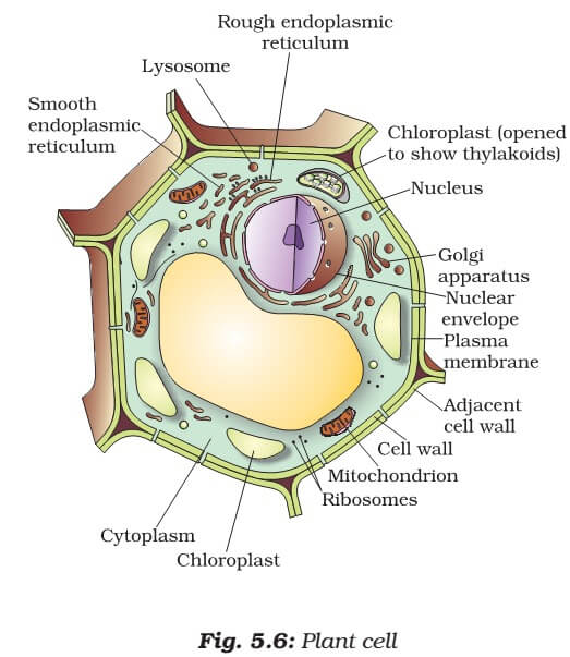
Cell Wall
• Cell wall is another rigid outer covering in addition to the plasma membrane found in plant cell.
The cell wall lies outside the plasma membrane.
• The plant cell wall is mainly composed of cellulose. Cellulose is a complex substance which
provides structural strength to plants.
→ Function of Cell Wall
• Cell walls permit the cells of plants, fungi and bacteria to withstand very dilute (hypotonic) external
media without bursting.
• In such media the cells tend to take up water by osmosis. The cell swells, building up pressure
against the cell wall. The wall exerts an equal pressure against the swollen cell.
• Because of cell wall, cells can withstand much greater changes in the surrounding medium than
animal cells.
Plasmolysis
• When a living plant cell loses water through osmosis there is shrinkage or contraction of the
contents of the cell away from the cell wall. This phenomenon is known as plasmolysis.
cell organelles
- Books Name
- Science Made Easy Science Book
- Publication
- Science Made Easy
- Course
- CBSE Class 9
- Subject
- Science
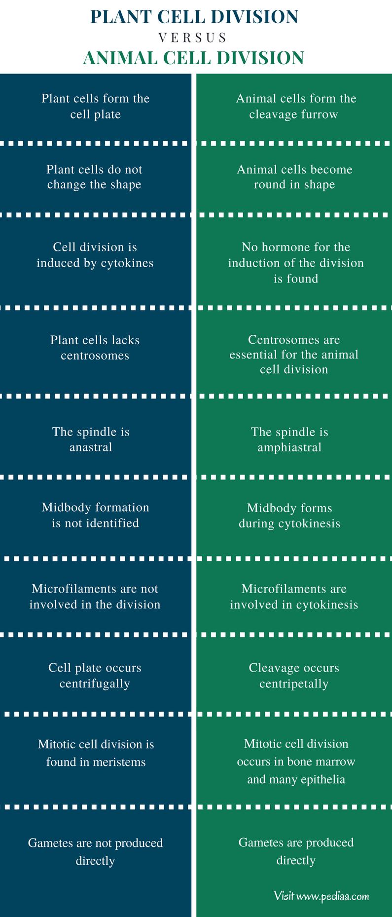
Difference between Animal cell and Plant cell

chloroplast, mitochondria, vacuoles, endoplasmic reticulum, Golgi apparatus
- Books Name
- Science Made Easy Science Book
- Publication
- Science Made Easy
- Course
- CBSE Class 9
- Subject
- Science
Cytoplasm
• The cytoplasm is the fluid content inside the plasma membrane.
• It also contains many specialised cell organelles. Each of these organelles performs a specific
function for the cell.
→ Function of Cytoplasm
• It helps in exchange of material between cell organelles.
• It act as store of vital chemicals such as amino acid, glucose, vitamins and iron etc.
• It is the site of certain metabolic pathways such as glycolysis.
Endoplasmic Reticulum (ER)
• The endoplasmic reticulum (ER) is a large network of membrane-bound tubes and sheets.
• It looks like long tubules or round or oblong bags (vesicles).
• The ER membrane is similar in structure to the plasma membrane. It is also made up of lipid and
proteins.
→ Types of Endoplasmic Reticulum
(i) Rough endoplasmic reticulum (RER)
(ii) Smooth endoplasmic reticulum (SER)
→ Functions of Endoplasmic Reticulum
• RER looks rough under a microscope because it has particles called ribosomes attached to its
surface. The ribosomes, which are present in all active cells, are the sites of protein manufacture.
The manufactured proteins are then sent to various places in the cell depending on need, using the
ER.
• The SER helps in the manufacture of fat molecules, or lipids, important for cell function.
• Some of these proteins and lipids help in building the cell membrane. This process is known as
membrane biogenesis.
• Some other proteins and lipids function as enzymes and hormones.
• Although the ER varies greatly in appearance in different cells, it always forms a network system.
• One function of the ER is to serve as channels for the transport of materials (especially proteins)
between various regions of the cytoplasm or between the cytoplasm and the nucleus.
• The ER also functions as a cytoplasmic framework providing a surface for some of the biochemical
activities of the cell.
• In the liver cells of the group of animals called vertebrates, SER plays a crucial role in detoxifying
many poisons and drugs.
Golgi Apparatus
• The Golgi apparatus consists of a system of membrane-bound vesicles arranged approximately
parallel to each other in stacks called cisterns.
• These membranes often have connections with the membranes of ER and therefore constitute
another portion of a complex cellular membrane system.
→ Function of Golgi Body
• The material synthesised near the ER is packaged and dispatched to various targets inside and
outside the cell through the Golgi apparatus.
• Its functions include the storage, modification and packaging of products in vesicles. In some
cases, complex sugars may be made from simple sugars in the Golgi apparatus.
• The Golgi apparatus is also involved in the formation of lysosomes.
Lysosomes
• Lysosomes are a kind of waste disposal system of the cell.
• It helps to keep the cell clean by digesting any foreign material as well as worn-out cell organelle
• Lysosomes have membrane-bounded structure whose sacs are filled with digestive enzymes.
→ Functions of Lysosomes
• Lysosomes break foreign materials entering the cell, such as bacteria or food as well as old
organelles into small pieces.
• They contain powerful digestive enzymes which are made in RER which is capable of breaking
down all organic material made in RER.
• During the disturbance in cellular metabolism such as when the cell gets damaged, lysosomes
may burst and the enzymes digest their own cell. Therefore, lysosomes are also known as the
‘suicide bags’ of a cell.
Mitochondria
• Mitochondria are known as the powerhouses of the cell.
→ Structure of mitochondria
• Mitochondria have two membrane coverings.
• The outer membrane is very porous while the inner membrane is deeply folded.
• These folds create a large surface area for ATP-generating chemical reactions.
→ Functions of mitochondria
• The energy required for various chemical activities needed for life is released by mitochondria in
the form of ATP (Adenosine triphopshate) molecules.
• ATP is known as the energy currency of the cell. The body uses energy stored in ATP for making new
chemical compounds and for mechanical work.
• Mitochondria have their own DNA and ribosomes. Therefore, mitochondria are able to make some
of their own proteins.
Plastids
• Plastids are present only in plant cells.
• There are three types of plastids:
(i) Chromoplasts (coloured plastids).
(ii) Leucoplasts (white or colourless plastids).
(iii) Chloroplasts (contains the pigment chlorophyll).
→ Structure of Plastids
• The internal organisation of the plastids consists of numerous membrane layers embedded in a
material called the stroma.
• Plastids also have their own DNA and ribosomes like mitochondria and similar to its structure.
→ Function of Plastids
• Chloroplasts are important for photosynthesis in plants.
• Chloroplasts also contain various yellow or orange pigments in addition to chlorophyll.
• Leucoplasts are primarily organelles in which materials such as starch, oils and protein granules
are stored.
Vacuoles
• Vacuoles are storage sacs for solid or liquid contents.
• They are small sized in animal cells while plant cells have very large vacuoles.
→ Function of vacuoles
• The central vacuole of some plant cells may occupy 50-90% of the cell volume.
• In plant cells vacuoles are full of cell sap and provide turgidity and rigidity to the cell.
• Many important substance in the life of the plant cell are stored in vacuoles which include amino
acids, sugars, various organic acids and some proteins.
• In single-celled organisms like Amoeba, the food vacuole contains the food items that the Amoeba
has consumed.
• In some unicellular organisms, specialised vacuoles also play important roles in expelling excess
nucleus, chromosomes - basic structure, number
- Books Name
- Science Made Easy Science Book
- Publication
- Science Made Easy
- Course
- CBSE Class 9
- Subject
- Science
Nucleus

• It is called the brain of the cell as it controls all the activities of cell.
→ Composition of Nucleus
• The nucleus has a double layered covering called nuclear membrane.
• The nuclear membrane has pores which allow the transfer of material from inside the nucleus to
the cytoplasm.
• The nucleus contains chromosomes, which are visible as rod-shaped structures only when the cell
is about to divide.
→ Functions of chromosomes
• Chromosomes contain information for inheritance of features from parents to next generation in
the form of DNA (Deoxyribo Nucleic Acid) molecules. Chromosomes are composed of DNA and
protein.
• DNA molecules contain the information necessary for constructing and organising cells.
• Functional segments of DNA are called genes.
• In non-dividing cell, this DNA is present as part of chromatin material.
• Chromatin material is visible as entangled mass of thread like structures. Whenever the cell is
about to divide, the chromatin material gets organised into chromosomes and perform cell division
→ Functions of Nucleus
• The nucleus plays a central role in cellular reproduction. It is the process by which a single cell
divides and forms two new cells.
• It also plays a crucial part, along with the environment, in determining the way the cell will develop
and what form it will exhibit at maturity, by directing the chemical activities of the cell.
Nucleoid
• In some organisms like bacteria, the nuclear region of the cell may be poorly defined due to the
absence of a nuclear membrane.
• Such an undefined nuclear region containing only nucleic acids is called a nucleoid.
basic issues in scientific naming, basis of classification
- Books Name
- Science Made Easy Science Book
- Publication
- Science Made Easy
- Course
- CBSE Class 9
- Subject
- Science
Classification
→ The method of arranging organisms into groups or sets on the basis of similarities anddifferences is called classification.
→ It makes the study of wide variety of organisms easy and in systematic manner.
→ It helps to understand how the different organisms have evolved with
time.
→ It helps to understand the inter-relationships among different groups of organisms.
→ It forms a base for the study of other biological sciences, like biogeography.
Basis of Classification
→ There are certain features or properties used for the classification of living organisms which areknown as characteristics.
→ Organisms with same characteristics are placed in same groups.
Classification System
• Two kingdom classification:
Carolus Linnaeus in 1758 classified the living organisms into twogroups as plants and animals.
• Five kingdom classification:
H. Whittaker in 1959 further classified the organisms into fivekingdoms as Kingdom Monera, Kingdom Protista, Kingdom Fungi, Kingdom Plantae and Kingdom Animalia.
→ Carl Woese in 1977 further divided Kingdom Monera into archaebacteria (or Archae) andEubacteria (or Bacteria).
Hierarchy of categories / groups
- Books Name
- Science Made Easy Science Book
- Publication
- Science Made Easy
- Course
- CBSE Class 9
- Subject
- Science
• Hierarchy of Classification:
Linnaeus proposed a classification system by arranging organismsinto taxonomic groups at different levels according to the characteristics they have.
Groups or Levels from top to bottom
→ The major characteristics considered for classifying all organisms into five major kingdoms.
• Type of cellular organization
(i) Prokaryotic cells: These are primitive and incomplete cells without well defined nucleus.
(ii) Eukaryotic cells: These are advanced and complete cells with well-defined nucleus.
• Body organization
(i) Unicellular organisms: These are organisms made up of single cell with all activities performed the single cell.
(ii) Multicellular organisms: These are organisms made up of large number of cells with differentfunctions performed by different cells.
• Mode of obtaining food
(i) Autotrophs: These are the organisms that make their own food by photosynthesis.
(ii) Heterotrophs: These are the organisms which depend on other organisms for food.
Five Kingdom Classification
R. H. Whittaker taxonomist was the first one to propose five kingdom classification.
Monera
(i) Type: Unicellular Prokaryotic
(ii) Mode of nutrition: Autotrophic or heterotrophic
(iii) Body: Lack well-defined nucleus and cell organelles
(iv) Examples: Bacteria, Blue-green algae
Protista
(i) Type: Unicellular Eukaryotic
(ii) Mode of nutrition: Autotrophic or Heterotrophic
(iii) Body: Some organisms use pseudopodia or cilia or flagella for movement
(iv) Examples: Amoeba, Paramecium, Euglena
Fungi
(i) Type: Multicellular Non-green Eukaryotic
(ii) Mode of nutrition: Saprophytic or Parasitic Sometimes symbiotic
(iii) Body: Fungus is made up of long filaments called hyphae. The network of hyphae is mycelium.
(iv) Examples: Yeast, Rhiozpus, Mushrooms moulds
Plantae
(i) Type: Multicellular Eukaryotic
(ii) Mode of nutrition: Autotrophic
(iii) Body: Exhibits high level of tissue differentiation and have specialized body organs.
(iv) Examples: Trees, Plants, Shrubs
Animalia
(i) Type: Multicellular Eukaryotic
(ii) Mode of nutrition: Heterotrophic
(iii) Body: Exhibits high level of tissue differentiation and have specialized body organs. They havewell developed nervous system.
(iv) Examples: Fish, Insects, Animals, Humans, Birds
Kingdom I: Monera
(i) Prokaryotic, unicellular.
(ii) Can be autotrophs or heterotrophs.
(iii) May or may not have cell wall.
(iv) Examples: Anabaena and Bacteria (heterotrophic), Cyano-bacteria or Blue-green algae(autotrophic).
Kingdom II: Protista
(i) Eukaryotic, unicellular.
(ii) Can be autotrophic or heterotrophic.
(iii) May have cilia, flagella or pseudophodia for locomotion.
(iv) Examples: Plants like unicellular algae, diatoms; animals like protozoans (Amoeba, Paramecium,Euglena); fungi like slime molds and water molds.
Kingdom III: Fungi
(i) Eukaryotic.
(ii) Mostly multicellular but sometimes unicellular (yeast).
(iii) Source of food:
• Mostly saprophytes:
These organisms use decaying material for food.
• Some parasitic:
These organisms live inside body of other living organism to have food and canbe disease causing.
• Symbiotic relation:
These are relations between two organisms in which they live together forbenefit of one or both.
→ Lichens are a symbiotic relation between fungi and cyanobacteria.
→ Here fungi gets food from cyanobacteria and in return cyanobacteria gets water and protectiofrom sunlight through fungi.
(iv) Cell wall is made of chitin.
(v) Examples: Mushrooms (Agaricus), green mold (Penicillium), smut (Aspergilus).
Kingdom IV: Plantae
(i) Eukaryotic, multicellular.
(ii) Autotrophs.
(iii) Cell wall present.
Major groups of plants (salient features) (Bacteria, Thalophyta, Bryo phyta, Pteridophyta, gymnosperms and Angiosperms).
- Books Name
- Science Made Easy Science Book
- Publication
- Science Made Easy
- Course
- CBSE Class 9
- Subject
- Science
Basis of division in Kingdom Plantae
(i) Differentiated body parts:
Body is differentiated into leaves, stems, roots, flowers, etc.
(ii) Presence of vascular tissue:
There are two types of vascular tissues present in the plants.
• Xylem: Helps in transport of water.
• Phloem: Helps in transport of food.
(iii) Reproduction through seeds or spores:
• Phanerogamae : Plants with seeds are called phanerogamae. They contains embryo with stored food and are multicellular.
• Cryotogamae: Plants with spores are called cryptogamae. They contains only naked embryo and are generally unicellular.
(iv) Seeds are inside the fruit or naked:
• Angiospermae: These are plants with seeds inside the fruit and bears flowers.
• Gymnospermae: These are plants with naked seeds and do not bear flowers.
Division 1: Thallophyta
(i) Basic and elementary plants with undifferentiated body parts.
(ii) Generally called algae.
(iii) No vascular tissue present.
(iv) Reproduce through spores.
(v) Mainly found in water.
(vi) Example: Ulva, Spirogyra, Ulothrix, Cladophora, Chara.
Division 2: Bryophyte
(i) Body structure differentiated but not fully developed.
(ii) No vascular tissues present.
(iii) Reproduce through spores.
(iv) Found on both land and water therefore known as ‘ Amphibians of Plantae Kingdom’.
(v) Example : Liverwort (Marchantia, Riccia), Mosses (Funaria), Hornwort (Dendrocerous).
Division 3: Pteridophyta
(i) Differentiated body structure – leaves, stems, roots, etc.
(ii) Vascular tissues present.
(iii) Reproduce through spores.
(iv) Examples : Marsilea, fern, horsetails.
Division 4: Gymnosperms
(i) Differentiated body parts.
(ii) Vascular tissues.
(iii) Naked seeds without fruits or flowers.
(iv) Perennial, evergreen and woody.
(v) Examples : Pines (deodar), Cycus, Ginkgo.
Division 5: Angiosperms
(i) Also known as flower-bearing plants.
(ii) Later on flower becomes fruit.
(iii) Seeds are inside the fruit.
(iv) Embryos in seeds have structure called cotyledons. They are also called seed leaves becausemany plants they emerge and become green when they germinate.
Angiosperms are further divided on the basis of number of cotyledons into two parts i.e. Monocotsand Dicots.
Major groups of animals (salient features) (Non-chordates upto phyla and chordates upto classes).
- Books Name
- Science Made Easy Science Book
- Publication
- Science Made Easy
- Course
- CBSE Class 9
- Subject
- Science
Kingdom V: Animalia
Basis of classification of Animalia kingdom:
• Symmetry
(i) Bilateral symmetry:
It is when an organism can be divided into right and left halves, identical bumirror images, by a single vertical plane.
(ii) Radial symmetry:
It is when an organism is equally spaced around a central point, like spokes oa bicycle wheel.
• Germ layers
→ In embryonic stages there are different layers of cells called germ cells.
• The three different types of germ cells are:
(i) Ectoderm:
It is the outermost layer which forms nail, hair, epidermis, etc.
(ii) Endoderm:
It is the innermost layer which forms stomach, colon, urinary, bladder, etc.
(iii) Mesoderm:
It is the middle layer between ectoderm and endoderm which forms bones,cartilage, etc.
• According to the number of germ layers present in embryonic stage, animal could be:
(i) Diploblastic:
Organisms which are derived from two embryonic germ layers (ecto and endo).
(ii) Triploblastic:
Organisms which are derived from all the three embryonic germ layers.
• Coelom
→ Body cavity or coelom is important for proper functioning of various organs.
→ For example, heart which has to contract and expand needs some cavity or empty space, which is provided by the coelom.
• On the basis of presence or absence of coelom, organisms are divided into:
(i) Acoelomates: These are the simple organisms having no body cavity.
(ii) Coelomates:
These are complex organisms having true cavity lined by mesoderm from all side
→ These are further sub-divided into schizocoelomates or protostomes (coelom formed due to splitting or mesoderm) and enterocoelomates or dueterostomes (coelom formed from pouchespinched off from endoderm).
(iii) Pseudo coelamate:
These are organisms having false coelom. They have pouches of mesoder scattered between endoderm and ectoderm.
• Notochord
→ It is a long rod like structure, which runs along the body between nervous tissues and gut and provides place muscle to attach for ease of movement.
→ Organisms could be:
• without notochord
• with notochord
• with notochord in initial embryonic stages and vertebral column in adult phase.
Phylum 1: Porifera or Sponges
(i) Cellular level of organization
(ii) Non-motile animals
(iii) Holes on body which led to a canal system for circulation of water and food
(iv) Hard outside layer called as skeletons
(v) Examples: Sycon, spongilla, euplectelia
Phylum 2: Coelenterata
(i) Tissue level of organization
(ii) No coelom
(iii) Radial symmetry, diploblastic
(iv) Hollow gut
(v) Can move from one place to another
(vi) Examples: Hydra, sea anemone, jelly fish (solitary), corals (colonies)
Phylum 3: Platyhelminthes
(i) Also called flat worms
(ii) No coelom present
(iii) Bilateral symmetry, triploblastic
(iv) Free living or parasite
(v) Digestive cavity has one opening for both ingestion and egestion
(vi) Examples: Planaria (free living), liver fluke (parasitic)
Phylum 4: Mollusca
(i) Coelom present
(ii) Triploblastic, bilateral symmetry
(iii) Soft bodies sometimes covered with shell
(iv) Generally not segmented
(v) No appendages present
(vi) Muscular foot for movement
(vii) Shell is present
(viii) Kidney like organ for excretion
(ix) Examples: Chiton, octopus, pila, unio
Phylum 5: Annelida
(i) Second largest phylum
(ii) Coelom present
(iii) Bilateral, triploblastic
(iv) Segmented (segments specialized for different functions)
(v) Water or land
(vi) Extensive organ differentiation
(vii) Examples: Earthworm, leech, nereis
Phylum 6: Arthropoda
(i) Largest phylum (consist of 80% of species)
(ii) Generally known as insects
(iii) Coelom present
(iv) Bilateral, triploblastic
(v) Segmented, sometimes fused
(vi) Tough exo-skeleton of chitin
(vii) Joing appendages like feet, antenna
(viii) Examples : Prawn, scorpio, cockroach, housefly, butterfly, spider
Phylum 7: Echinodermata
(i) Spiny skin, marine
(ii) No notochord
(iii) Coelom present, bilateral symmetry, triploblastic
(iv) Endoskeleton of calcium carbonate
(v) Water vascular system for locomotion
(vi) Bilateral symmetry before birth and radial symmetry after birth
(vii) Examples : Antedon, sea cucumber, star fish, echinus
Phylum 8: Protochordata
(i) Marine animals.
(ii) Bilaterally symmetrical, triploblastic and have a coelom.
(iii) Gills present at some phase of life
(iv) Notochord is present which is a long rod-like support structure (chord=string) that runs along back of the animal separating the nervous tissue from the gut.
(v) Notochord provides a place for muscles to attach for ease of movement.
(vi) Examples : Balanoglossus, Herdmania and Amphioxus
Phylum 9: Nematoda
(i) Bilaterally symmetrical and triploblastic.
(ii) Body is cylindrical rather than flattened.
(iii) Tissues, but no real organs.
(iv) Sort of body cavity or a pseudocoelom, is present.
(v) Familiar as parasitic worms causing diseases.
(vi) Worms causing elephantiasis (filarial worms) or the worms in the intestines (roundworm orpinworms).
(vii) Examples: Ascaris, Wuchereria
Phylum 10: Vertebrata
(i) Notochord converted to vertebral column
(ii) 2, 3, 4 chambered heart
(iii) Organs like kidney for excretion
(iv) Pair appendages
(v) Examples: Humans (4-chambered), frog (3-chambered), fishes (2-chambered)
→ Vertebrates are divided into five classes namely Pisces, Amphibia, Reptilia, Aves and Mammalia
• Warm blooded organisms:
These are organisms which maintain same body temperatureirrespective of outside temperature.
Example: Humans. Human’s body temperature is approximately 37º.
• Cold blooded organisms:
These are organisms which change their body temperature as persurrounding temperature.
Example : Frog.
Pisces (Fishes)
→ They are fishes living in water.
→ Their skin is covered with scales or plates.
→ They respire using gills.
→ They have streamlined body and fins which help them to move in water.
→ They are cold blooded and their heart has only two chambers.
→ They lay eggs from which the young ones hatch out.
• Fishes are divided into two categories on the basis of skeleton:
(i) Fishes with cartilage skeleton called cartilaginous fishes. Example : Shark, Rays etc.
(ii) Fishes with bony skeleton called bony fishes. Example : Tuna, Rohu etc.
Amphibia (Amphibians)
→ They are found in land and water.
→ They do not have scales but have mucous glands on their skin.
→ They are cold blooded and the heart is three chambered.
→ Respiration is through gills or lungs. They lay eggs in water.
Example: Frogs, Toads, Salamanders etc.
Reptilia (Reptiles)
→ They have scales and breathe through lungs.
→ They are cold blooded.
→ Most of them have three chambered heart but crocodiles have four chambered heart.
→ They lay eggs with hard covering in water.
→ Example: Snakes, Turtles, Lizards, Crocodiles etc.
Aves (Birds)
→ They are warm blooded animals.
→ They have four chambered heart.
→ They breathe through lungs.
→ They have an outer covering of feathers.
→ Their two fore limbs are modified into wings for flying. They lay eggs.
→ Example: Crow, Sparrow, Pigeon, Duck, Stork, Ostrich etc.
Mammalia (Mammals)
→ They are warm blooded animals.
→ They have four chambered heart.
→ They have mammary glands for production of milk to nourish their young ones.
→ The skin has hairs and sweat glands. Most of them give birth to their young ones.
→ Some of them lay eggs (like Platypus and Echidna).
→ Example: Cat, Rat, Dog, Lion, Tiger, Whale, Bat, Humans etc.
Nomenclature
→ An organism can have different names in different languages. This creates confusion in naming
organism.
→ A scientific name is needed which is same in all languages.
→ Binomial nomenclature system given by Carolus Linnaeus is used naming different organisms.
Some conventions in writing the scientific names:
(i) Genus should be written followed by the species.
(ii) First letter of the genus should be capital and that of the species should be in small letter.
(iii) When printed the name should be written in italics and when written with hands genus and
species should underlined separately.
Example : Homo sapiens for humans, Panthera tigris for tiger.
Infectious and Non-infectious diseases, their causes and manifestation
- Books Name
- Science Made Easy Science Book
- Publication
- Science Made Easy
- Course
- CBSE Class 9
- Subject
- Science
Infectious and Non-infectious Diseases
(i) Infectious Diseases: The diseases which spread due to infection by micro-organisms are called
infectious diseases.
→ It is communicated from diseased person to healthy person, caused by some biological
agents/pathogens like viruses, bacteria, fungi, protozoans, fungi worms.
(ii) Non-infectious Diseases: The disease which does not spread by contact between infected and
healthy person through air and water, is called non-infectious disease.
Example: Arthritis, heart disease.
Different Micro-organisms
SARS Viruses
→ SARS viruses are coming out of the surface of an infected cell (see the arrows for example).
→ 500 nanometer = 0.5 micrometer = 0.001 millimeter.
Trypanosoma
→ Trypanosoma is a protozoan organism.
→ It causes sleeping sickness.
→ The saucer-shaped substance lying next tothe protozoa, is a red blood cell.
Staphylococcus bacteria
→ The Staphylococcus bacteria causes acne.
→ The scale is indicated at the line at the top left of the picture. It is 5 micrometers long.
Adult roundworm
→ Adult roundworm is found in the small intestine.
→ Its technical name is Ascaris Lumbricoides.
→ The ruler next to it shows 4 centimeter to give an idea of the scale.
Leishmania
→ Leishmania, the protozoan organism causes kala-azar.
→ The organisms are oval-shaped, and each has one long whip-like structure.
→ The immune cell is about ten micrometres in diameter.
Antibiotics
→ Antibiotics blocks biochemical pathways important for bacteria. Hence, they are effective against
them. Example: Penicillin, tetracycline.
→ Many bacteria make a cell wall to protect themselves, the antibiotics (Penicillin) blocks the
bacterial process that builds cell wall.
→ Antibiotics works only against the bacteria and not against the viruses.
Means of Spread of Infectious Diseases
→ Infectious diseases spread from an infected person to a healthy person through air, water, food,
vectors, physical contact and sexual contact.
• Through air: By sneezing and coughing, the microbes spread into air and enter into the body of
healthy person, like common cold, tuberculosis, pneumonia etc.
• Through water : The microbes enter into our body by drinking/eating polluted and contaminate
water/food, like cholera, amoebic dysentery etc.
• Vectors: Some organisms like female anopheles mosquito also work as a vector of disease, like
malaria, dengue, yellow fever etc.
• Through sexual contact: Syphilus, AIDS spread by sexual contact with infected person. AIDS virus
can also spread through blood transfusion and from the mother to her child during pregnancy an
through breast feeding.
Diseases caused by microbes (Virus, Bacteria and protozoans) and their prevention
- Books Name
- Science Made Easy Science Book
- Publication
- Science Made Easy
- Course
- CBSE Class 9
- Subject
- Science
Principles of treatment and prevention
- Books Name
- Science Made Easy Science Book
- Publication
- Science Made Easy
- Course
- CBSE Class 9
- Subject
- Science
Principles of Treatment
→ The treatment of infectious diseases consists of two steps. They are to reduce the effects of the
disease (symptoms) and to kill the microbes which caused disease.
(i) To reduce the effects of the disease: This can be done by taking medicines to bring down the
effects of the disease like fever, pain or loose motions etc. and by taking bed rest to conserve our
energy.
(ii) To kill the microbes: This can be done by taking suitable antibiotics and drugs which kills the
microbes and the disease is cured.
Principles of Prevention
• There are two ways of prevention of infectious diseases. They are general ways and specific way
(i) General ways of prevention: Public hygiene is most important for prevention of infectious
diseases. Proper and sufficient food for everyone will make people healthy to resist the infection.
→ Air borne diseases can be prevented by living in conditions that are not crowded. Water borne
diseases can be prevented by providing safe drinking water.
→ Vector borne diseases can be prevented by providing clean environment.
(ii) Specific ways of prevention: There are disease specific measures which are used to fight them
It is done by Immunisation.
→ This is the process of introducing a weakened pathogen inside the body of the host to fool his/h
immune system to produce antibodies against that particular disease.
→ Not only does our immune system fight the disease (feeble pathogen), but also keeps a memo
of the incident by keeping those antibodies in blood.
→ Thus, next time even if the disease will strike the host’s body with full vigor, the body will be able
protect itself with the help of these antibodies.
→ This is also the basic law followed by vaccination programmes done for infants.

Pulse Polio programmes.
- Books Name
- Science Made Easy Science Book
- Publication
- Science Made Easy
- Course
- CBSE Class 9
- Subject
- Science
Pulse polio programme-
Pulse polio immunization programme forms the largest single day public health project. Pulse means a dose of a substance (here polio vaccine) especially when applied for a short period of time. It was conducted for the first time in 1995. The program uses oral polio vaccine or OPV. As per the national immunization Schedule (NIS), a dose of 3 drops is given orally to the child, i.e. one dose each at 1.5, 2.5 and 3.5 months age. Finally a booster dose si given at the age of 1.5 years. After oral administration. Virus particles in the vaccine begin to live in the intestine of the human body and multiply. It leads to production of protective molecules in the intestine and the blood.
