 ACME SMART PUBLICATION
Human Neural System
- Books Name
- ACME SMART COACHING Biology Book
- Publication
- ACME SMART PUBLICATION
- Course
- CBSE Class 11
- Subject
- Biology
Human Neural System
The neural system of all animals is composed of highly specialised cells called neurons, which can detect, receive and transmit different kinds of stimuli.
The neural organisation is very simple in lower invertebrates. For example, in Hydra it is composed of network of neurons.
Neural system is better organised in insects, where a brain is present along with number of ganglia and neural tissues.
The vertebrates have a more developed neural system.
Human Neural System Human neural system is divided into two parts:
(i) The Central Nervous System (CNS)
(ii) The Peripheral Nervous System (PNS)
The CNS includes the brain and the spinal cord and is the site of information processing and control.
The PNS comprises of all the nerves of the body associated with CNS (brain and spinal cord).
The nerve fibres of the PNS are of two types
(a) Afferent Fibres (b) Efferent Fibres
Afferent fibres transmit impulses from tissues/organs to the CNS and the efferent fibres transmit regulatory impulses from the CNS to concerned peripheral tissues/organs.
PNS is divided into two divisions called somatic neural system and autonomic nervous system.
Somatic neural system relays impulses from the CNS to skeletal muscles while the autonomic neural system transmits impulses from CNS to the involuntary organs and smooth muscles of the body.
The autonomic neural system is further classified into sympathetic neural system and parasympathetic neural system.
structure of neuron
- Books Name
- ACME SMART COACHING Biology Book
- Publication
- ACME SMART PUBLICATION
- Course
- CBSE Class 11
- Subject
- Biology
A neuron is a microscopic structure composed of three major parts : (i) cell body , (ii) dendrites and (iii) axon . Thell contains cytoplasm , cell organelles and certain granular bodies called Nissl's granules . Dendrites are shon branch repeatedly and project out of the cell body . They also contain Nissl's granules .
Dendrites transmit impulses towards the cell body . The axon is a long fibre , the distal end of which is bra branch terminates into a bulb - like structure called synaptic knob . Synaptic knob possess synaptic vesicle con chemicals called neurotransmitters . The axons transmit nerve impulses away from the cell body to a synapse ar neuromuscular junction.
Types of Neurons
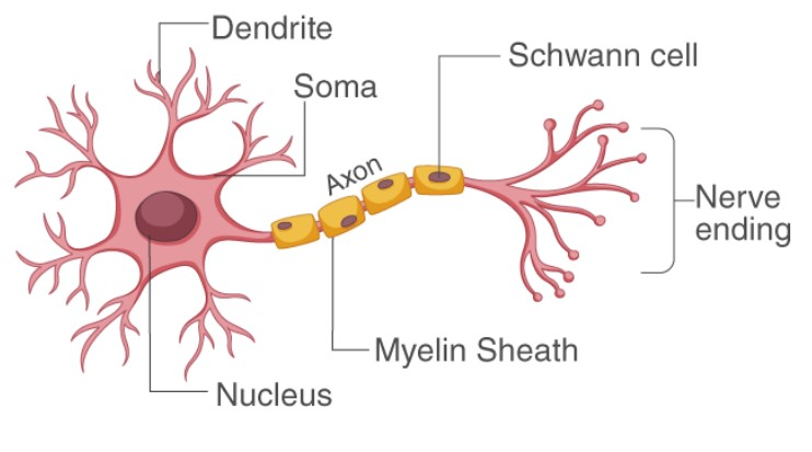
Based on the number of axon and dendrites , the neurons are divided into three types :
Unipolar neurons have cell body with one axon only . They are found usually in the embryonic stage.
Bipolar neurons are with one axon and one dendrite . They are found in the retina of eye .
Multipolar neurons are with one axon and two or more dendrites . These are found in the cerebral cortex
On the basis of function , neurons are of two types :
Afferent neurons : These neurons carry impulses from receptors CNS . They are sensory in nature . The terminals of dendrites become modified to form receptors .
Efferent neurons : They carry impulses from CNS to effectors like muscles or glands . They are motor in nature . The axon terminals come in contact with the motor end plate to form neuromuscular junction .
Types of Axons
There are two types of axons : myelinated and non - myelinated .
The myelinated nerve fibres are enveloped with Schwann cells , which form a myelin sheath around the axon . The gaps between two adjacent myelin sheaths are called nodes of Ranvier . Myelinated
nerve fibres are found in spinal and cranial nerves .
Unmyelinated nerve fibre is enclosed by a Schwann cell that does form a myelin sheath around the axon . It is commonly found autonomous and the somatic neural systems .
generation and conduction of impulse
- Books Name
- ACME SMART COACHING Biology Book
- Publication
- ACME SMART PUBLICATION
- Course
- CBSE Class 11
- Subject
- Biology
Generation and conduction of impulse
Nerve cell have polarised membrane i.e., have electrical potential difference or membrane potential.
This is because of a variety of ion channels (pores formed by proteins) specific for a particular type of ions.
Some remain open while most ion channels open under one condition, but closes under other condition.
Because of such regulated or voltage gated channel, membrane become excitable as these channels respond to different type of stimuli e.g., Light, touch, sound etc.
When a neuron is not sending any signal, it is said to be at rest and its membrane has resting membrane potential.
Resting Membrane Potential
In the resting nerve fibre, the cytoplasm just beneath its membrane is electronegative relative to the layer of extracellular fluid (ECF) just outside the membrane.
If the two sides of the membrane are connected by galvanometer (double beam cathode ray oscilloscope) the inner side is seen to possess a negative potential of about 70 mV relative to the outerside.
This is called the resting membrane potential. This results from two factors:
(i) The resting membrane has a poor permeability for Na+ although it has a higher permeability for K+. Therefore, K+ can cross more easily while Cl– and Na+ have more difficulty in crossing.
(ii) Negatively charged protein molecule inside the neuron cannot cross the plasma membrane because of its semipermeability.
The differential flow of the positively charged ions and the fact that the negatively charged organic ions within the nerve fibre cannot pass out cause an increasing positive charge on the outside of the membrane and negative charge on the inside of the membrane.
This makes the membrane of the resting nerve fibre polarized (i.e., its outside being positively charged with respect to the inside.)
Such electrochemical gradients are maintained by the active transport of ions involving Na+ – K+ ion transmembrane pump.
It pumps out 3Na+ for every 2K+ ions passed inwardly.
K+ concentration is 30 times more inside neuron than outside and Na+ concentration is 10 times more in interstitial fluid as compared to inside of neuron.
Conduction of Nerve Impulse
It involves initiation of impulse followed by conduction along the axon so as to be transferred to target muscle/tissue.
Initiation of Impulse:
When stimulated, voltage gated Na+ channel open which causes a rapid, very localised, temporary inflow of Na+ into the cell which causes development of net positive charge on the inner side of membrane in that area.
This is called depolarisation.
It occurs at a particular region of neuron called trigger zone.
Voltage gated ion channels are clustered in the area of trigger zone.
Stimulus of threshold value causes stoppage of Na+ – K+ ATP-ase pump.
Continued passage of Na+ ions into inside of neuron creates a reverse potential of +20 mV to +30 mV, rarely to +60 mV.
The total change occurs in spike-like fashion which is also called spike potential.
Na+ ion channels open for about 0.5 m sec.
It creates a potential that sets in a wave of depolarisation through the nerve fibre.
The membrane potential which sets in a wave of depolarisation is called action potential.
For most excitable cells, the threshold is about -55 mV to -60 mV.
Conduction of Impulse:
In the area of depolarisation, the potential difference across the membrane is small while its nearby region has large difference in membrane potential.
This produces a small local current in the area.
The local current becomes a stimulus and causes the gated Na+ channels of next region to open and depolarise the area to produce fresh action potential.
The process will continue till the impulse reaches the end of neuron.
Repolarisation:
As the Na+ channels close after 0.5 m sec the membrane becomes extra permeable to K+ ions due to opening of K+ ion gates.
With the pumping out of K+ ions, the neuron interior becomes negative and the potential falls back to resting potential.
The phenomenon of change of membrane potential from excited state to resting state is called repolarisation.
However, K+ ion channels remain open for a bit longer period so that the membrane potential becomes more negative than -70 mV.
It is called hyperpolarisation.
It takes about 1 -5 m sec for repolarisation.
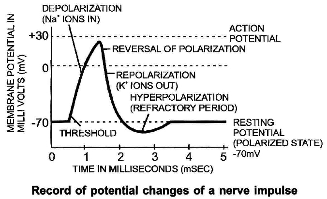
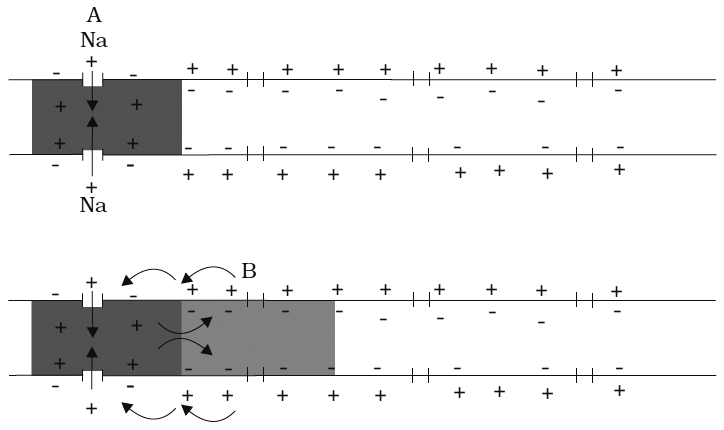
Figure: Diagrammatic representation of impulse conduction through an axon (at points A and B)
transmission of impulse
- Books Name
- ACME SMART COACHING Biology Book
- Publication
- ACME SMART PUBLICATION
- Course
- CBSE Class 11
- Subject
- Biology
Transmission of impulse
Synapes are the neuroneuronal junctions through which information from one neuron can pass to the other.
There are mainly two types of synapses depending upon the nature of transfer of information across the synapse. (1) Electrical, (2) Chemical
1.Electrical Synapses:
At an electrical synapse, ionic current spreads directly from one cell to another through gap junctions.
Each gap junction contains is hundred or so tubular protein structures called connexons that from tunnel to connect the cytosol of the two cells.
This provides a path for ionic current flow.
Gap junctions are common in visceral (single-unit) smooth muscle, cardiac muscle, and a developing embryo.
They also occur in the CNS.
Electrical synapses have three obvious advantages:
(i) They allow faster communication than do chemical synapses, since impulses conduct across gap junction.
(ii) They can synchronize the activity of a group of neurons or muscle fibers. The value of synchronized action potentials in the heart or in visceral smooth muscles is to achieve coordinated contraction of these fibers.
(iii) They may allow two-way transmission of impulses in contrast to chemical synapses, which function as one way points of communication.
2. Chemical Synapses:
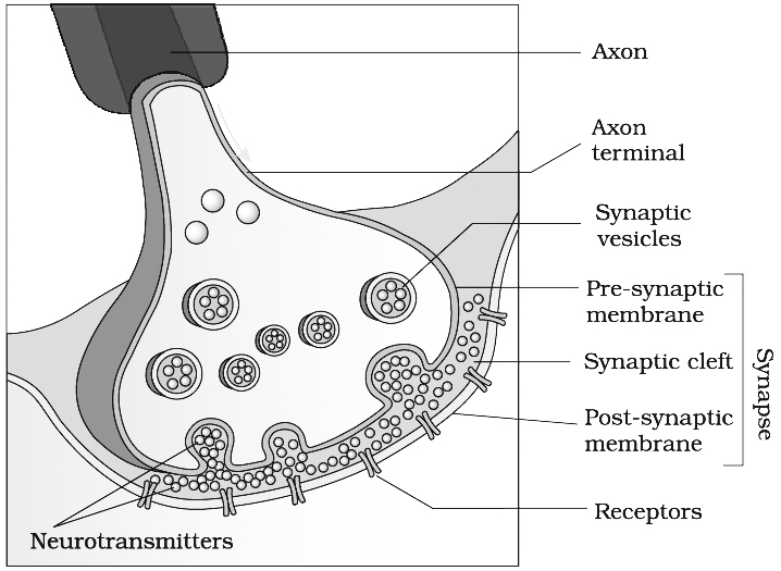
Chemical synapse have 10 to 20 nm gap which is too great a distance for such direct electrical coupling.
Chemical synapses are commonest type of synapse, these consist of a bulbous expansion of a nerve terminal, called synaptic knob, lying in close proximity to the membrane of a dendrite or other part of neuron.
The cytoplasm of the synaptic knob contains numerous tiny, round sacs, called synaptic vesicles.
Each vesicle has a diameter of approximately 50 nm, and contains as many as 10,000 molecules of a neurotransmitter substance responsible for the transmission of nerve impulse across the synapse.
The membrane of the synaptic knob on the axon side, thickened as a result of cytoplasmic condensation, is called presynaptic membrane.
Mechanism of transmission of nerve impulse through chemical synapse
Wave of depolarisation reaches the presynaptic membrane.
3
Voltage-gated calcium channels open, Ca++ ions diffuses into the axon terminal from the surrounding fluid.
3
Ca++ stimulates fusion of synaptic vesicle with pre-synaptic membrane, and release of neurotransmitter by
exocytosis into synaptic cleft.
3
Neurotransmitter bind with specific receptor molecules of post-synaptic membrane.
3
This binding opens sodium ion channels allowing the entry of Na+ ions
which can generate a new potential in the post-synaptic neuron.
3
The new potential developed may be either excitatory or
inhibitory depends upon neurotransmitter.
Concept Builder
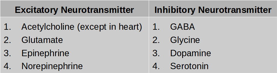
1. Acetylcholinestrase is present in the muscle cell or post synaptic neuron. It breaks down acetylcholine into acetate and choline and terminate the action of the transmitter.
2. Nor epinephrine secreted by sympathetic neural system and also by some neurons of central neural system is inactivated by enzyme monamine oxidase.
Central Neural System
- Books Name
- ACME SMART COACHING Biology Book
- Publication
- ACME SMART PUBLICATION
- Course
- CBSE Class 11
- Subject
- Biology
Central Nervous System
The structures of the CNS arise from its embryological components.
Prosencephalon
(i) Becomes the thalamus and hypothalamus (diencephalon) and
(ii) The cerebral cortex, corpus striatum, hippocampus and amygdala (telencephalon).
Mesencephalon
Becomes mid brain.
Rhombencephalon develops into
(i) The medulla (myelencephalon)
(ii) The pons and cerebellum (metencephalon).
BRAIN
Meninges :
The brain is surrounded by three protective coats of connective tissue besides the bony cranium. These are known as meninges (singular, meninx).
(i) Piamater : It is the inner meninx. It is very thin, highly vascular, and closely invests the brain. It is covered by simple squamous epithelium.
(ii) Arachnoid Mater or Membrane: It is the middle meninx. It is also thin but is nonvascular. It is covered with simple squamous epithelium on both (intemal and extemal) surfaces. There is a narrow space between the pia mater and the arachnoid membrane. It is called the subarachnoid space. It contains cerebrospinal fluid and is crossed by a number of connective tissue strands.
(iii) Duramater : It is the outer meninx. It is thick, tough and lines the cranial cavity. Its internal surface is covered with simple squamous epithelium. A very narrow space also exists between the dura mater and the arachnoid membrane. It is called the subdural space. It contains a little fluid which is not the cerebrospinal fluid.
The adult human brain contains more than 100 billion neurons and almost 10 times neuroglia cells. The brain is divided into three main sections:
(i) Fore brain; (ii) Mid brain; (iii) Hind brain
Fore brain: It consists of two main parts, the cerebrum and the diencephalon.
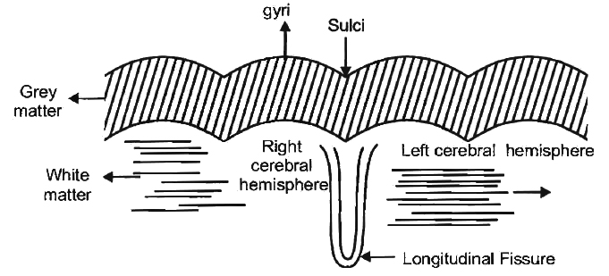
(a) Cerebrum. By far the largest and most highly developed part of the brain is cerebrum. It is divided into two hemispheres by a prominent longitudinal fissure. The two hemispheres are connected by a bundle of transverse fibres called corpus callosum. The anterior part of corpus callosum is curved and is called genu while the posterior part is called splenium. Each cerebral hemisphere is divided into four lobes. These are the frontal at the front, the parietal towards the top of the head, the temporal on the side and the occipital at the rear.
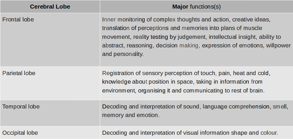
Cerebral cortex :
The outer surface of cerebrum, called the cortex, is a layer only 2-4 millimetres thick.
Because the six layers of it are packed with ten billion (109) pyramidal, spindle and stellate neurons with a greyish brown appearance, it is referred to as grey matter.
The cerebral cortex contains roughly 10 percent of all neurons of brain.
Much of the neural activities occur here, e.g., from the touch of a feature to the movement of an arm.
Unlike mouse brain, human brain is greatly convoluted.
These convolutions or folds consist of sulci (sing. Sulcus : small groove), fissures (large grooves), and gyri (sing. Gyrus : buldge between adjacent sulci or fissures).
These greatly enlarge the surface area of the cortex.
In fact, two-thirds of the surface of the cortex is hidden in the sulci and fissures.
Thus, their presence triples the area of the cerebral cortex.
Beneath this run millions of axons comprising nerve fibre tracts, connecting the neurons of cerebral cortex with those located elsewhere in the brain.
The large concentration of myelin gives this tissue an opaque white appearance.
Hence, they are referred by the term white matter.
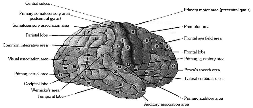
By examining the effect of injuries or lesions and the effect of electrical stimulation on the behaviour, it has been possible to map roughly the location of its various associative activities on the cerebral cortex.
Each area is referred to as a specialised cortex.
There are three general kinds of cortex : sensory, motor and associative.
(b) Diencephalon:
The diencephalon contains the epithalamus, thalamus and hypothalamus.
Epithalamus is thin, non nervous roof of diencephalon.
Its anterior region is folded and fused with piamater to form anterior choroid plexus.
This is responsible for formation of CSF.
Above it is present pineal stalk bearing the pineal body at the top of it.
Pineal body is endocrine gland and also taken as vestige of 3rd eye.
The thalamus directs sensory impulses from the lower parts of the brain and spinal cord to appropriate parts of the cerebrum.
Limited sensory awareness of pain, temperature, touch and pressure is provided by the thalamus.
Hypothalamus:
As the name implies, hypothalamus nestles at the base of the thalamus, and so of the brain.
Although relatively small, just 4 grams, about 1/300 of the total brain mass is highly vascularised.
It integrates and controls the visceral activities.
The hypothalamus, through its connection with brain stem, maintains homeostasis and the body's internal equilibrium, specialising in involuntary behaviour control.
The nuclei in it signal the body to eat, drink, get angry, keep cool, and make love and so on.
Hyothalamus organises behaviour related to survival of species: fighting, feeding, fleeing and mating.
It keeps body temperature at roughly 37ºC by means of a complex thermostat system.
It also influences respiration and heartbeat and sends out signals to correct them when they are wrong.
Through connections with the pituitary gland, the hypothalamus controls growth and sexual behaviour.
It also controls many more functions.
Basal Ganglia:
The inside of human brain is not so densely packed, but there are all kinds of different collections of neurons, called nuclei, each with its specific functions.
These control different body activities automatically.
Basal ganglia is a collection of subcortical nuclei in the forebrain, at the base of the cortex.
The largest nucleus in it is the corpus striatum.
It regulates planning and execution of stereotyped movements.
Other basal ganglia perform at subconscious level learned pattern of movements like slow and fast pedalling; slow and fast writing/typing etc.
Destruction of dopamine secreting pars compacta part of basal nucleus called substantia nigra leads to paralysis agitansl parkinson's disease. Huntington's chorea is due to degeneration of GABA secreting neurons of corpus striatum and acetylcholine secreting neurons of other parts.
Limbic System :
Flared like a wish bone a ring or fork, through extensive neural links with the cerebrum and the brain stem below, constitute what is called limbic (meaning lip-like) system.
This system sends out signals to the rest of the brain and the body which have great effect on your behaviour.
Limbic system includes hypothalamus, amygdala, hippocampus, septum, anterior nucleus of thalamus and portion of basal ganglia.
Amygdala :
Above the hypothalamus, attached to the interior lips of both forks, is almond-shaped amygdala.
This bulge of neurons is like a defense castle controlling the moods, especially anger and rage.
Various regions of the amygdala play important role in emotional behaviour, such as aggression and remembering fear.
Hippocampus:
Taking its name from the Greek for "sea horse", whose shape roughly resembles, the hippocampus make the swollen lower lip of the limbic fork.
The remarkable organ deals with a strange mix of signals about smells and memories.
The hippocampus functions as a kind of index for recall of an event with its associated memory.
The hippocampus converts information from short-term to long-term memory, essential in learning.
The septum linked to the hypothalamus contains yet another emotion centre for sexual arousal.
Mid Brain
It has two structures -corpora quadrigemina and cruracerebri.
(a) Corpora Quadrigemina :
It contains 4 lobes, therefore, corpora quadrigemina.
Its principal structures are superior colliculi and inferior colliculi.
The superior pair of colliculi receive sensory impulses from the eyes and muscles of the head and control visual reflexes.
For example, they control and coordinate the movement of the head and eyes, to fix and focus on an object.
The inferior pair of colliculi receive sensory impulses from the ears and muscles of the head and control auditory reflexes such as the movement of the head to locate and detect the source of a sound.
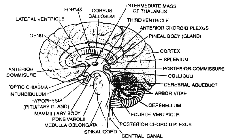
(b) Crura cerebri (Cerebral peduncle) :
These are two heavy fibrous tracts on the inferior side of mid brain and connect hind brain with fore brain.
Crura cerebri is involved in controlling muscle tone and modifying some motor activities.
These relay sensory as well as motor impulses between fore brain and hind brain.
Hind Brain
Consist of cerebellum, Pons and Medulla.
Cerebellum:
To the rear of the brain and placed under the cerebrum, is the second largest part of the brain, called the cerebellum that means simply "little cerebrum".
Wedged between cerebral hemispheres and brainstem, cerebellum is made up of two cerebellar hemispheres.
Like the cerebrum, the cerebellum has its grey matter on the outside, comprising of three layers of cells and fibres.
The middle layer contains characteristically large-flask-shaped Purkinje cells.
Tree-like themselves with myriad of dendrites, purkinje cells rank among the most complex of all neurons.
The white and grey matter form arbor vitae.
Central portion of the cerebellum has worm like appearance as it is narrow and furrowed.
It is called Vermis.
Three paired bundles of myelinated nerve fibres, called cerebellar peduncles, form communication pathways between the cerebellum and other parts of the CNS.
The superior cerebellar peduncles connect the cerebellum to the midbrain, the middle cerebellar peduncles communicate with the pons, and the inferior cerebellar peduncles consist of pathways between the cerebellum and the medulla oblongata, as well as spinal cord.
Cerebellum does not initiate movement but modulates or reorganises motor commands.
Cerebellum's unconscious directions and cerebrum's conscious instructions determine how and when to move body parts.
The cerebellum is vital to the control of rapid muscular activities, such as running, typing and even talking.
All the activities of the cerebellum are involuntary, but may involve learning in their early stages.
Pons, (Latin meaning : the bridge) forms the floor of the brain stem.
It serves as a neuronal link between the cerebral cortex and the cerebellum.
It has pneumotaxic centre, the switch off centre for inspiration.
Medulla oblongata, literally meaning oblong marrow, is the posterior most part that connects the spinal cord and various parts of the brain.
It has with it breathing centre, cardiovascular centre, vomiting centre.
Vagus nerve arises from medulla.
Its roof is thin and non-nervous and constitute posterior choroid plexus.
Below the plexus, the roof has three opening, a pair of lateral apertures called foramina luschka and a single median foramina Magendie.
These apertures connect external and internal components of CSF of brain.
Most of the sensory as well as motor nerve tracts cross over to the other side in medulla, therefore, right half of cerebrum controls left half of body and vice a versa.
Reticular formation that connects to the thalamus and major nerves in the spinal cord, is the gatekeeper to consciousness.
Brain Stem: It is the area of the brain between the thalamus and the spinal cord and includes medulla, pons and midbrain. Diencephalon mayor may not be included.
Ventricles of the Brain and Cerebrospinal Fluid
The ventricles consist of four hollow, fluid filled spaces inside the brain.
A lateral ventricle lies inside each hemisphere of the cerebrum.
Each lateral ventricle is connected to the third ventricle by an interventricular formen (foramen of Monro).
The third ventricle consists of a narrow channel between the hemispheres through the area of the thalamus.
It is connected by the cerebral aqueduct or aqueduct of Sylvius or iter in the midbrain portion of the brain stem to the fourth ventricle in the pons and medulla.
The fourth ventricle continues with the central canal of the spinal cord.
Three openings in the roof of the fourth ventricle, a pair of lateral apertures (foramina of Luschka) and a median aperture (foramen of Magendie) allow cerebrospinal fluid to move upward to the subarachnoid space that surrounds the brain and spinal cord.
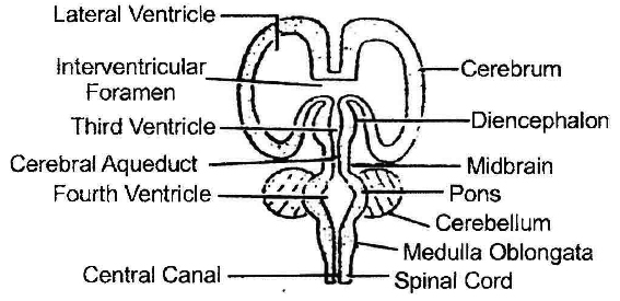
The cerebrospinal fluid is secreted by anterior choroid plexus and posterior choroid plexus and is found inside the ventricles of the brain, the central canal of the spinal cord.
The cerebrospinal fluid acts as a shock absorber for the brain and spinal cord and may also contribute to nourish brain tissue it contains protein, glucose, chloride and water.
Spinal Cord
It is an elongated cylindrical structure which lies in the neural canal of the vertebral column and is continued with the \ medulla oblongata through foramen magnum of the skull.
It measures about 45 cm in length.
It extends down upto first lumbar vertebra where it tapers to a point called conus medularis/conus terminalis.
However, the meninges of the spinal cord continues as filum terminale starts from the conus, and runs upto coccygeal region.
The spinal cord shows two enlargements
(i) Brachial swelling -from 4th cervical to 1st thoracic vertebrae.
(ii) Lumbar swelling -from 9th thoracic to 12th thoracic vertebrae.
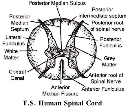
The spinal cord possesses an anterior and a posterior median fissure running along its length.
The grey matter of spinal cord is internal and present around the central canal.
It is produced into a posterior and anterior pairs of grey columns/roots.
Each dorsal root has a ganglion called dorsal root ganglion.
Dorsal root is sensory and ventral root is motor in nature. Both get combine with each other before coming out of vertebral column through intervertebral foramina.
The white mater is outer, and divided into four funiculi one dorsal, one ventral and two lateral.
Spinal cord conducts impulses to and from the brain.
Dorsal funiculus has ascending nerve tract for conducting sensory impulses towards brain.
Lateral and ventral funiculus conduct motor impulse from brain to spinal cord.
It controls most of the reflex activities.
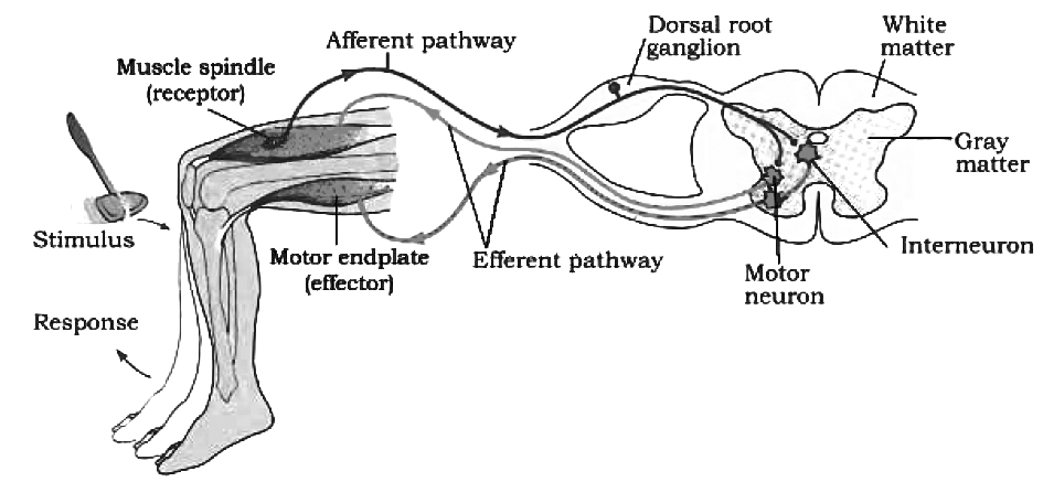
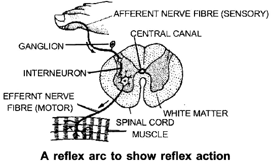
Reflex Action and Reflex Arc
- Books Name
- ACME SMART COACHING Biology Book
- Publication
- ACME SMART PUBLICATION
- Course
- CBSE Class 11
- Subject
- Biology
Reflex Action and Reflex Arc
The reflex action is the simplest kind of activity which can be defined as an integrated activity occurring involuntarily in response to a stimulus applied to a receptor.
The reflex arc is composed of the following:
(i) receptor organ; (ii) an afferent neuron; (iii) synapse involving some cells in the CNS (modulator); (iv) an efferent neuron; and (v) an effector organ.
(A) Reflexes can be classified in a number of ways as follows:
(a) Unconditioned reflexes: These are inborn, e.g., knee jerk, salivation on tasting the food, peristalsis, closing of eyes on being approached by an object.
(b) Conditioned reflexes: These are acquired, i.e., developed after birth through conditioning or learning e.g., secretion of saliva after seeing tasty food, playing a musical instrument, knitting without looking, writing as well as reading.
(B) According to the number of synapses in the reflex path
(a) Monosynaptic reflexes: When there is only one synapse in the reflex path. e.g., Knee jerk.
(b) Polysnaptic reflexes: When there are more than one synapse in the reflex path.
Concept Builder
Salivation on seeing or hearing or smelling delicious food; is an example of cerebral reflex action and withdrawl of legs; when a drop of HCl drop over the sciatic nerve of decapitated frog is an example of spinal reflex action.
Characteristics of Reflexes
Although the reflexes are involuntary functions, they have certain features which make them highly complicated. Some important characteristics are:
(i) Predictability: Once the response of an organ to a specific stimulus is observed, one can predict that the same stimulus will always elicit the same response.
(ii) Purposefulness: Generally all reflex actions are useful to the organism and are performed with a definite purpose.
(iii) Localization : In performing a reflex action, a specific effector is involved in response to the stimulus applied to a specific receptor.
(iv) Delay: Reflex time is the interval between the application of the stimulus to a receptor and the beginning of a response by an effector organ. A synaptic delay occurs due to latent period and reflex time at the synapse. This depends upon the number of synapses in the nerve pathway.
(v) Unlearned: In order to activate spinal effector mechanisms, no experience is needed to bring them in operation.
(vi) Adjustive and Protective: Reflexes serve adjustive and protective purposes and become an important part of the animal behaviour.
(vii) Fatigue: Reflex responses are readily fatigued after prolonged and continuous work. As a consequence, the latent period of contraction becomes longer and the rise of tension smaller and more gradual.
Sensory Reception and processing
- Books Name
- ACME SMART COACHING Biology Book
- Publication
- ACME SMART PUBLICATION
- Course
- CBSE Class 11
- Subject
- Biology
Sensory Reception and processing
The stimuli are received by certain structures in the body.
These are called receptors or sense organs.
A receptor may be extremely simple, such as those of touch, taste and smell, or they may be highly complex in their structure as well as working e.g., the sense organs of sight and hearing.
CLASSIFICATION OF SENSE ORGANS
(1) According to their position:
(i) Exteroreceptors -The external sense organs which receive the stimuli from outer environment.
(ii) Proprioreceptors -Simple receptors present in joints, skeletal muscles, tendons etc. They are not in direct contact with environment but are affected by the changes in the environment.
(iii) Visceroreceptors or internal receptors -The receptors present within the visGera. They receive stimuli originating within the body itself. They are simple and mostly represented by free nerve endings. Perception is conscious awareness and interpretation of sensation.
(2) According to the form of stimulus they receive, the sense organs are classified into the following types given in the Table.
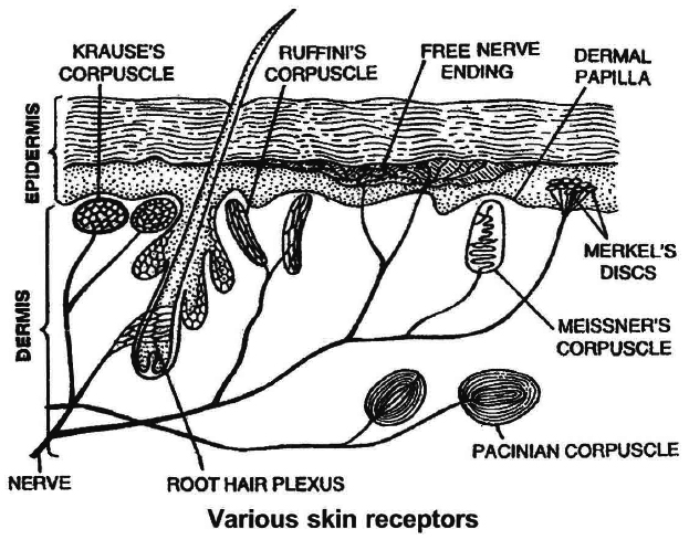
Tangoreceptors-
These are the sense organs for touch, pressure, pain, heat or cold. They are located in the skin and include:
(i) Meissner's corpuscles : They are present immediately below the epidermis, and receive the stimulus of touch/gentle pressure.
(ii) Pacinian corpuscles : Situated deep in the dermis of skin, joints, tendons and muscles. Each corpuscle has a nerve ending surrounded by connective tissue. They respond to pressure changes.
(iii) Merkel's disc occurs in the epidermis and are responsible for touch.
Skin is often called hypothermio because it has more cold receptors. The regulation of temperature in human body is mediated by hypothalamus which has a 'set point' (96.4°F or 37°C) around which the core temperature ossilates.
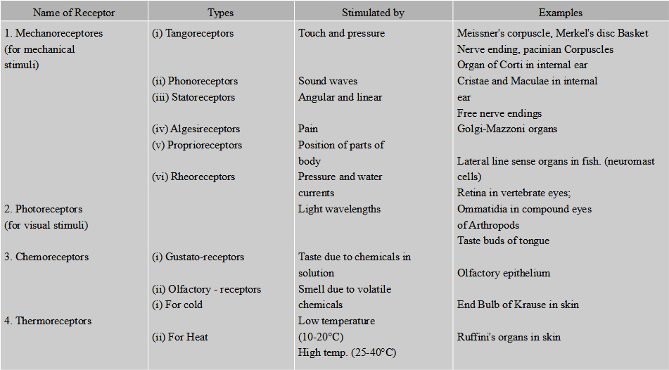
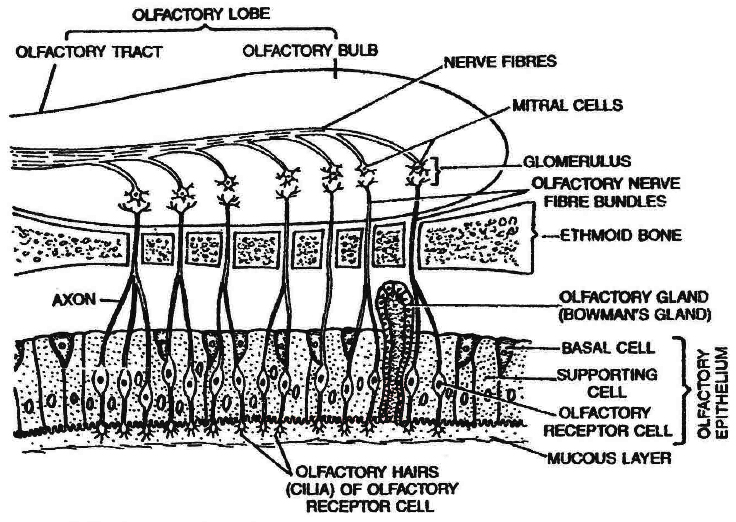
SMELL RECEPTORS (Olfactoreceptors)
Location:The receptors of smell occur in a small patch of olfactory epithelium (pseudostratified epithelium) located in the roof of the nasal cavity.
(i) Olfactory Receptor Cells: They act as sensory receptors as well as conducting neurons. The olfactory receptor cells are "unusual" bipolar neurons. Each cell is spindle shaped and has a thin apical dendrite that terminates in a knob which bears non motile cilia called olfactory hairs. Olfactory receptor cells are unique in that they are the only neurons that undergo turnover throughout adult life.
(ii) Supporting Cells: These are columnar cells which lie between the olfactory receptor cells to support them. They have brownish yellow pigment (similar to lipofuscin) which gives the olfactory epithelium its yellowish colour.
(iii) Basal Cells: These are small cells that do not reach the surface. They give rise to new olfactory receptor cell to replace the worn out ones. This is an exception to the fact that neurons are not formed in the postnatal (after birth) life. The olfactory receptor cells survive only for about two months.
Olfactory glands (Bowman's glands): Many olfactory glands occur below the olfactory epithelium that secrete mucus to spread over the epithelium to keep it moist. The mucus also protects the cells from dust and bacteria.
Working: The dissolved chemicals stimulate the olfactory receptors by binding to protein receptors in the olfactory hairs (cilia) membranes and opening specific Na+ and K+ channels. This leads ultimately to an action potential that is conducted to the first relay station in the olfactory bulb. The fibres of the olfactory nerves synapse with mitral cells (second = order neurons) in complex structures called glomeruli (balls of yarn). When the mitral cells are activated, impulses from the olfactory bulbs via olfactory tracts to main destinations (e.g., temporal lobe of the cerebrum).
Women often have a keener sense of smell than men, especially at the time of ovulation.
Smoking damages the olfactory receptors.
With ageing the sense of smell deteriorates.
Hyposmia (hypo-less, osmi-smell) is a reduced ability to smell.
Concept Builder
In addition to smell receptors nose, mouth and tongue contain a network of nerves that form trigeminal nerve (fifth cranial nerve) also known as dentist's nerve reacts to message of path.
Brain combines the trigeminal signals with those of smell to identify some odours.
When exposed to irritants such as ammonia or vinegar.
The trigeminal can protect by warning about harmful chemicals in the air. Bowman's glands inside the nose release fluids to get rid of the irritating substances.
TASTE RECEPTORS (Gustatoreceptors)
Location: The receptors for taste are found in the taste buds, mostly located on the tongue but also found on the palate, pharynx and epiglottis and even in the proximal part of oesophagus. The number of taste buds declines with age.
Structures : Each taste bud is an oval body consisting of three kinds of cells.
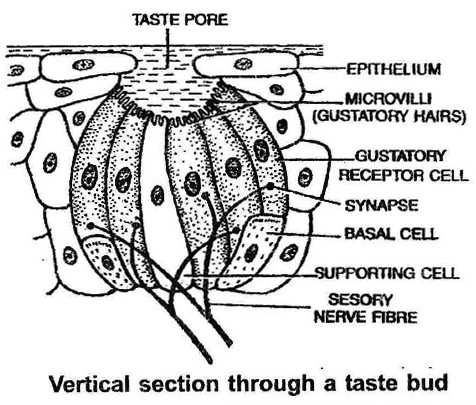
(i) Gustatory Receptor Cells: They bear at the free end microvilli projecting into the taste pore. The microvilli have special protein receptor sites for taste-producing molecules and come in contact with the food being eaten. Nerve fibres of the cranial nerves VII (facial), IX (glossopharyngeal) or X (Vagus) end around the gustatory receptor cells, forming synapses with them. The gustatory receptor cells (taste cells) survive only about 10 days and are then replaced by new cells.
(ii) Supporting cells: These cells lie between the gustatory receptor cells in the taste bud. They bear microvilli but lack nerve endings.
(iii) Basal Cells: These cells are found at the periphery of the taste bud. They produce supporting cells, which then develop into gustatory receptor cells.
Working: Specific chemicals in solution pass into the taste bud through the taste pore to come in contact with the protein receptor sites on the microvilli of the gustatory receptor cells. The latter set up nerve impulses in the sensory nerve fibres. The nerve fibres transmit the impulses to the taste centre in the brain (e.g., parietal lobe of the cerebrum) where the sensation of taste arises.
The facial nerve (VII) serves the anterior two-thirds of the tongue, the glossopharyngeal nerve (IX) serves the posterior one-third of the tongue and the vagus nerve (X) serves the pharynx and epiglottis but not the tongue.
Eye and Ear
- Books Name
- ACME SMART COACHING Biology Book
- Publication
- ACME SMART PUBLICATION
- Course
- CBSE Class 11
- Subject
- Biology
Eye and Ear
EYE
Organs of sight in man are a pair of eyes located in the eye orbits of the skull.
The exposed part of the eye is protected by a upper and a lower eyelid which are provided with eye lashes.
Each eye is represented in the form of a spherical eye ball which is moved in the eye orbit by the help of six eye muscles namely superior oblique, inferior oblique, superior rectus, inferior rectus, external rectus and internal rectus.
Eyeball measures about 2.5 cm. in diameter and is hollow.
Its wall is formed of three layers or coats-the outermost is called fibrous coat, the middle one as vascular coat and the inner one as retina.
(i) Fibrous coat : The outer coat of the eyeball is thick and tough. It provides form and shape to the eyeball. Fibrous coat consists of two parts, the sclera and cornea.
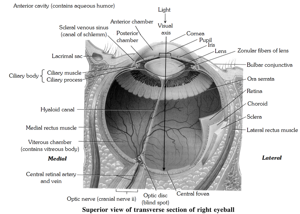
Sclera constitutes about five-sixth of the outer coat. It is white (made up of tough but elastic sheath of fibrous connective tissue containing collagen fibres) and opaque, and popularly called white of the eye. Most part of sclera is concealed in the orbit.
Cornea is the anterior transparent part of sclera and constitutes about 1/6 th of the fibrous coat. It is non-vascular and convex anteriorly. The cornea is covered by a thin and transparent membrane called conjunctiva composed of stratified epithelium and continued over the inner surface of the lids.
(ii) Vascular coat:
The middle coat of the eyeball is differentiated into three regions namely choroid, ciliary body and iris.
(a) Choroid is delicate, highly vascular and pigmented part which lies in contact with the sclera. It provides dark colour to the interior of the eyeball, it is black in colour. It prevents internally reflected light within the eye. The blood vessels of choroid nourish the retina.
(b) Ciliary body. It is the part of vascular coat immediately behind the peripheral margin of the iris. Ciliary body is thicker and less vascular than choroid. Its inner surface is folded to form ciliary processes. Present within the ciliary body are ciliary muscles.
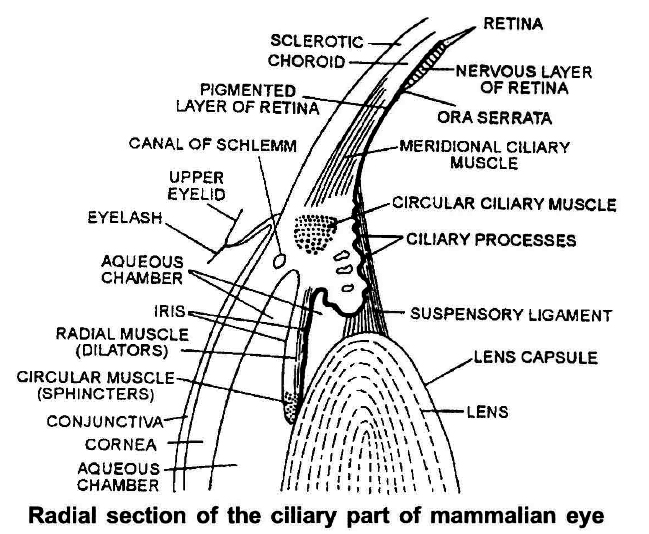
(c) Iris is the anterior part of vascular coat which lies behind the cornea. It is centrally perforated by pupil, the size of which is regulated by the iridial muscles arranged in radial and circular manner. The iris, being pigmented, provides colour to eye.
Concept Builder
Mirror like tapetum layer of carnivores like cats, dogs increases sensitivity by reflecting unabsorbed light back through photoreceptor layer to shine in dark.
(iii) Retina (Nervous Tunic):
The third and inner coat of the eyeball, the retina (nervous tunic), lines the posterior three-quarters of the eyeball and is the beginning of the visual pathway.
The optic disc is the site where the optic nerve exits the eyeball.
Bundled together with the optic nerve are the central retinal artery, a branch of the ophthalmic artery, and central retinal vein.
Branches of the central retinal artery fan out to nourish the anterior surface of the retina.
The central retinal vein drains blood from the retina through the optic disc.
The retina consists of a pigment epithelium (nonvisual portion) and a neural portion (visual portion).
The pigment epithelium is a sheet of melanin-containing epithelial cells that lies between the choroid and the neural portion of the retina some histologists classify it as part of the choroid rather than the retina.
Melanin in the choroid and the pigment epithelium absorbs stray light rays, which prevents reflection and scattering of light within the eyeball.
This enables that the image cast on the retina by the cornea and lens remains sharp and clear.
The pigmented layer is continuous over choroid, ciliary body and Iris while the nervous layer terminates just before ciliary body.
This point is called Orra serrata.
Albinos lack melanin pigment in all parts of the body, including the eye.
The neural portion of the retina is a multilayered out-growth of the brain.
It processes visual data extensively before transmitting nerve impulses to the thalamus, which then relays nerve impulses to the primary visual cortex.
Three distinct layers of retinal neurons, are separated by two zones where synaptic contacts are made, the inner and outer synaptic layers.
The three layer of retinal neurons, in the order in which they process visual input, are the photoreceptor layer, bipolar cell layer, and ganglion cell layer.
Note that light passes through the ganglion and bipolar cell layers before reaching the photoreceptor layer.
Two other types of cells present in the retina are called horizontal cells and amacrine cells. These cells form laterally directed pathways that modify the signals being transmitted along the pathway from photoreceptors to bipolar cells to ganglion cells.
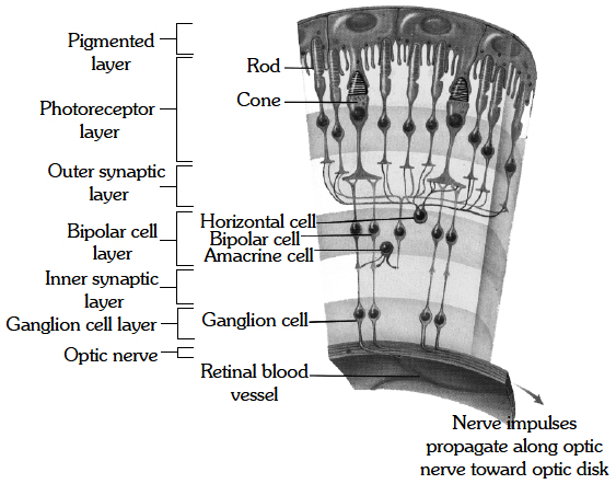
retina and main structures therein.
Photo receptors are specialized to transduce light rays into receptor potentials.
The two types of photoreceptors are rods and cones.
Each retina has about 6 million cones and 120 million rods.
Rods are most important for seeing shades of gray in dim light.
They also allow us to see shapes and movement.
Cones provide color vision in bright light.
The visual pigments for colour vision are: erythropsin (sensitive to red), chloropsin (sensitive to green) and cyanopsin (sensitive to blue).
In moonlight we cannot see colors because only the rods are functioning.
Due to the low light level cones are not functioning.
The macula lutea is in the exact center of the posterior portion of the retina, at the visual axis of the eye.
The central fovea, a small depression in the center of the macula lutea, contains only cone photoreceptors.
In addition, the layers of bipolar and ganglion cells, which scatter light to some extent, do not cover the cones here; these layers are displaced to the periphery of the fovea.
As a result, the central fovea is the area of highest visual acuity or resolution (sharpness of vision).
Rods are absent from the fovea and macula and increase in number towards the periphery of the retina.
From photoreceptors, information flows to bipolar cells through the outer synaptic, layer and then from bipolar cells through the inner synaptic layer to ganglion cells.
The axons of ganglion cells extend posteriorly to the optic disc and exit they eyeball as the optic nerve.
The optic disc is also called the blind spot.
Since it contains no rods or cones.
Accomodation
Accommodation (focussing) is the reflex mechanism by which light rays from objects at various locations in the near visual field are brought to focus on the retina.
Altering the shape of the lens does this. In bright light the circular muscle of the iris contracts, the radial muscle relaxes, the pupil becomes smaller and less light enters the eye, preventing damage to the retina.
In dim light, the opposite muscular contractions and relaxations occur.
In the dark of night", your pupil may become up to 16 times bigger.
The added advantage of reducing the pupil size is that it increases the depth of focus of the eye, so that any displacement of the photosensors in the retina will not impair the focus.
Light rays from distant objects (>6 metres) are parallel when they strike the eye.
Light rays from near objects (<6 metres) are diverging when they reach the eye.
In both cases, the light rays must be refracted or bent to focus on the retina and refraction must be greater for light from near objects.
The normal eye is able to accommodate light from objects from about 25 cm to infinity.
With the involuntary ciliary muscles at rest, the flatter lens has the correct optical properties to focus distant images on the retina, but not close images.
The state of contraction of the ciliary muscles changes the tension of suspensory ligaments.
This acts on the natural elasticity of the lens, which causes it to change its radius of curvature, and thus, the degree of refraction.
As the radius of curvature of the lens decreases it becomes thicker, round up and amount of refraction increases.
It is the tension of the suspensory ligaments applied to the lens which determines the shape of the lens.
When the circular ciliary muscles are relaxed and the suspensory ligament becomes tout, the lens is pulled into a flattened shape suitable for focussing distant objects, decreasing the refraction.
When the tension is decreased, the circular ciliary muscles are contracted and the suspensory ligaments slack, consequently the lens becomes a more spherical shape suitable for focussing objects.
The image produced by the lens of eye on the retina is inverted and reversed.
However, objects are perceived the right way up because of the way in which the brain interprets the images.
The region of the environment from which each eye collects light is called the visual field.
Since both our eyes are frontally placed, there is an overlap between the visual fields of each eye. This is called binocular vision.
Concept Builder
Image formation is refractive process, maximum refraction takes place on cornea.
Extra ocular muscle of eye
Eye is rotated in the orbit by six strap shaped muscles inserted on the sclera. These are arranged in two groups, rectus and oblique.
Rectus: Superior rectus – Oculomotor nerve
Inferior rectus – Oculmotor nerve
Internal (medial) rectus – Oculomotor nerve
External (lateral) rectus – Abducens nerve
Oblique: Superior oblique – Trochlear nerve
Inferior oblique – Oculomotor nerve
CHAMBERS OF EYEBALL
The lens and suspensory ligament divide the interior of eyeball into two chambers, the anterior small aqueous chamber containing a watery fluid, the aqueous humour; and the posterior larger vitreous chamber containing viscous fluid, the vitreous humour.
Aqueous humour maintain intra ocular pressure mainly where as vitreous humour responsible for shaping of eye ball.
Mechanism of Vision
Light rays in visible wavelength focussed on the retina
3
Activation of photopigment of rods and cones
3
Light induces dissociation of the retinal form opsin resulting in the changes in the structure of the opsin.
3
Membrane permeability of rods and cones changes.
3
Potential difference generated in the photoreceptor cell.
3
If generate action potential in the ganglionic cell s through the bipolar cells.
3
Action potentials are transmitted by the optic nerve to the visual cortex of brain.
3
Image formed on the retina is recognised based on earlier memory and experience.
Protective devices of Eye
(1) The Eyebrows: Two arched eminences of skin having numerous hairs project obliquely from the surface of the skin. The function of the eyebrows is to protect the anterior aspect of the eyeball from sweat, dust and other foreign bodies.
(2) The Eyelids (Palpebrae) and Eyelashes: The eyelids are two movable folds and have on their free edges, hairs -the eyelashes. The third eyelid is vestigial and is called plica semilunaris (nictitating membrane). The inner surface of each eyelid and parts of the eyeball are covered with mucous membrane, called the conjunctiva.
(3) Glands of Zeis: These are modified sebaceous glands which are associated with the follicles of eye lashes. They open into the follicles of eye lashes. Meibomian or tarsal glands are also modified sebaceous glands (oil glands) which are present along the edges of the eyelids. They produce an oily secretion which serves to lubricate the corneal surface and hold a thin layer of tears over the cornea. Glands of Moll are modified sweat glands at the edge of the eye lid.
(4) Conjunctiva: The palpebral conjunctiva is very vascular and has numerous papillae. Over the sclera the ocular conjunctiva is loosely connected to the eye ball; here it is thin, transparent, without papillae and slightly vascular. Reaching the cornea it continues as the corneal epithelium. The epithelium of the palpebral conjunctiva near the margin of the lids is non-keratinized squamous stratified epithelium. The conjunctiva helps to protect the eye ball and keeps it moist. It is this membrane that becomes inflamed in conjunctivitis or "pink eye".
(5) The Lacrimal Apparatus: The lacrimal apparatus of each eye consists of a lacrimal gland and its numerous ducts, the superior and inferior canaliculi, a lacrimal sac and nasolacrimal duct. The lacrimal gland secretes tears which are composed of water, salts and bactericidal protein called lysozyme. Lysozyme destroys microorganisms present on the front of the eyeball.
(6) Adipose Tissue (fat):A layer of adipose tissue surrounds the eyeball in the orbit. It serves as soft, shockproof pad.
Disorders of Eye:
(1) Myopia or Nearsightedness : In this case, the eyeball is antero-posteriorly elongated so that the image of distant objects is formed in front of yellow spot. The defect can be removed by using concave glasses.
(2) Hypermetropia or Long sightedness : The person can see distant objects clearly, but not those which are closer. This is due to antero-posterior shortening of the eyeball, so that the image is formed behind the yellow spot. The defect can be overcome by using convex lens.
(3) Presbyopia : A common defect in old age people due to the loss of elasticity of lens and reduced power of accomodation. The disorder can be corrected by convex lenses.
(4) Astigmatism : The disorder due to rough curvature of cornea or lens which can be corrected by the use of cylindrical glasses.
(5) Cataract: The sight is impaired due to the lens becoming opaque (Safaid Motia). The defect can be cured by surgical removal of the defective lens.
(6) Glaucoma: It occurs due to increase in intra-occular pressure as may develop due to blockage of canal of schlemn. It exerts pressure on optic nerve causing its damage. It leads to permanent blindness (Kala Motia)
(THE EARS)
The organs (Phonoreceptors) in man are a pair of ears situated on the head. Apart from their auditory function, the ears are also the organs of balancing.
Each ear has three portions-the external ear, the middle ear and internal ear.
(1) External Ear:
It consists of pinna and external auditory canal.
The latter is a curved passage which is lined by profusion of hair and about 4,000 ceruminous glands.
The glands secrete cerumen, a waxy material which entraps dust and also lubricates tympanum.
Tympanum or ear drum is a circular membrane present on the inner end the external auditory canal and partitions it from the tympanic cavity.
(2) Middle Ear:
The middle ear is represented by air-filled tympanic cavity which communicates with the pharynx by a passage called eustachian canal.
Present in the inner wall of the tympanic cavity are two openings, the upper fenestra ovalis and the lower, fenestra rotunda, each covered by a membrane.
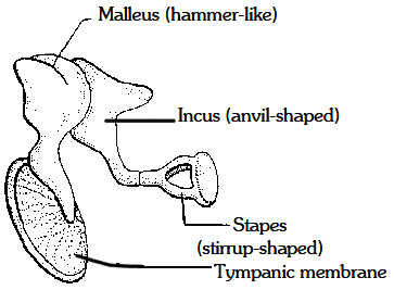
The tympanic cavity contains three small bones, the ear ossicles which from outside to inside include malleus, incus and stapes.
Malleus is hammer shaped; incus is anvil shaped and the stapes is stirrup shaped.
The outer arm of malleus is in contact with inner surface of tympanum, while the inner end of stapes forms contact with the membrane on fenestra ovalis.
Middle ear is responsible for amplification of signal due to leverage system of ossicle (10 times) by ear ossicles and 2.2 times by smaller size of membrane covering fenestra ova lis. The oval window is the door to internal ear.
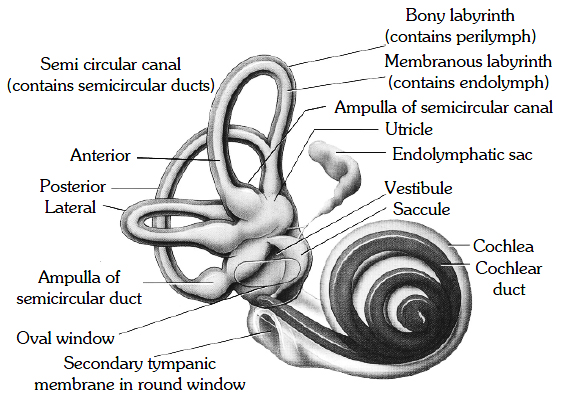
(3) Internal Ear:
It is also called membranous labyrinth and is surrounded by bony labyrinth of almost similar shape.
The space between the membranous labyrinth and bony labyrinth is filled by a watery fluid, the perilymph.
The membranous labyrinth contains endolymph.
The internal ear is a delicate organ and differentiated into vestibule, semicircular canals and cochlear duct.
The vestibule is the central body and is formed of two chambers, the upper utriculus and the lower sacculus.
Semicircular canals are three arched structures which emerge from utriculus and open back into it.
They include anterior and posterior vertical canals and a horizontal canal.
The vertical canals join to form a common passage crus commune, before they open into utriculus.
Each semicircular canal is dilated at the base to form ampulla which contains sensory spot called crista formed of receptor cells and supporting cells.
The receptor cells bear sensory hair, which are embedded into a gelatinous cupule above.
The vestibule also contains two sensory spots called maculae, one in sacculus and another in utriculus.
Maculae are similar to cristae, but there is no cupule.
The sensory hair are embedded in otolith membrane containing calcareous bodies called otoliths.
The cristae and maculae are the receptors of balance.
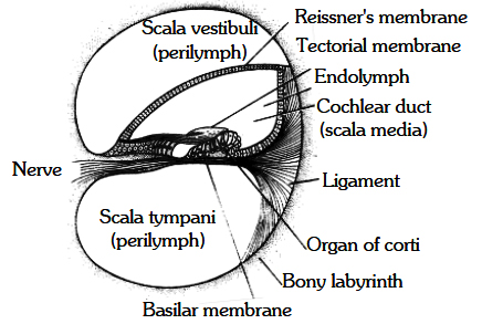
The auditory region of intern al ear is represented by a spirally coiled structure called cochlea.
It consists of cochlear duct arising from the sacculus, which is surrounded by similarly shaped cochlear canal, a part of bony labyrinth.
The cochlear duct is fused with cochlear canal on lateral sides, but is free laterally therefore, in T.S., the cochlea shows three chambers, the upper scala vestibuli, the middle scala media and the scala tympani.
The scala media is partitioned from the scala vestibuli by Reissner's membrane and from the scala tympani by basilar membrane.
Scala vestibuli and scala tympani contain perilymph while scala media is filled with endolymph.
The upper and lower chambers communicate through helicotrema, a narrow opening present at the distal end of cochlea.
The basilar membrane, sensory hair cells and tectorial membrane make up the smallest unit of the ear, called the organ of Corti, first described by Italian microscopist, Alfonso Corti (1822-1888).
Sensory hair cells inside the ear resemble tracts left in the sand by truck tires.
The cochlea contains 16,000 to 24,000 hair cells arranged in four rows.
In three of the rows, the hairs form V-shaped patterns. In the fourth row, the hair stand in a straight line.
Each hair cell has up to 100 hairs.
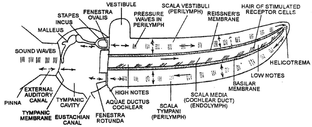
When sound vibrations pass through the oval window, they create waves in the lymph fluid of the cochlea, like sea wave in a tidal current.
The waves cause the basilar membrane to ripple.
This movement bends the hair cells, pressing against the tectorial membrane and setting off nerve impulses in their associated afferent neurons.
More than 30,000 neurons and nerve fibres emerging from these, convey the electrical Signals to the brain, just 2 cm away via auditory (vestibulocohlear) nerve.
The basal ends of hair cells synapse with fibres of cochlear branch.
When the waves reach the round windows of the cochlea, they die away.
Mechanism of Hearing
External ear receives sound waves
3
Directed towards the ear drum
3
When the waves strike the tympanicmembrane the alternate compression and
decompression of the air causes the membrane to vibrate
3
Vibrations are transmitted through the ear ossicles (M -7 I -7 S) to oval window
3
The movement of the oval window set up wave in the perilymph of scala vestibuli
3
Vibration of endolymph of scala media
3
The waves in the endolymph induced a ripple in the basilar membrane
3
Basilar movements bend the hair
cells pressing them against the tectorial membrane
3
Nerve impulse generated in the associated afferent neurons
3
Impulse transmitted to auditory region of brain via auditory nerve
3
Impulse get analysed and sound is recognised
Concept Builder
The high frequency resonance of the basilar membrane occurs near the base, where the sound waves enter the cochlea while low frequency resonance occurs near the apex mainly because of stiffness of fibres of basilar membrane.
The three internal ossicles of ear are malleus incus and stapes.
In case of non-mammals (amphians, reptiles, birds) there is just one bone called columella auris.
Physiology of Equilibrium
There are two kinds of equilibrium (balance).
One, called static equilibrium, refers to the maintenance of the position of the body (mainly the head) relative to the force of gravity.
The second king, dynamic equilibrium, is the maintenance of body position (mainly the head) in response to sudden movements such as rotation, acceleration, and deceleration.
Collectively, the receptor organs for equilibrium are called the vestibular apparatus, which includes the saccule, utricle, and semicircular ducts.
Static Equilibrium
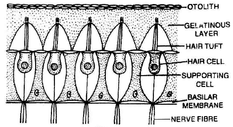
The wall of both the utricule and saccule contains a small, thickened region called macula (Pleural maculae).
The maculae are the receptors for static equilibrium and also contribute to some aspects of dynamic equilibrium.
For static equilibrium they provide sensory informations on the position of head and essential for maintaining appropriate posture and balance.
For dynamic equilibrium they linear acceleration and deceleration.
For example the sensation you feel while in an elevator or a car that is speeding up or slowing down.
Dynamic Equilibrium
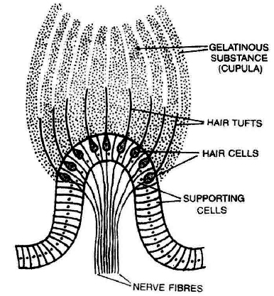
Vestibular apparatus contains three semicircular canals positioned at right angles to one another.
The dilated portion of each duct, ampulla, contains a small elevation called the crista.
Each crista is composed of a group of hair cells, supporting cells covered by a mass of gelatinous material called the cupula.
The cristae in the three semicircular canals maintain dynamic equilibrium.
Diseases of the Ear
1. Meniere's diseases: due to increased amount of the fluid of internal ear, loss of hearing.
2. Myringitis: Inflammation of tympanic membrane.
3. Otitis media: Acute infection in middle ear.
4. Vertigo: Type of dizziness where there is feeling of motion when one is stationary.
5. Cobyrinthine diseases: Improper functioning of internal ear.
Some Important Points
1. Most domestic mammals and sharks lack colour vision.
2. Tapetum lucidum. It is a part of choroid adjacent to the retina in the eyes of large number of elasmobranchs (cartilaginous fish). It possesses cells containing light-reflecting guanine crystals. It reflects light and causes the eyes to shine in dark. It also reflects additional light on the retinal cells to enable the fish to see in water where light is poor.
3. Accommodation : Fishes are able to see objects at different distances by changing the size of eyeball.
4. Pecten. It is a remarkable, highly vascular and pigmented structure projecting into the vitreous chamber from the blind spot normally. The pecten occurs in all birds except Kiwi (Apteryx). It is also found in some reptiles (e.g. Uromastix) but is absent in mammals. In Uromastix it is like cushion however, in pigeon it is comb-like and folded like a fan. The actual function of pecten is unknown but possibly it aids in the nutrition of the eye ball. In birds, it also helps in accommodation which is remarkably will developed in birds, by pressing the lens forward.
5. Phaco-emulsification technique in cataract surgery -"stitchless" technique. Foldable IOL (intraocular lens) is used.
6. Most birds have only day vision as their retina has mainly cones.
7. Owls have much better night vision as they contain a large number of rods and few cones in their retina.
8. Taste of chillies, is not true sensation. It is mainly sensation of burning pain produced by the stimulation of pain receptors of the tongue.
9. Hordeolum. Inflammation of sebaceous glands of eye lid.
10. Owls and cats see only with the help of available light from the stars or moon at night.
11. Frog is short sighted in air and long sighted in water.
12. Many insects like honeybees possess the gustatory receptors on their feet.
13. Largest cranial nerve-Trigeminal. .
14. Smallest/Thinnest cranial nerve-Pathetic/Trochlear.
15. Other names of various parts of brain
(i) Fore Brain = Prosencephalon
(ii) Mid Brain = Mesencephalon
(iii) Hind Brain = Rhombencephalon
(iv) Olfactory lobes = Rhinencephalon
(v) Cerebrum = Telencephalon
(vi) Diencephalon = Thalamencephalon
(vii) Cerebellum and Pons = Metencephalon
(viii) Medulla oblongata = Myelencephalon
(ix) Fourth ventricle = Metacoel
(x) Third ventricle = Diocoel
(xi) Iter = Mesocoel and aqueduct of sylvius.
(xii) Lateral ventricle = Paracoel
(xiii) Spinat canal = Myelocoel
(xiv) Cavity of olfactory lobe = Rhinocoel (absent in human)
16. Origin of CNS-develops from neural tube that is formed by infolding of ectoderm in early embryo.
17. Neopallium-Dorsal wall of cerebrum/cerebral cortex of brain.
18. Monosynaptic/simple reflex involves a single sensory fibre and a single motor fibre e.g., knee jerk. No interneuron. Polysynaptic/compound reflex involves one (or more) sensory and more than one motor nerve fibres. A number of interneurons are present. Polysynaptic reflexes are more common. All our visceral reflexes are polysynaptic
