 ACME SMART PUBLICATION
- Books Name
- ACME SMART COACHING Biology Book
- Publication
- ACME SMART PUBLICATION
- Course
- CBSE Class 11
- Subject
- Biology
Sensory Reception and processing
The stimuli are received by certain structures in the body.
These are called receptors or sense organs.
A receptor may be extremely simple, such as those of touch, taste and smell, or they may be highly complex in their structure as well as working e.g., the sense organs of sight and hearing.
CLASSIFICATION OF SENSE ORGANS
(1) According to their position:
(i) Exteroreceptors -The external sense organs which receive the stimuli from outer environment.
(ii) Proprioreceptors -Simple receptors present in joints, skeletal muscles, tendons etc. They are not in direct contact with environment but are affected by the changes in the environment.
(iii) Visceroreceptors or internal receptors -The receptors present within the visGera. They receive stimuli originating within the body itself. They are simple and mostly represented by free nerve endings. Perception is conscious awareness and interpretation of sensation.
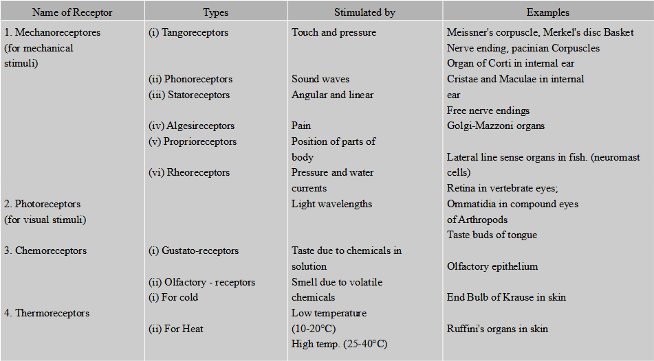
(2) According to the form of stimulus they receive, the sense organs are classified into the following types given in the Table.
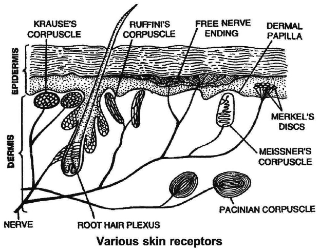
Tangoreceptors-
These are the sense organs for touch, pressure, pain, heat or cold. They are located in the skin and include:
(i) Meissner's corpuscles : They are present immediately below the epidermis, and receive the stimulus of touch/gentle pressure.
(ii) Pacinian corpuscles : Situated deep in the dermis of skin, joints, tendons and muscles. Each corpuscle has a nerve ending surrounded by connective tissue. They respond to pressure changes.
(iii) Merkel's disc occurs in the epidermis and are responsible for touch.
Skin is often called hypothermio because it has more cold receptors. The regulation of temperature in human body is mediated by hypothalamus which has a 'set point' (96.4°F or 37°C) around which the core temperature ossilates.
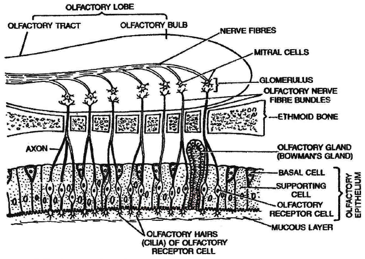
SMELL RECEPTORS (Olfactoreceptors)
Location:The receptors of smell occur in a small patch of olfactory epithelium (pseudostratified epithelium) located in the roof of the nasal cavity.
(i) Olfactory Receptor Cells: They act as sensory receptors as well as conducting neurons. The olfactory receptor cells are "unusual" bipolar neurons. Each cell is spindle shaped and has a thin apical dendrite that terminates in a knob which bears non motile cilia called olfactory hairs. Olfactory receptor cells are unique in that they are the only neurons that undergo turnover throughout adult life.
(ii) Supporting Cells: These are columnar cells which lie between the olfactory receptor cells to support them. They have brownish yellow pigment (similar to lipofuscin) which gives the olfactory epithelium its yellowish colour.
(iii) Basal Cells: These are small cells that do not reach the surface. They give rise to new olfactory receptor cell to replace the worn out ones. This is an exception to the fact that neurons are not formed in the postnatal (after birth) life. The olfactory receptor cells survive only for about two months.
Olfactory glands (Bowman's glands): Many olfactory glands occur below the olfactory epithelium that secrete mucus to spread over the epithelium to keep it moist. The mucus also protects the cells from dust and bacteria.
Working: The dissolved chemicals stimulate the olfactory receptors by binding to protein receptors in the olfactory hairs (cilia) membranes and opening specific Na+ and K+ channels. This leads ultimately to an action potential that is conducted to the first relay station in the olfactory bulb. The fibres of the olfactory nerves synapse with mitral cells (second = order neurons) in complex structures called glomeruli (balls of yarn). When the mitral cells are activated, impulses from the olfactory bulbs via olfactory tracts to main destinations (e.g., temporal lobe of the cerebrum).
Women often have a keener sense of smell than men, especially at the time of ovulation.
Smoking damages the olfactory receptors.
With ageing the sense of smell deteriorates.
Hyposmia (hypo-less, osmi-smell) is a reduced ability to smell.
Concept Builder
In addition to smell receptors nose, mouth and tongue contain a network of nerves that form trigeminal nerve (fifth cranial nerve) also known as dentist's nerve reacts to message of path.
Brain combines the trigeminal signals with those of smell to identify some odours.
When exposed to irritants such as ammonia or vinegar.
The trigeminal can protect by warning about harmful chemicals in the air. Bowman's glands inside the nose release fluids to get rid of the irritating substances.
TASTE RECEPTORS (Gustatoreceptors)
Location: The receptors for taste are found in the taste buds, mostly located on the tongue but also found on the palate, pharynx and epiglottis and even in the proximal part of oesophagus. The number of taste buds declines with age.
Structures : Each taste bud is an oval body consisting of three kinds of cells.
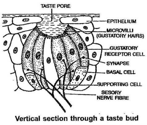
(i) Gustatory Receptor Cells: They bear at the free end microvilli projecting into the taste pore. The microvilli have special protein receptor sites for taste-producing molecules and come in contact with the food being eaten. Nerve fibres of the cranial nerves VII (facial), IX (glossopharyngeal) or X (Vagus) end around the gustatory receptor cells, forming synapses with them. The gustatory receptor cells (taste cells) survive only about 10 days and are then replaced by new cells.
(ii) Supporting cells: These cells lie between the gustatory receptor cells in the taste bud. They bear microvilli but lack nerve endings.
(iii) Basal Cells: These cells are found at the periphery of the taste bud. They produce supporting cells, which then develop into gustatory receptor cells.
Working: Specific chemicals in solution pass into the taste bud through the taste pore to come in contact with the protein receptor sites on the microvilli of the gustatory receptor cells. The latter set up nerve impulses in the sensory nerve fibres. The nerve fibres transmit the impulses to the taste centre in the brain (e.g., parietal lobe of the cerebrum) where the sensation of taste arises.
The facial nerve (VII) serves the anterior two-thirds of the tongue, the glossopharyngeal nerve (IX) serves the posterior one-third of the tongue and the vagus nerve (X) serves the pharynx and epiglottis but not the tongue.
