 ACME SMART PUBLICATION
Cell cycle
Chapter 10
Cell Cycle and cell division
Cell Cycle
Cells, and all living things for that matter, exhibit growth and reproduction. Each parental cell produces two daughter cells every time it divides, which is how all cells reproduce. A new cell population can be created by the growth and division of a single parental cell and its offspring, which is accomplished by the newly generated daughter cells. In other words, repeated cycles of growth and division enable the formation of structures made up of millions of cells from a single cell.
All living things go through the process of cell division. DNA replication and cell proliferation also happen when a cell divides. To ensure proper division and the production of offspring cells with complete genomes, processes like cell division, DNA replication, and cell development are coordinated. Cell cycle refers to the series of actions that a cell takes to reproduce its genome, synthesise the other components of the cell, and ultimately divide into two daughter cells. DNA synthesis only takes place during one particular stage of the cell cycle, despite the fact that cell growth (as measured by cytoplasmic expansion) is a constant process.During cell division, a complicated chain of processes transfers the replicated chromosomes (DNA) to the daughter nuclei. These occurrences are genetically determined.
In order to form two genetically identical cells, cells go through a series of carefully timed and regulated steps of growth, DNA replication, and division. Interphase and the mitotic phase are the two main stages of the cell cycle. The cell develops and DNA replication occurs during interphase. The cell divides and the replicated DNA and cytoplasm are separated during the mitotic phase.
The replication and reproduction of cells, whether in eukaryotes or prokaryotes, happens during the cell cycle. Although it serves several purposes for organisms, it ultimately ensures their survival. Prokaryotes can continue to exist by dividing into two new daughter cells thanks to a process termed binary fission in the cell cycle.
Reproduction, growth, and gamete creation are the three primary purposes of cell division. For asexual reproduction, growth, repair, and regeneration, mitosis is necessary. The bodies must create new cells—and permit the death of old cells—in order to expand and develop. The process of healing an injury also requires cell division.If cells were unable to divide and produce new cells, living organisms would never be able to regenerate skin cells to treat rashes or regrow a fingernail.
Phases of the Cell Cycle
Phases of Cell Cycle
Human cells in culture provide an example of a normal eukaryotic cell cycle. Every 24 hours or so, these cells divide once. The length of the cell cycle can, however, differ from organism to organism and from cell type to cell type. For instance, yeast may complete the cell cycle in about 90 minutes.
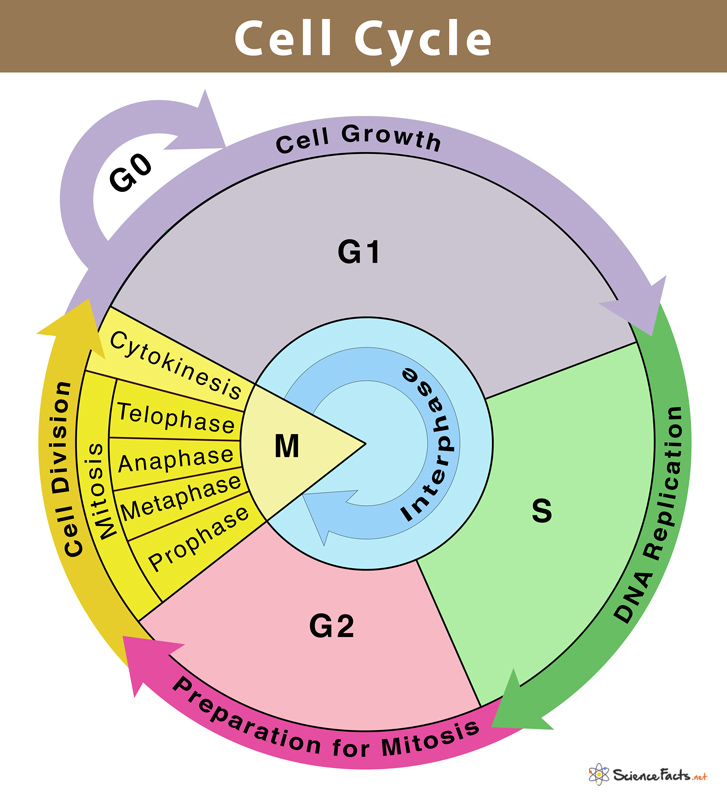
Interphase and M Phase(Mitosis phase) are the two fundamental phases of the cell cycle.
Interphase
A cell spends the majority of its time in what is known as interphase, where it develops, duplicates its chromosomes, and gets ready to divide. The cell then exits interphase, goes through mitosis, and finishes dividing. The phases of interphase are G1 phase (cell growth), S phase (DNA synthesis), and G2 phase (cell growth). The mitotic phase, which consists of mitosis and cytokinesis and produces two daughter cells, begins after interphase. In the cell cycle, the interphase is a protracted resting phase during which , DNA is replicated, RNA is synthesised, and proteins are produced.
The transitional period between mitosis and the start of DNA replication is known as the G1 phase. The cell is metabolically active and continues to develop during the G1 phase but does not duplicate its DNA. The time when DNA synthesis or replication occurs is known as the "S" phase. The amount of DNA in each cell doubles throughout this period. DNA grows from 2C (the initial amount) to 4C (the final amount). However, the number of chromosomes does not grow; if the cell had diploid or 2n chromosomes at G1, the number of chromosomes continues to be 2n even after S phase.In animal cells, the centriole doubles in the cytoplasm and DNA replication starts in the nucleus during the S phase. Proteins are created during the G2 phase as cells continue to expand in preparation for mitosis.Thus interphase is vital in the cell cycle as it permits the cell to grow and develop into a mature cell before it is able to reproduce.
Heart cells are one type of cell that does not appear to divide in adult animals, and many other cells only sporadically divide when it is necessary to replenish cells lost to damage or cell death. These cells leave the G1 phase and enter the quiescent stage (G0), which is an inactive phase of the cell cycle. While still metabolically active, cells in this stage no longer divide unless specifically instructed to do so by the organism. Only the diploid somatic cells in animals undergo mitotic cell division. In contrast, both haploid and diploid cells in the plants can exhibit mitotic divisions.
M Phase
It is the most dramatic phase of the cell cycle and involves a significant reorganisation of almost all cell components. It is also known as equational division since both the parent and progeny cells have the same number of chromosomes. Although mitosis has been conveniently split into four stages of nuclear division, it is crucial to realise that cell division is a progressive process, and extremely obvious distinctions between different stages cannot be made. Two processes make up the M phase: cytokinesis (or cell division), in which a cell's cytoplasm splits in half to create two different daughter cells, and mitosis, in which the cell's chromosomes are divided equally between the two daughter cells.
Mitosis and it's significance
Mitosis and its significance
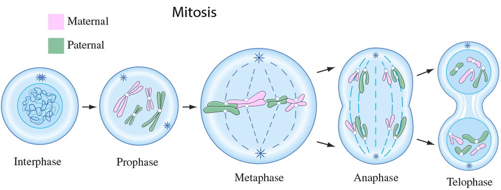
A cell prepares for cell division by replicating its chromosomes, segregating them, and creating two identical nuclei during the mitotic phase. The cell's contents are often evenly divided into two daughter cells with identical genomes after mitosis.Mitosis is divided into the following four stages:
1. Prophase:
Interphase's S and G2 phases are followed by prophase, the initial step of mitosis. The newly synthesised DNA molecules are not distinct but rather entangled in the S and G2 phases. The beginning of chromosomal material condensing characterises prophase. During the process of chromatin condensation, the chromosomal material is untangled. The centriole, which underwent duplication during interphase's S phase, now starts to migrate in the direction of the cell's opposing poles.Thus, the following distinctive occurrences can indicate the end of prophase:
- Compact mitotic chromosomes are created when chromosomal material condenses.Two chromatids are observed to be joined together at the centromere to form chromosomes.
- The beginning of the mitotic spindle's construction, the microtubules, and the proteinaceous elements of the cell cytoplasm aid in the process.
- The golgi complex, endoplasmic reticulum, nucleolus, and nuclear envelope are absent from cells near the end of prophase when they are observed under a microscope.
2. Metaphase:
The second phase of mitosis begins when the nuclear envelope completely disintegrates, and as a result, the chromosomes are dispersed throughout the cell's cytoplasm. The chromosome condensation process is now complete, and the chromosomes may be seen clearly under a microscope. Therefore, this is the period at which it is easiest to study the shape of chromosomes. The two sister chromatids that make up the metaphase chromosome at this point are joined by the centromere. Kinetochore refers to a little disc-shaped structure at the centromere surface. These structures act as the points of attachment for the spindle fibres, which are created by the spindle fibres, to the chromosomes that are placed at the cell's centre.Thus, all of the chromosomes align at the equator during the metaphase, with one chromatid of each chromosome attached by its kinetochore to spindle fibres from one pole and its sister chromatid connected by its kinetochore to spindle fibres from the opposite pole. The term "metaphase plate" refers to the chromosomes' alignment plane during metaphase. Metaphase's primary characteristics are:
- Spindle fibres adhere to chromosomal kinetochores.
- Chromosomes are transferred to the spindle equator, where they are positioned along the metaphase plate and along the spindle fibres to both poles.
3. Anaphase:
Each chromosome on the metaphase plate splits simultaneously at the start of anaphase, and the two daughter chromatids, which are now known as the chromosomes of the future daughter nuclei, start to move in opposite directions. The centromere of each chromosome is at the pole and, as a result, at the leading edge, with the arms of the chromosome trailing behind as they advance away from the equatorial plate. Events that define the anaphase stage are:
- Centromeres divide, and chromatids dissociate.
- Chromatids shift to the polar opposites
4. Telophase:
The chromosomes that have reached their respective poles decondense and lose their identity at the beginning of the last stage of mitosis, known as telophase. The individual chromosomes are no longer visible, and the two poles tend to accumulate a mass of chromatin material. The essential events at this stage include:
- Chromosomes cluster at opposing spindle poles, losing their identity as distinct elements.
- The nucleolus, Golgi complex, and ER remodel themselves
- The nuclear envelope forms around the chromosomal clusters.
Cytokinesis:
In addition to segregating duplicated chromosomes into daughter nuclei (karyokinesis), mitosis also divides the cell into two daughter cells by a separate process known as cytokinesis, marking the completion of cell division. This occurs when a furrow forms in the plasma membrane of an animal cell. The cytoplasm of the cell is split in half by the furrow, which eventually merges in the centre. Plant cells, on the other hand, are surrounded by a cell wall that is rather inextensible; as a result, they go through cytokinesis using a different process. In plant cells, wall construction begins in the cell's middle and extends outward to meet the lateral walls that already exist.The basic precursor known as the cell-plate, which symbolises the middle lamella between the walls of two neighbouring cells, is formed before the new cell wall can be fully formed. Organelles like mitochondria and plastids are dispersed between the two daughter cells during cytoplasmic division. In some organisms, cytokinesis does not follow karyokinesis, which results in a multinucleate state and the development of syncytium.
Significance of Mitosis :
Only diploid cells often undergo mitosis, also known as equational division. However, haploid cells can also divide through mitosis in some lower plants and social insects. Understanding the importance of this division in an organism's life is crucial. Typically, mitosis produces daughter cells that are diploid and have the same genetic makeup. Mitosis is responsible for multicellular organisms' growth. The ratio of the nucleus to the cytoplasm is disturbed as a result of cell expansion. Therefore, cell division is required to re-establish the nucleo-cytoplasmic ratio. Cell repair is one of mitosis' most important functions.Blood cells, stomach lining cells, and the outermost layer of the epidermis all undergo continuous replacement. Plants grow continuously throughout their lives as a result of mitotic divisions in the meristematic tissues known as the apical and lateral cambium.
meiosis and it's significance
Meiosis and its significance
Meiosis is a type of cell division that results in the production of four gamete cells and a 50% reduction in the number of chromosomes in the parent cell. To develop egg and sperm cells for sexual reproduction, this process is necessary. There are four haploid daughter cells formed during meiosis (containing half as many chromosomes as the parent cell).Following are the main characteristics of meiosis:
- Meiosis requires just one cycle of DNA replication but two successive cycles of nuclear and cell division called meiosis I and meiosis II.
- After the paternal chromosomes have duplicated to form identical sister chromatids at the S phase, meiosis I begins.
- Meiosis involves the pairing and recombination of homologous chromosomes.
- At the end of meiosis II, four haploid cells are produced.
Events of Meiosis are classified as:
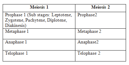
Meiosis 1
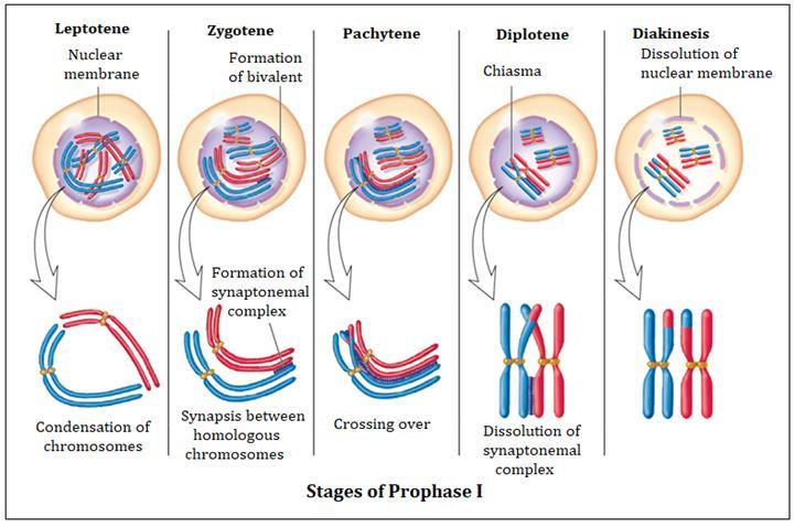
Prophase I: When compared to the prophase of mitosis, the prophase of the first meiotic division is often longer and more complex. Based on chromosomal behaviour, it has been further split into the following five phases: Leptotene, Zygotene, Pachytene, Diplotene, and Diakinesis.
Leptotene: The chromosomes begin to condense at this stage and are connected to the nuclear membrane via their telomeres.
Zygotene: A synaptonemal complex forms between homologous chromosomes at the start of synapsis.
Pachytene - During this stage, genetic material is transferred between chromatids that are not sisters. This is called crossing over.
Diplotene - At this stage, homologous pairs are still connected at the chiasmata after synapsis has ended and the synaptonemal complex has vanished.
Diakinesis: Before metaphase 1, the nuclear membrane finally breaks down and the chromosomes are entirely condensed.
Metaphase I: The bivalent chromosomes line up on the equatorial plate during metaphase I. The spindle's opposing poles' microtubules connect to the pair of homologous chromosomes.
Anaphase I: Sister chromatids are still connected at their centromeres during anaphase I, but homologous chromosomes split.
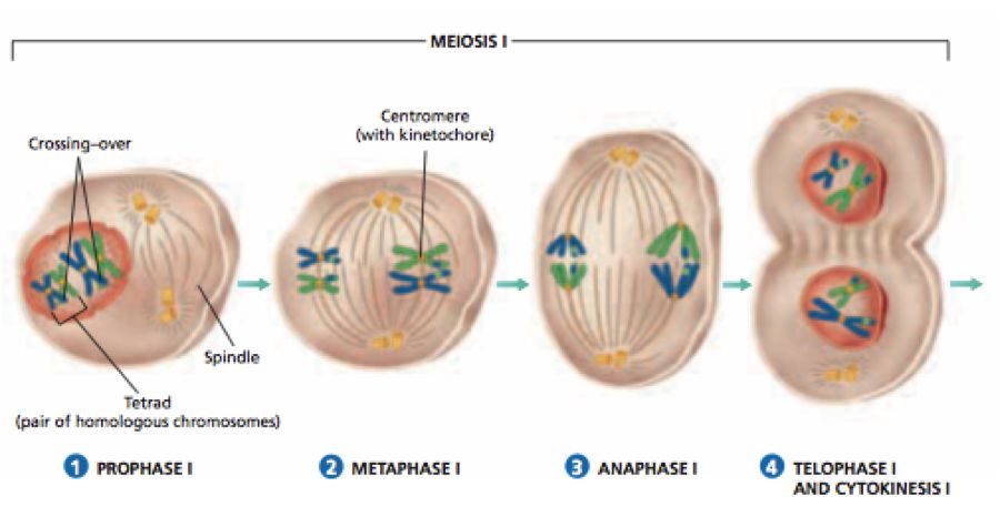
Telophase I: Cytokinesis occurs after the nuclear membrane and nucleolus re-emerge, and this is known as the "diad of cells." Even though the chromosomes do experience some dispersion in many instances, they rarely reach the interphase nucleus's very stretched state. Interkinesis, which occurs between the two meiotic divisions, is typically a transient stage. Prophase II, which is substantially less complex than prophase I, comes after interkinesis.
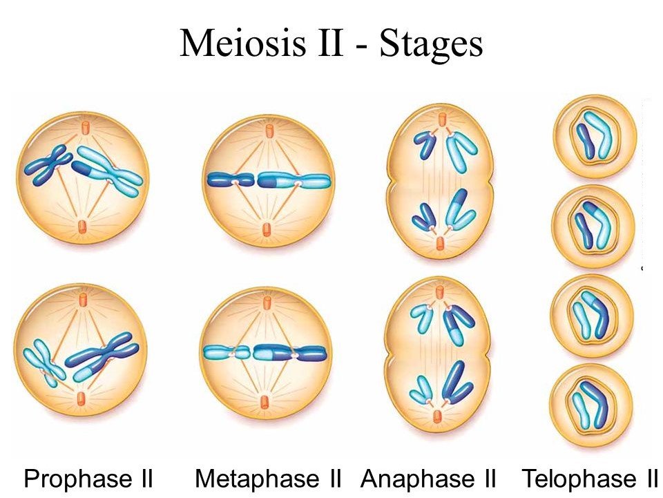
Meiosis 2:
Prophase II: Following cytokinesis, meiosis II begins right away, typically before the chromosomes have fully expanded. Meiosis II resembles a typical mitosis in contrast to meiosis I. By the end of prophase II, the nuclear membrane is gone. Chromosomes once more become condensed.
Metaphase II: Chromosomes align at the equator during metaphase II, and sister chromatids' kinetochores get attachments of microtubules from the spindle's opposing poles.
AnaphaseII: Beginning with the simultaneous division of each chromosome's centromere (which had been binding the sister chromatids together), anaphase II allows the chromosomes to migrate toward their respective poles of the cell.
Telophase II: Telophase II marks the completion of meiosis, during which the two chromosomal groups are once more encased in a nuclear membrane. Cytokinesis then takes place, culminating in the production of a tetrad of cells, or four haploid daughter cells.
Significance of Meiosis:
Even though the process itself paradoxically reduces the number of chromosomes by half, meiosis is the mechanism that allows sexually reproducing animals to maintain the particular chromosomal number of each species throughout generations. From one generation to the next, it also makes the population of organisms more genetically variable. For the process of evolution, variations are crucial.
