 ACME SMART PUBLICATION
Cell cycle
- Books Name
- ACME SMART COACHING Biology Book
- Publication
- ACME SMART PUBLICATION
- Course
- CBSE Class 11
- Subject
- Biology
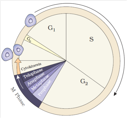
CELL CYCLE AND ITS PHASES
The total duration of cell cycle varies from organism to organism and also from cell type to cell type.
Yeast for example, can progress through the cell cycle in only about 90 minutes.
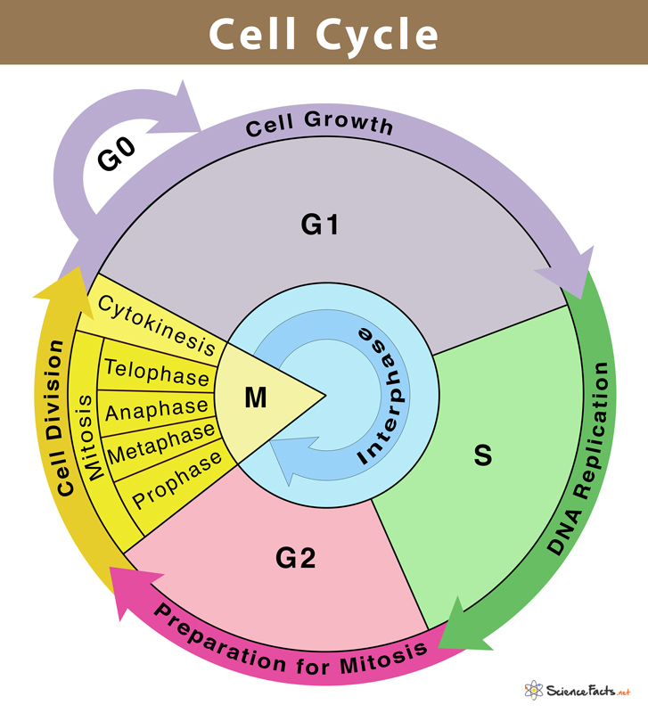
The cell cycle is divided into two basic phases :
1. Interphase
2. M-Phase (Mitosis phase)
1. Interphase :
It is also called as preparatory phase and a period of great metabolic activity.
It is the stage, between two successive cell divisions in which no division of chromosomes or cytoplasm occurs.
'In this stage the nucleus and cytoplasm remain metabolically and synthetically very active.
It generally covers over 95% of the total duration of cell cycle.
During this phase, replication of DNA, synthesis of nuclear histones, division of centrioles to form a new pair of centrioles, synthesis of energy rich compounds, RNA and proteins occur. Nuclear envelope remains intact.
Chromosomes occur in the form of long, coiled, indistinctly visible chromatin fibres.
The size of nucleolus is greatly increased due to accumulation of rRNA and ribosomal proteins.
Interphase is divided into three phases:
(a) G1 Phase (b) S or Synthetic Phase
(c) G2 Phase
(a) G1 Phase (Post-mitotic gap phase) : It corresponds to the interval between mitosis and initiation of DNA replication. Following biochemical changes occur during this sub-stage.
(i) The cell grows to its maximum size due to normal metabolic activity for the preparation of DNA replication, but no change occurs in the DNA contents of the cell.
(ii) It undergoes synthesis of new proteins and RNA. Transcription of rRNA, tRNA and mRNA occurs during this phase.
(iii) Nucleotides, amino acids and energy rich compounds (e.g., ATP) are formed.
(iv) It takes maximum time of all the stages. It is most variable in length, due to which time of cell division differs in cell to cell. G1 can be terminated by various stimuli, but once a cell has completed G1 and entered the 'S' phase to start replication of DNA, it cannot be terminated.
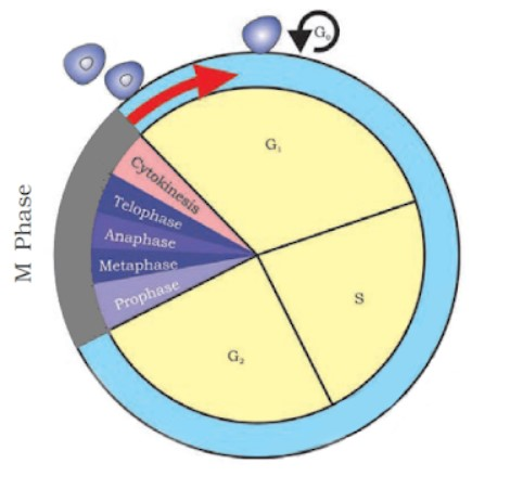
(v) Some cells in the adult animals do not appear to exhibit division (e.g., heart cells) and many other cells divide only occasionally, as needed to replace cells that have been lost because of injury or cell death. These cells do not divide further, exit G1 phase to enter an inactive stage called quiescent stage (G0) of the cell cycle. Cells in this stage remain metabolically active, but no longer proliferate unless called on to do so, depending upon the requirement of the organism. Hence, this exit may be temporary or permanent.
Antephase is the end of G1 when the cell reaches a stage whereby, it will divide even under stress condition.
(b) S or Synthetic Phase
(i) In this phase, the synthesis or replication of DNA occurs on the template of existing DNA.
(ii) During this phase, the amount of DNA per cell doubles (means the organism will have duplicate set of genes). However, there is no increase in the chromosome number (ploidy level remains same). If the initial amount of DNA is denoted as 2C then, it increases to 4C, and if the cell had 2n number of chromosomes at G1, even after S phase the number remains same, i.e., 2n.
(iii) In animal cells, the replication occurs in nucleus, and the centriole duplicates in cytoplasm.
(iv) Histone proteins are synthesised in S-phase. S-phase is called invisible phase of cell cycle as replicated chromosomes are not visible at this stage.
(c) G2 Phase (pre-mitotic gap phase)
(i) In this phase, the cytoplasmic organelles such as mitochondria, chloroplast and golgi complex are doubled.
(ii) Synthesis of RNA and protein continues. Spindle protein (tubulin) synthesis and aster formation occurs.
(iii) A cell contains double the amount (4C) of DNA present in the original diploid (2N) cell.
(iv) The cell prepares itself to enter into "M" or Mitotic phase.
(v) It is also signified by the synthesis of some protein kinases for regulation of cell division.
2. M-Phase :
It represents the phase when the actual cell division or mitosis occurs.
It starts with the nuclear division, corresponding to the separation of daughter chromosomes (Karyokinesis) and usually ends with division of cytoplasm (Cytokinesis).
A diagrammatic view of cell cycle indicating formation of two cells from one cell
Regulation of Cell Cycle
Decision of a cell to divide occurs in G1 phase. If a cell is not to divide it will enter into G0 phase or Quiescent phase. When the conditions change, the cell can enter back into G1 phase. G1 S transition in the cell cycle is called as Restriction point or check point. This is the major check point. Once the cell crosses the restriction point rest of the cell cycle is completed. Another minor check point is G2 M transition.
Concept Builder
(i) Cell cycle is regulated by cyclin-dependent protein kinase.
(ii) Cyclins are proteins that activate protein kinases to regulate eukaryotic cell cycle.
(iii) G1 to S transition is carried out by G1 cyclin + cdc 2 kinase.
(iv) G2 to M transition is triggered by maturation promoting factor (MPF) formed by mitotic cyclin + cdc2 kinase, Nucleus attains the maximum size.
(v) The factors which determine whether a cell has to divided or not are
(a) Surface area: Volume ratio. A cell should have high surface area : volume ratio.
(b) Karyoplasmic index.
(vi) Onion root tips or other meristematic tissues are used to study mitosis.
(vii) Mitogens are substances which induce mitosis. e.g., Auxin, Cytokinin, Gibberellin, Insulin etc.
(viii) In animal cell, mitosis is called as Amphiastral (Spindle is associated with 2 asters).
(ix) In plant cells, the mitosis is called as Anastral (no aster, no centriole).
(x) If mitosis is extranuclear, it is Eumitosis.
(xi) If mitosis is intranuclear, it is called as Premitosis. If centrioles are present then it is called as centric.
The cell division is of three types
I. Mitosis II. Meiosis III. Amitosis
Cell cycle
Chapter 10
Cell Cycle and cell division
Cell Cycle
Cells, and all living things for that matter, exhibit growth and reproduction. Each parental cell produces two daughter cells every time it divides, which is how all cells reproduce. A new cell population can be created by the growth and division of a single parental cell and its offspring, which is accomplished by the newly generated daughter cells. In other words, repeated cycles of growth and division enable the formation of structures made up of millions of cells from a single cell.
All living things go through the process of cell division. DNA replication and cell proliferation also happen when a cell divides. To ensure proper division and the production of offspring cells with complete genomes, processes like cell division, DNA replication, and cell development are coordinated. Cell cycle refers to the series of actions that a cell takes to reproduce its genome, synthesise the other components of the cell, and ultimately divide into two daughter cells. DNA synthesis only takes place during one particular stage of the cell cycle, despite the fact that cell growth (as measured by cytoplasmic expansion) is a constant process.During cell division, a complicated chain of processes transfers the replicated chromosomes (DNA) to the daughter nuclei. These occurrences are genetically determined.
In order to form two genetically identical cells, cells go through a series of carefully timed and regulated steps of growth, DNA replication, and division. Interphase and the mitotic phase are the two main stages of the cell cycle. The cell develops and DNA replication occurs during interphase. The cell divides and the replicated DNA and cytoplasm are separated during the mitotic phase.
The replication and reproduction of cells, whether in eukaryotes or prokaryotes, happens during the cell cycle. Although it serves several purposes for organisms, it ultimately ensures their survival. Prokaryotes can continue to exist by dividing into two new daughter cells thanks to a process termed binary fission in the cell cycle.
Reproduction, growth, and gamete creation are the three primary purposes of cell division. For asexual reproduction, growth, repair, and regeneration, mitosis is necessary. The bodies must create new cells—and permit the death of old cells—in order to expand and develop. The process of healing an injury also requires cell division.If cells were unable to divide and produce new cells, living organisms would never be able to regenerate skin cells to treat rashes or regrow a fingernail.
Phases of the Cell Cycle
- Books Name
- ACME SMART COACHING Biology Book
- Publication
- ACME SMART PUBLICATION
- Course
- CBSE Class 11
- Subject
- Biology
Phases of Cell Cycle
Cell division cycle or the cell cycle is a 4-stage process in a somatic cell during which two significant molecular processes occur – parent chromosome duplication (occurring in S phase) and equal detachment of the chromosome to the daughter cells (occurring during M phase)
In eukaryotic cells, the cell cycle phases are split into two significant phases – interphase and the mitotic phase. While in interphase, the cell significantly grows and replicates a DNA copy, in the mitotic phase or the M phase, the cell splits its DNA into two sets and hence the division of the cytoplasm to form two daughter cells.
Interphase
Interphase is the time lapse between two successive M phases of cell division. The cell prepares for division, grows and DNA replication takes place. Interphase is further divided into three phases: G1, S, G2
G1 (Gap 1) Phase
This is the primary stage of the interphase, known as the G1 or first gap phase as diminutive changes are observed due to the hyperactivity of the cell at the biochemical degree
This phase is characterized by changes in the chromosomes from the condensed to the extended state in addition to a range of metabolic activities leading to the initiation of replication of DNA.
Characteristics of the chromatin fibres in this phase are – less coiled and slender, extended fully and ready for transcription. The process of transcription results in the production of RNAs and also a sequence of protein molecules vital for DNA replication to be initiated
G1 phase is lengthier than the other three phases and varies from cell to cell
This is a significant phase as cell grows and assembles building blocks of chromosomal DNA and the linked proteins. In addition, it also reserves adequate energy to accomplish chromosome replication
DNA synthesis in this phase is initiated at a distinct checkpoint. The cell progresses towards division once all the biochemical events at this particular point have concluded.
Synthesis (S) phase
It is an active DNA synthesis and histone synthesis phase of the interphase
Here the chromosomes replicate, enabled by linked proteins and DNA replication. Most of the histone protein synthesis occurs in this phase, though some of it occurs in G1 phase
Identical pair of DNA molecules are formed as the process of DNA replication is discontinuous and semi-conservative
Even after the chromosomes have doubled, the sister chromatids are securely attached to the centromeric region. The chromosome count of the cell remains the same
Centrosomes of animal cells at the centre of each animal cell is linked with centrioles positioned perpendicular to each other. The centrioles are functional in organizing the cell division process
During this phase, the centrosome is duplicated, producing the mitotic spindle, the apparatus which liaises chromosomal movement while mitosis is taking place
G2 (Gap 2) Phase
This phase is succeeded by the S phase. Here the chromosomes comprise two chromatids thus cell has double the quantity of DNA
Here, the cell restores its energy, producing proteins essential for chromosomes to manipulate
Few of the cell organelles are replicated. Cytoskeleton dismantles to render resources for mitosis
Additional growth of cell may be observed. Before the cell enters the first phase of mitosis, the concluding preparations of the mitotic phase must be done.
M (Mitotic) Phase
This phase is succeeded by the G2 phase. Here the cell divides into two daughter cells along with equal distribution of chromosomes between the daughter cells. Once the M phase steps into the G1 phase, the next cell cycle is initiated to be repeated. Some cells, however, do not enter into the G1 phase. These are referred to as G0 cells
It comprisesq the following sub-phases –
Prophase – in this stage, the nucleus disappears, spindle fibres are formed, DNA condenses into sister chromatids
Metaphase – the sister chromatids orient alongside the cell-equator by linking their centromeres to the spindle fibres
Anaphase – separation of sister chromatids at the centromere, being pulled towards the opposite poles of the cell by mitotic spindle
Telophase – At the opposite poles, the chromosomes arrive to unwind into fine DNA strands. Spindle fibres vanish. Nuclear membrane resurfaces
Cytokinesis – cell membrane splits, animal cells drift away. Plant cells form a cell plate which turns into a new cell wall
Cells arriving at the G0 phase, which is the inactive phase once they exit the cell cycle when they are not preparing actively to divide. Few of these cells tend to remain in this stage permanently.
Phases of the Cell Cycle
Phases of Cell Cycle
Human cells in culture provide an example of a normal eukaryotic cell cycle. Every 24 hours or so, these cells divide once. The length of the cell cycle can, however, differ from organism to organism and from cell type to cell type. For instance, yeast may complete the cell cycle in about 90 minutes.
Interphase and M Phase(Mitosis phase) are the two fundamental phases of the cell cycle.
Interphase
A cell spends the majority of its time in what is known as interphase, where it develops, duplicates its chromosomes, and gets ready to divide. The cell then exits interphase, goes through mitosis, and finishes dividing. The phases of interphase are G1 phase (cell growth), S phase (DNA synthesis), and G2 phase (cell growth). The mitotic phase, which consists of mitosis and cytokinesis and produces two daughter cells, begins after interphase. In the cell cycle, the interphase is a protracted resting phase during which , DNA is replicated, RNA is synthesised, and proteins are produced.
The transitional period between mitosis and the start of DNA replication is known as the G1 phase. The cell is metabolically active and continues to develop during the G1 phase but does not duplicate its DNA. The time when DNA synthesis or replication occurs is known as the "S" phase. The amount of DNA in each cell doubles throughout this period. DNA grows from 2C (the initial amount) to 4C (the final amount). However, the number of chromosomes does not grow; if the cell had diploid or 2n chromosomes at G1, the number of chromosomes continues to be 2n even after S phase.In animal cells, the centriole doubles in the cytoplasm and DNA replication starts in the nucleus during the S phase. Proteins are created during the G2 phase as cells continue to expand in preparation for mitosis.Thus interphase is vital in the cell cycle as it permits the cell to grow and develop into a mature cell before it is able to reproduce.
Heart cells are one type of cell that does not appear to divide in adult animals, and many other cells only sporadically divide when it is necessary to replenish cells lost to damage or cell death. These cells leave the G1 phase and enter the quiescent stage (G0), which is an inactive phase of the cell cycle. While still metabolically active, cells in this stage no longer divide unless specifically instructed to do so by the organism. Only the diploid somatic cells in animals undergo mitotic cell division. In contrast, both haploid and diploid cells in the plants can exhibit mitotic divisions.
M Phase
It is the most dramatic phase of the cell cycle and involves a significant reorganisation of almost all cell components. It is also known as equational division since both the parent and progeny cells have the same number of chromosomes. Although mitosis has been conveniently split into four stages of nuclear division, it is crucial to realise that cell division is a progressive process, and extremely obvious distinctions between different stages cannot be made. Two processes make up the M phase: cytokinesis (or cell division), in which a cell's cytoplasm splits in half to create two different daughter cells, and mitosis, in which the cell's chromosomes are divided equally between the two daughter cells.
Mitosis and it's significance
- Books Name
- ACME SMART COACHING Biology Book
- Publication
- ACME SMART PUBLICATION
- Course
- CBSE Class 11
- Subject
- Biology
MITOSIS
It was first observed by Strasburger in plant cells and by Flemming in animal cells.
It may be defined as, "The exact replication of a parent cell into two identical daughter cells having the identical number, and kind of chromosomes, the identical DNA and identical hereditary instructions as found in the parental cell".
It mainly occurs in somatic cells of animals and in meristematic tissue cells of plants for multiplication of undifferentiated cells.
In mitosis the cell cycle involves series of changes which occur in a newly formed cell to become fully grown and to be ready for cell division.
It has two phases :
(1) Interphase (as described earlier)
(2) Mitotic phase or M-phase.
It consist of the following four sub-stages :
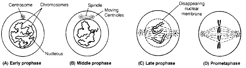
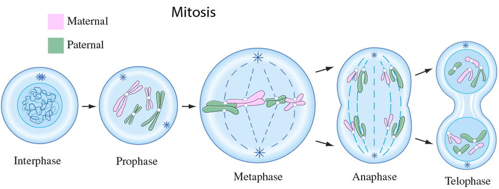
(a) Prophase: It is the first phase of mitosis which is longest of the M-phase.
Following changes occur during this stage:
(i) The chromatin fibres become shorter and thicker due to the process of coiling and folding and get condensed into distinct thread like chromosomes in late prophase.
(ii) Each chromosome appears double and consists of two coiled sister chromatids joined by a centromere. Their ends are not visible in early prophase. Therefore, the chromosomes appear like a ball of wool. It is also called spireme stage.
(iii) Cells at the end of prophase, when viewed under the microscope, do not show golgi complex, endoplasmic reticulum, nucleolus and the nuclear envelope.
(iv) The centriole now begins to move towards opposite poles.
(v) Fine radiating microtubules appear around each pair of centrioles to form the astral rays. A pair of centrioles and astral rays constitute a star like aster body.
(b) Metaphase
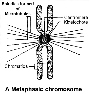
 (i) The complete disintegration of the nuclear envelope marks the start of the second phase of mitosis. By this stage condensation of chromosomes is completed. At this stage, metaphase chromosome appears to be made up of two sister chromatids, which are held together by the centromere.
(ii) Small disc shaped structures called kinetochores, serve as the sites of attachment of spindle fibres to the chromosomes that are moved into position at the centre of the cell.
Hence, the metaphase is characterised by all the chromosomes coming to lie at the equator and getting aligned along metaphase plate or equatorial plate through spindle fibres to both the poles.This chromosomal movement to equator is called as congression.
(iii) Spindle fibres which connect the pole to the chromosomes at the kinetochores are called chromosomal fibres (Tractile fibres) and those which extend without interruption from one pole to other are the continuous fibres.
(iv) The morphology of chromosomes can be easily studied at this stage as they are in a highly condensed stage and are distinctly visible. Chromosomes can be counted at this stage.
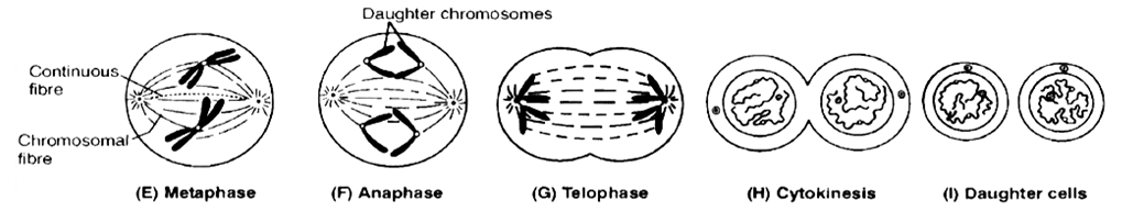
(c) Anaphase
(i) It is of shortest duration.
(ii) The centromere of each chromosome splits into two, resulting into daughter chromatids (chromosome of future nuclei).
(iii) These begin to migrate towards respective poles. The centromeres always lead the daughter chromosomes (towards the poles) and the arms trail behind.
(iv) As the daughter chromosomes move apart, fine microtubules in the form of interzonal fibres appear in between them.
(v) Expansion of interzonal fibres and dissolution of microtubules of chromosomal fibres is supposed to be the possible reason for anaphasic movement.
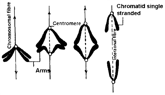
(vi) The daughter chromosomes now assume V, J and I shapes depending upon the position of centromere on them. Thus shape of chromosomes can be observed during this phase.
(vii) Finally, the polar migration of daughter chromosomes after reaching the opposite poles end, which mark the end of anaphase.
(d) Telophase :
(i) Daughter chromosomes undergo decondensation and uncoiling at each pole, so they lose their individuality.
(ii) The chromatin gets surrounded by discontinuous segments of nuclear membrane from elements of ER, which become fully developed at the end of telophase.
(iii) New nucleoli. ER and golgi complex reform.
(iv) The spindle fibres and astral rays gradually disintegrate and disappear. These become absorbed in the cytoplasm.
(v) Each daughter nucleus now enters into the interphase of cell cycle. The end of telophase marks the end of karyokinesis.
Cytokinesis : Karyokinesis results in formation of two nuclei inside a cell and now it is followed by division of cytoplasm (Cytokinesis), thus forming two cells (daughter cells).
(a) Cytokinesis occurs by two methods :
(i) Cell furrow method : This is characteristic of animal cells. Due to absence of rigid cell wall here, the more flexible plasma membrane forms the outer layer of cell. A furrow or invagination appears in plasma membrane at centre of equator, which deepens gradually and finally two daughter cells are separated.
(ii) Cell plate method : This is characteristic of plant cells. Here, vesicles provided by Golgi apparatus unite to form phragmoplasts, which join to form cell plate. Cell plate is first laid down in centre and then proceeds towards periphery (i.e., centrifugal plate formation). Cell wall materials are now laid down on both sides of cell plate, resulting in two daughter cells.
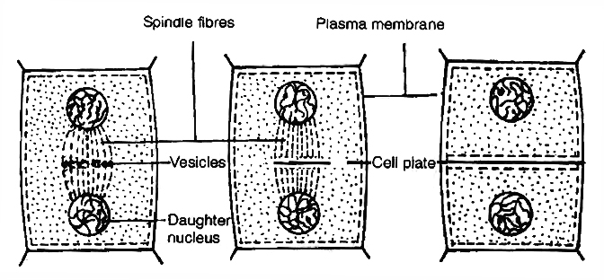
Cytokinesis by cell plate method in plant cells
Concept Builder
Mitotic Poisons
All those substances or chemicals which affect the cells during mitosis or prevent them from entering into normal mitotic divisions are called mitotic poisons. The various mitotic poisons are:
(i) The enzyme ribonuclease acts as poison at prophase. Azide and cyanide also inhibit prophase.
(ii) Mustard gas causes agglutination of chromosomes.
(iii) Chalones also inhibit mitosis. They are small peptides or glycoproteins in the extracellular fluid.
(iv) The alkaloid colchicine inhibits the formation of mitotic spindle (inhibits polymerisation of microtubules) and holds the cells in metaphase. The chromosomes and DNA undergo replication but remain within the same cell. The nucleus does not divide. This increases the number of chromosome sets per cell. This process leads to endopolyploidy or endomitosis in which nucleus contains multiple sets of chromosomes instead of the normal two sets as found in a diploid cell. Such cells are called polyploid cells.
(v) X-rays cause uncontrolled mitosis and induces breakage of chromosomes.
Abnormal Mitosis
(i) Intranuclear mitosis (Premitosis) : In Amoeba, Yeast, other fungi and a number of algae, the nuclear envelope does not degenerate during mitosis. Spindle is intranuclear.
(ii) Dinomitosis : Dinoflagellates possess condensed chromosomes even in non-dividing nuclei. Nuclear envelope does not degenerate. Division of chromosomes occurs with the help of special channels that develop in the nucleus.
(iii) Free Nuclear Division : Sometimes, repeated mitosis occur without subsequent cytokinesis. It produces multinucleated condition, e.g.:-Rhizopus, Vaucheria, Slime moulds, etc.
Significance of Mitosis
(i) It is an equational division since the number of chromosomes and the genetic constitution of the daughter cells remain the same as found in parent cells. Thus, mitosis helps in the survival of a species and continuation of its race. It provides a complete set of genetic information to each cell.
(ii) Mitosis occurs in somatic cells (n or 2n) and gonad cells for the multiplication of cell number. Thus, mitosis is related to the growth of an individual from zygote to adult stage. It also provides an opportunity for the growth and development of organs and body of organisms.
(iii) Mitosis produces new cells to replace the old worn out and injured cells. Thus, it helps in repairing of cells, healing of wounds and regeneration of body cells. Blood cells, intestinal cells and skin cells are regularly regenerated and replaced by mitosis.
(iv) It is a method of restoring nucleocytoplasmic ratio.
(v) Mitosis helps the organisms in both sexual and asexual reproduction.
Mitosis and it's significance
Mitosis and its significance
A cell prepares for cell division by replicating its chromosomes, segregating them, and creating two identical nuclei during the mitotic phase. The cell's contents are often evenly divided into two daughter cells with identical genomes after mitosis.Mitosis is divided into the following four stages:
1. Prophase:
Interphase's S and G2 phases are followed by prophase, the initial step of mitosis. The newly synthesised DNA molecules are not distinct but rather entangled in the S and G2 phases. The beginning of chromosomal material condensing characterises prophase. During the process of chromatin condensation, the chromosomal material is untangled. The centriole, which underwent duplication during interphase's S phase, now starts to migrate in the direction of the cell's opposing poles.Thus, the following distinctive occurrences can indicate the end of prophase:
- Compact mitotic chromosomes are created when chromosomal material condenses.Two chromatids are observed to be joined together at the centromere to form chromosomes.
- The beginning of the mitotic spindle's construction, the microtubules, and the proteinaceous elements of the cell cytoplasm aid in the process.
- The golgi complex, endoplasmic reticulum, nucleolus, and nuclear envelope are absent from cells near the end of prophase when they are observed under a microscope.
2. Metaphase:
The second phase of mitosis begins when the nuclear envelope completely disintegrates, and as a result, the chromosomes are dispersed throughout the cell's cytoplasm. The chromosome condensation process is now complete, and the chromosomes may be seen clearly under a microscope. Therefore, this is the period at which it is easiest to study the shape of chromosomes. The two sister chromatids that make up the metaphase chromosome at this point are joined by the centromere. Kinetochore refers to a little disc-shaped structure at the centromere surface. These structures act as the points of attachment for the spindle fibres, which are created by the spindle fibres, to the chromosomes that are placed at the cell's centre.Thus, all of the chromosomes align at the equator during the metaphase, with one chromatid of each chromosome attached by its kinetochore to spindle fibres from one pole and its sister chromatid connected by its kinetochore to spindle fibres from the opposite pole. The term "metaphase plate" refers to the chromosomes' alignment plane during metaphase. Metaphase's primary characteristics are:
- Spindle fibres adhere to chromosomal kinetochores.
- Chromosomes are transferred to the spindle equator, where they are positioned along the metaphase plate and along the spindle fibres to both poles.
3. Anaphase:
Each chromosome on the metaphase plate splits simultaneously at the start of anaphase, and the two daughter chromatids, which are now known as the chromosomes of the future daughter nuclei, start to move in opposite directions. The centromere of each chromosome is at the pole and, as a result, at the leading edge, with the arms of the chromosome trailing behind as they advance away from the equatorial plate. Events that define the anaphase stage are:
- Centromeres divide, and chromatids dissociate.
- Chromatids shift to the polar opposites
4. Telophase:
The chromosomes that have reached their respective poles decondense and lose their identity at the beginning of the last stage of mitosis, known as telophase. The individual chromosomes are no longer visible, and the two poles tend to accumulate a mass of chromatin material. The essential events at this stage include:
- Chromosomes cluster at opposing spindle poles, losing their identity as distinct elements.
- The nucleolus, Golgi complex, and ER remodel themselves
- The nuclear envelope forms around the chromosomal clusters.
Cytokinesis:
In addition to segregating duplicated chromosomes into daughter nuclei (karyokinesis), mitosis also divides the cell into two daughter cells by a separate process known as cytokinesis, marking the completion of cell division. This occurs when a furrow forms in the plasma membrane of an animal cell. The cytoplasm of the cell is split in half by the furrow, which eventually merges in the centre. Plant cells, on the other hand, are surrounded by a cell wall that is rather inextensible; as a result, they go through cytokinesis using a different process. In plant cells, wall construction begins in the cell's middle and extends outward to meet the lateral walls that already exist.The basic precursor known as the cell-plate, which symbolises the middle lamella between the walls of two neighbouring cells, is formed before the new cell wall can be fully formed. Organelles like mitochondria and plastids are dispersed between the two daughter cells during cytoplasmic division. In some organisms, cytokinesis does not follow karyokinesis, which results in a multinucleate state and the development of syncytium.
Significance of Mitosis :
Only diploid cells often undergo mitosis, also known as equational division. However, haploid cells can also divide through mitosis in some lower plants and social insects. Understanding the importance of this division in an organism's life is crucial. Typically, mitosis produces daughter cells that are diploid and have the same genetic makeup. Mitosis is responsible for multicellular organisms' growth. The ratio of the nucleus to the cytoplasm is disturbed as a result of cell expansion. Therefore, cell division is required to re-establish the nucleo-cytoplasmic ratio. Cell repair is one of mitosis' most important functions.Blood cells, stomach lining cells, and the outermost layer of the epidermis all undergo continuous replacement. Plants grow continuously throughout their lives as a result of mitotic divisions in the meristematic tissues known as the apical and lateral cambium.
meiosis and it's significance
- Books Name
- ACME SMART COACHING Biology Book
- Publication
- ACME SMART PUBLICATION
- Course
- CBSE Class 11
- Subject
- Biology
MEIOSIS
The term meiosis was first introduced by Farmer and Moore. It can be defined as, "The reductional division occurring only in diploid cells for the formation of haploid cells in which the number of chromosomes and the nuclear DNA content are reduced to half and there is recombination of hereditary material."
The key features of meiosis are as follows:
(a) It involves two sequential cycles of nuclear and cell division called meiosis I and meiosis II, but only a single cycle of DNA replication.
(b) Meiosis I can initiate only after the S phase (where parental chromosomes have replicated to produce identical sister chromatids)
(c) It involves pairing of homologous chromosomes and recombination between them.
(d) Four haploid cells are formed at the end of meiosis II.
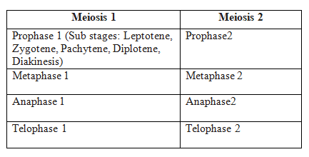
(e) Meiotic events can be grouped under the following phases:
Meiosis-I Meiosis-II
Prophase-I Prophase-II
Metaphase-I Metaphase-II
Anaphase-I Anaphase-II
Telophase-I Telophase-II
Process of Meiosis
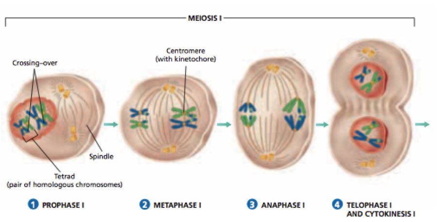
1. Meiosis I or Reductional or Heterotypic division : It starts after the interphase of the cell cycle where DNA duplication occurs in S phase. It results in the reduction of chromosome number to half.
Meiosis I consists of four stages, i.e., Prophase-I, Metaphase-I, Anaphase-I and Telophase-I.
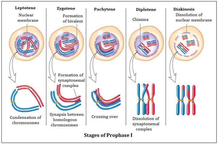
(a) Prophase-I : It is very complex and of very long duration. It is divided into following substages on the basis of chromosome behaviour :
(i) Leptotene or leptonema :
The duplicated centrioles start moving apart. Aster formation occurs.
The chromatin fibres undergo progressive condensation, coiling, shortening and thickening to appear in the form of long, thin condensed filamentous chromosomes.
These possess darkly stained bead like structures called chromomeres along their entire length.
The chromosomes are replicated but the chromatids are not distinguishable due to the presence of nuclear protein between them.
The chromosomes form loops whose ends are attached to the nuclear membrane at attachment plate.
This specific arrangement of chromosomes is often called the "bouquet stage".
(ii) Zygotene or Zygonema or Synaptic stage:
The homologous chromosomes come to lie in pairs.
In each homologous pair of chromosomes, one chromosome comes from mother through ova and is called maternal chromosome and the other chromosome comes from father through sperm and is called paternal chromosome.
This pairing of homologous chromosomes is called synapsis and these paired chromosomes are called bivalents.
Homologous chromosomes in a pair are of the same length, carry the same genes in the same sequence.
Electron micrographs of this stage indicate that chromosome synapsis is accompanied by the formation of complex, tripartite, protienaceous structure called synaptonemal complex.
The number of bivalents in a cell is equal to the number of haploid chromosomes. The chromatids are still not visible.
(iii) Pachytene or Pachynema :
The bivalent chromosomes become more thickened, shortened and condensed.
Each chromosome of a bivalent consists of two chromatids and are called sister chromatids.
The two sister chromatids of one chromosome in a homologous pair with regard to the other chromosome of pair are called non-sister chromatids.
Four chromatids in a pair of homologous chromosomes constitute the tetrad.
This stage is characterised by the appearance of recombination nodules, the sites at which crossing over occurs between non-sister chromatids of the homologous chromosomes.
Crossing over is the exchange of genetic material between two homologous chromosomes and also an enzyme-mediated process and the enzyme involved is called recombinase.
The best theory to explain crossing over is Darlington's theory of breakage and union :
(a) The enzyme endonuclease will help in the development of breaks (nicking.)
(b) The formation of gaps in the nicks is done by exonuclease.
(c) The separation of chromatid segments in gaps is done by U-protein or unwindase.
(d) Re-annealing (rejoining) is done by R-protein or Re-annealing protein.
The newly constituted chromosomes, thus become different from the previous set of chromosomes.
This results in the formation of new characters (recombinants) and ultimately variations in the population. These variations form the basis of evolution.
(iv) Diplotene or Diplonema :
Longest phase of prophase-I. There is dissolution of synaptonemal complex, so the recombined homologous chromosomes will start separating i.e., desynapsis.
The separation of the homologous chromosomes is not completed.
They remain attached to one or more points where crossing over has occured.
These point of attachment are called as Chiasmata (X-shaped structures).
In oocytes of some vertebrates, the diplotene can last for months or years.
Suspended diplotene stage is called as Dictyotene stage.
At this stage the chromosomes decondense and are engaged in rapid synthesis of RNA. Terminalization of chiasmata starts.
(v) Diakinesis :
This stage is marked by complete terminalisation of chiasmata.
The nucleoli and the nucleolar membrane disintegrate and disappear.
The spindle fibres extend from one pole of paired centrioles to the other pole of paired centrioles.
Astral rays and asters become fully developed.
Centrioles are absent in plant cells and thus, no asters are formed.
This marks the end of prophase-I and transition to metaphase-I.
(b) Metaphase-I :
The bivalents arrange themselves on the equator of the bipolar spindle.
Since, there are two centromeres in each bivalent, the centromeres of all the bivalents produce a double equatorial or metaphasic plate.
Each plate will have half the number of diploid chromosomes.
The microtubule from the opposite poles of the spindle attach to the pair of homologous chromosomes.
(c) Anaphase-I :
One chromosome of each homologous pair moves to the opposite poles with recombined characters of both paternal and maternal chromosomes.
The movement of chromosomes occurs along the path of their tractile fibres (chromosome fibres).
Each chromosome consists of two chromatid threads joined by a centromere.
There is no division of centromere.
At the end of anaphase-I half of the chromosomes reach one pole and other half reach on to the other pole.
There occurs true reduction in the number of chromosomes at this stage.
(d) Telophase-I :
The haploid number of chromosomes which has reached at each pole, undergo elongation, uncoiling and decondensation and changes into chromatin network.
Although in many cases the chromosomes do undergo some dispersion, they do not reach the extremely extended state of the interphase nucleus.
The nuclear envelope develops from the elements of ER around the chromatin fibres. New nucleolus is formed.
The astral rays and spindle fibres disintegrate and disappear.
The two daughter nuclei, each containing haploid number of chromosomes are formed.
The end of telophase-I marks the end of karyokinesis-I also.
Karyokinesis-I may be followed by cytokinesis-I.
By a cleavage furrow in an animal cell and a cell plate in a plant cell, the diploid parental cell is divided into two haploid daughter cells each having half the number of chromosomes, but double content of DNA.
Interkinesis or intrameiotic interphase :
Each daughter cell formed from meiosis-I sometimes undergoes interkinesis, where there is no replication phase of chromosomes and no duplication of genes.
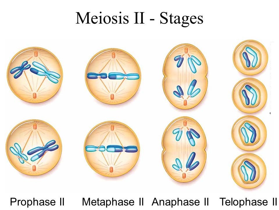
2. Meiosis II or Homotypic Division : It resembles mitotic division.
Meiosis-II is necessary because each daughter cell formed from meiosis-I contains chromosomes, each having two chromatids, each chromatid with 2C amount of DNA. This reduction in DNA from 2C to 1 C DNA occurs only in Meiosis-II.
It consists of following four phases :
(a) Prophase-II: One pair each of the centrioles migrate to the opposite poles. The nuclear membrane and the nucleolus disintegrate and disappear. The chromatin fibres undergo compaction to appear in the form of distinct chromosomes. Each chromosome consists of two chromatid threads, joined by a centromere.
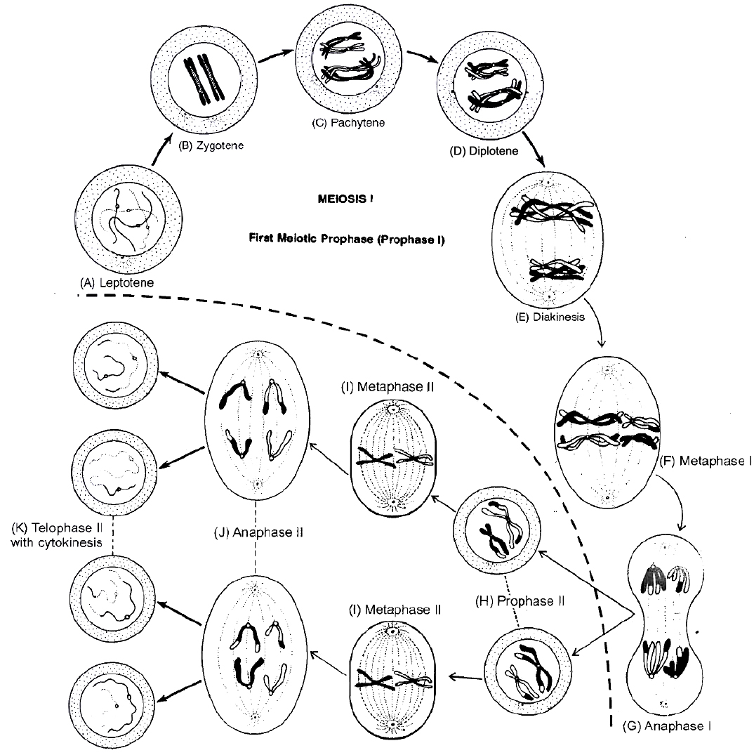
Different Stage of Meiosis
(b) Metaphase II : Chromosomes come to lie on the equator of the cell and thus, form a Single equatorial or metaphasic plate. The spindle fibres become attached to both kinetochores of the centromere of each chromosome.
(c) Anaphase-II: Begins with simultaneous splitting of the centromere of each chromosome that allows them to move towards opposite poles of the cell.
(d) Telophase-II: The daughter chromosomes on the opposite poles become decondensed to form chromatin fibres. The nuclear envelope develops from ER. New nucleolus is reorganised. The spindle fibres and astral rays disintegrate and disappear. It marks the end of the telophase-II and karyokinesis-II . It is followed by cytokinesis (similar in mitosis) which divides each cell into two daughter cells, resulting in formation of tetrad of cells.
Concept Builder
(i) Meiosis is commonly studied using onion buds.
(ii) Meiosis was first demonstrated by Van Benden and described by Winiwarter.
(iii) Gametic meiosis is also called as terminal meiosis.
(iv) Zygotic meiosis is also called as initial meiosis.
(v) Sporogenic meiosis is also called as intermediate meiosis.
(vi) Cytokinesis: Cytokinesis can be of two types, successive and simultaneous. In successive type, cytokinesis occurs after every nuclear division. The four cells formed by successive cytokinesis can be arranged either in a linear or isobilateral tetrad.
In the simultaneous type, cytokinesis takes place only at the end of both the divisions. The nuclei are generally arranged in the form of a tetrahedron.
Significance of Meiosis
1. Meiosis forms gametes that are essential for sexual reproduction.
2. Meiosis maintains the fixed number of chromosomes generation after generation in sexually reproducing organisms. It is essential since the chromosome number becomes double after fertilization.
3. Meiosis is the main cause of production of variations, which are very important for evolutionary process.
Some Major Differences between Mitosis and Meiosis

AMITOSIS OR DIRECT CELL DIVISION
It is the method of asexual reproduction, which occurs, in acellular organisms like bacteria, protozoans, diseased cells, old cells, mammalian cartilage cells and in foetal membranes.
it was first discovered by Remak. It is also called direct cell division.
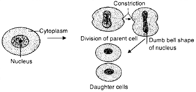
During amitosis, the nucleus of the cell elongates.
Then, a constriction appears in the nucleus which gradually deepens and divides the nucleus into two daughter nuclei.
Finally, a constriction appears in the cytoplasm which divides the cytoplasm and the nuclei into two daughter cells, each with a nucleus.
In this division, no spindle formation and no distinct chromosome formation occurs. Nuclear envelope remains intact.
The daughter cells are approximately the two equal halves of a parental cell.
Concept Builder
(i) Meiosis is commonly studied using onion buds.
(ii) Meiosis was first demonstrated by Van Benden and described by Winiwarter.
(iii) Gametic meiosis is also called as terminal meiosis.
(iv) Zygotic meiosis is also called as Initial meiosis.
(v) Sporogenic meiosis is also called as Intermediate meiosis.
(vi) If we have to calculate the number of mitotic divisions for the formation of n number of cell, it will be n-1 i.e. for getting 100 cells 99 mitotic divisions are required.
(vii) Number of generations of mitosis required can be calculated (Like mitosis generations required for producing 128 cells are 7) using 2n, n is number of generation.
(viii) In animal cell, mitosis is called as Amphlastral (Spindle is associated with 2 asters).
(ix) In plant cells, the mitosis is called as Anastral (no aster, no centriole).
(x) If mitosis is extranuelear, it is Eumitosls.
(xi) If mitosis is intranuclear, it is called as Premitosis. If centrioles are present then it is called as centric.
(xii) Cytokinesis: Cytokinesis can be of two types, successive and simultaneous. In successive type, cytokinesis occurs after every nuclear division. The four cells formed by successive cytokinesis can be arranged either in a linear or isobilateral tetrad.
In the simultaneous type, cytokinesis takes place only at the end of both the divisions. The nuclei are generally arranged in the form of a tetrahedron.
Formulae Chart :
1. Number of mitotic division for the formation of n number of cells.
Example : For getting 100 cells 99 mitotic division are required.
![]()
2. Number of generations (n) of mitosis for producing 'x' cells.

3. Number of meiosis for the formation of 'n' seeds/grains/fruits.

SUMMARY
1. According to the cell theory, cells arise from pre-existing cells. The process by which this occurs is called cell division.
2. Any sexually reproducing organism starts its life cycle from a single-celled zygote.
3. Cell division does not stop with the formation of the mature organism but continues throughout its life cycle.
4. The stages through which a cell passes from one division to the next is called the cell cycle.
5. Cell cycle is divided into two phases called (i) Interphase -a period of preparation for cell division, and (ii) Mitosis (M phase) -the actual period of cell division.
6. Interphase is further subdivided into G1, Sand G2. G1-phase is the period when the cell grows and carries out normal metabolism. Most of the organelle duplication also occurs during this phase.
7. S-phase marks the phase of DNA replication and chromosome duplication.
8. G2-phase is the period of cytoplasmic growth.
9. Mitosis is also divided into four stages namely prophase, metaphase, anaphase and telophase.
10. Chromosome condensation occurs during prophase.
11. Simultaneously, the centrioles move to the opposite poles.
12. The nuclear envelope and the nucleolus disappear and the spindle fibres start appearing.
13. Metaphase is marked by the alignment of chromosomes at the equatorial plate.
14. During anaphase, the centromeres divide and the chromatids start moving towards the two opposite poles.
15. Once the chromatids reach the two poles, the chromosomal elongation starts, nucleolus and the nuclear membrane reappear. This stage is called the telophase.
16. Nuclear division is then followed by the cytoplasmic division and is called cytokinesis.
17. Mitosis thus, is the equational division in which the chromosome number of the parent is conserved in the daughter cell.
18. In contrast to mitosis, meiosis occurs in the diploid cells, which are destined to form gametes. It is called the reduction division since it reduces the chromosome number by half while making the gametes.
19. In sexual reproduction when the two gametes fuse the chromosome number is restored to the value in the parent.
20. Meiosis is divided into two phases -meiosis-I and meiosis-II. In the first meiotic division the homologous chromosomes pair to form bivalents, and undergo crossing over.
21. Meiosis I has a long prophase, which is divided further into five phases. These are leptotene, zygotene, pachytene, diplotene and diakinesis.
22. During metaphase-I , the bivalents arrange on the equatorial plate.
23. In anaphase-I, homologous chromosomes move to the opposite poles with both their chromatids. Each pole receives half the chromosome number of the parent cell.
24. In telophase-I, the nuclear membrane and nucleolus reappear.
25. Meiosis-II is similar to mitosis.
26. During anaphase-II, the sister chromatids separate. Thus at the end of meiosis, four haploid cells are formed.
meiosis and it's significance
Meiosis and its significance
Meiosis is a type of cell division that results in the production of four gamete cells and a 50% reduction in the number of chromosomes in the parent cell. To develop egg and sperm cells for sexual reproduction, this process is necessary. There are four haploid daughter cells formed during meiosis (containing half as many chromosomes as the parent cell).Following are the main characteristics of meiosis:
- Meiosis requires just one cycle of DNA replication but two successive cycles of nuclear and cell division called meiosis I and meiosis II.
- After the paternal chromosomes have duplicated to form identical sister chromatids at the S phase, meiosis I begins.
- Meiosis involves the pairing and recombination of homologous chromosomes.
- At the end of meiosis II, four haploid cells are produced.
Events of Meiosis are classified as:
Meiosis 1
Prophase I: When compared to the prophase of mitosis, the prophase of the first meiotic division is often longer and more complex. Based on chromosomal behaviour, it has been further split into the following five phases: Leptotene, Zygotene, Pachytene, Diplotene, and Diakinesis.
Leptotene: The chromosomes begin to condense at this stage and are connected to the nuclear membrane via their telomeres.
Zygotene: A synaptonemal complex forms between homologous chromosomes at the start of synapsis.
Pachytene - During this stage, genetic material is transferred between chromatids that are not sisters. This is called crossing over.
Diplotene - At this stage, homologous pairs are still connected at the chiasmata after synapsis has ended and the synaptonemal complex has vanished.
Diakinesis: Before metaphase 1, the nuclear membrane finally breaks down and the chromosomes are entirely condensed.
Metaphase I: The bivalent chromosomes line up on the equatorial plate during metaphase I. The spindle's opposing poles' microtubules connect to the pair of homologous chromosomes.
Anaphase I: Sister chromatids are still connected at their centromeres during anaphase I, but homologous chromosomes split.
Telophase I: Cytokinesis occurs after the nuclear membrane and nucleolus re-emerge, and this is known as the "diad of cells." Even though the chromosomes do experience some dispersion in many instances, they rarely reach the interphase nucleus's very stretched state. Interkinesis, which occurs between the two meiotic divisions, is typically a transient stage. Prophase II, which is substantially less complex than prophase I, comes after interkinesis.
Meiosis 2:
Prophase II: Following cytokinesis, meiosis II begins right away, typically before the chromosomes have fully expanded. Meiosis II resembles a typical mitosis in contrast to meiosis I. By the end of prophase II, the nuclear membrane is gone. Chromosomes once more become condensed.
Metaphase II: Chromosomes align at the equator during metaphase II, and sister chromatids' kinetochores get attachments of microtubules from the spindle's opposing poles.
AnaphaseII: Beginning with the simultaneous division of each chromosome's centromere (which had been binding the sister chromatids together), anaphase II allows the chromosomes to migrate toward their respective poles of the cell.
Telophase II: Telophase II marks the completion of meiosis, during which the two chromosomal groups are once more encased in a nuclear membrane. Cytokinesis then takes place, culminating in the production of a tetrad of cells, or four haploid daughter cells.
Significance of Meiosis:
Even though the process itself paradoxically reduces the number of chromosomes by half, meiosis is the mechanism that allows sexually reproducing animals to maintain the particular chromosomal number of each species throughout generations. From one generation to the next, it also makes the population of organisms more genetically variable. For the process of evolution, variations are crucial.
