 Science Made Easy
 ACERISE INDIA
tructure and functions of animal and plant tissues (four types in animals; meristematic and permanent tissues in plants).
- Books Name
- Science Made Easy Science Book
- Publication
- Science Made Easy
- Course
- CBSE Class 9
- Subject
- Science
Introduction
→ A group of cells that are similar in structure and/or work together to achieve a particular function
forms a tissue.
→ Most of the tissues in plants are supportive, which provides them with structural strength.
→ These tissues are dead, since dead cells can provide mechanical strength as easily as live ones
and need less maintenance.
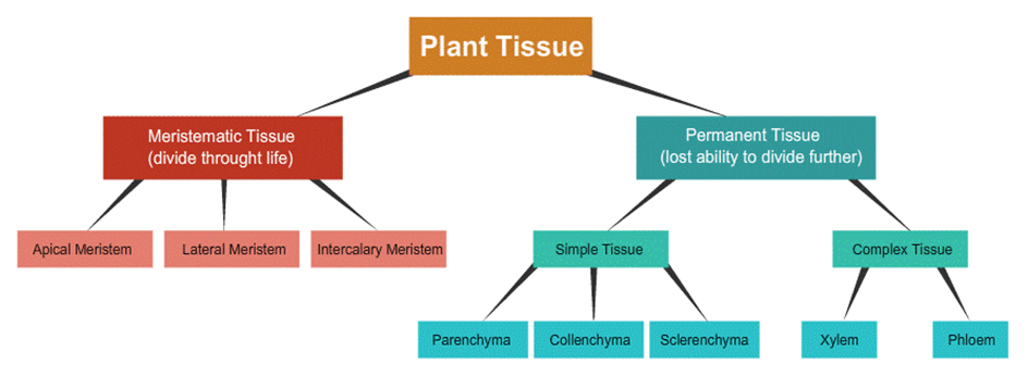
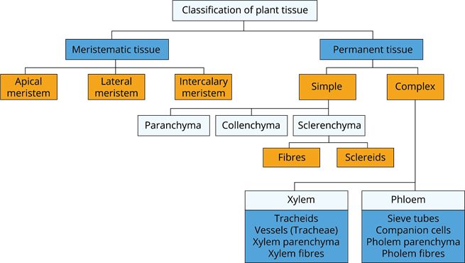
→ Plant Tissues are of two types Meristematic & Permanent tissues.
Meristematic Tissue
→ These are simple living tissues having thin walled compactly arranged immature cells which are
capable of division and formation of new cells.
Features of Meristematic tissues:
→ Thin primary cell wall (cellulosic).
→ Intercellular spaces are absent (compact tissue).
→ Generally vacuoles are absent, dense cytoplasm & prominent nuclei are present.
→ Large numbers of cell organelles are present.
→ Active metabolic state, stored food is absent.
→ Actively dividing cells are present in growing regions of plants, example: root & shoot tips.
Classification of Meristematic Tissues on the Basis of Origin
• Primary (Promeristem)
→ Derived directly from the meristems of embryo.
→ They consist of cells derived from primary meristem.
→ They add to primary growth of plants.
• Secondary Meristematic Tissues
→ Formed by permanent tissues.
→ These are having cells derived from primary permanent tissue.
→ They usually add to the diameter of plants.
Classification of Meristematic Tissues on the Basis of Location
• Apical Meristem
→ It is present at the growing tips of stems and roots.
→ Cell division in this tissue leads to the elongation of stem & root, thus it is involved in primary
growth of the plant.
• Intercalary Meristem
→ It is present behind the apex.
→ It is the part of apical meristem which is left behind during growth period.
→ These are present at the base of leaf & internode region.
→ These lead to the increase in the length of leaf (Primary), example: in grass stem, bamboo stem
mint stem etc.
• Lateral Meristem
→ It is also called as secondary meristem.
→ It occurs along the sides of longitudinal axis of the plant.
→ It gives rise to the vascular tissues.
→ Causes growth in girth of stem & root.
→ They are responsible for secondary growth.
Permanent Tissue
→ The permanent tissues are composed of those cells which have lost their capability to divide.
→ They have definite shape, size and thickness. The permanent tissue may be dead or living.
→ The division & differentiation of the cells of meristematic tissues give rise to permanent tissues.
→ In cell differentiation, developing tissue and organs change from simple to more complex form
to become specialized for specific functions.
→ The cells of permanent tissue loose the capacity to divide and attain a permanent shape, size
and function.
• Permanent tissues are classified into two types on the basis of Structure and Composition i.e.
Simple Permanent Tissues and Complex Permanent Tissues.
Simple Permanent Tissues
→ These are made up of same type of cells which are similar structurally and functionally.
→ They include two types of tissue Protective tissues and Supporting Tissues.
• Protective Tissues: These tissues are primarily protective in function.
→ They consist of Epidermis and Cork/Phellem.
(i) Epidermis
→ Epidermis forms one cell thick outermost layer of various body organs of plants such as leaves,
flowers, stems and roots.
→ Epidermis is covered outside by cuticle. Cuticle is a water-proof layer of waxy substance called
cutin which is secreted by the epidermal cells.
→ Cuticle is very thick in xerophytes.
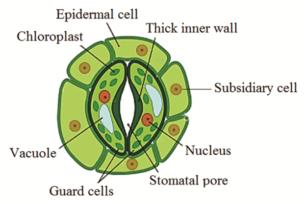
→ Cells of epidermis of leaves are not continuous at some places due to the presence of small
pores called as stomata.
→ Each stomata is guarded by a pair of bean-shaped cells called as guard cells. These are the one
epidermal cells which possess chloroplasts, the rest being colourless.
Functions of Epidermis
→ The main function of epidermis is to protect the plant from desiccation and infection.
→ Cuticle of epidermis cuts the rate of transpiration and evaporation of water and prevents wilting.
→ Stomata in epidermis allow gaseous exchange to occur during photosynthesis respiration.
→ Stomata also helps in transpiration.
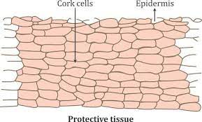
(ii) Cork or Phellem
→ In older roots and stems, tissues at the periphery become cork cells or phellem cells.
→ Cork is made up to dead cells with thick walls and do not have any intercellular spaces.
→ The cell walls in cork deposit waxy substance called as suberin.
→ The cells of cork become impermeable to water and gases due to the deposition of suberin.
→ The cork cells are without any protoplasm but are filled with resins or tannins.
Functions of Cork
→ Cork is protective in function. Cork cells prevent desiccation, infection and mechanical injury.
→ Imperviousness, lightness, toughness, compressibility and elasticity make the cork commercial
valuable.
→ Cork is used for insulation, as shock absorber in linoleum.
→ Cork is used in the making of a variety of sport goods such as cricket balls, table tennis, shuttle
cocks, wooden paddles etc.
• Supporting Tissues: These are supportive in function.
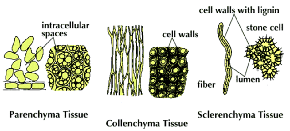
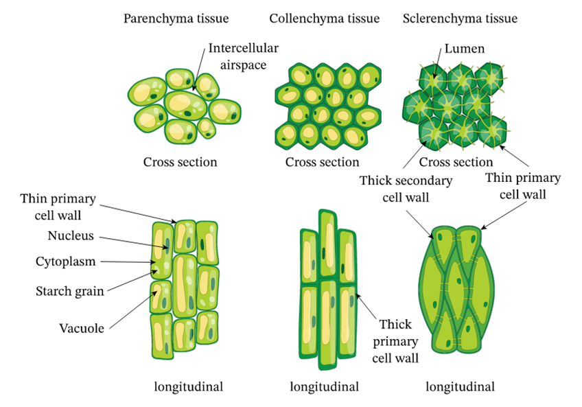
→ There are three types of Supporting tissues i.e. Parenchyma, Collenchyma and Sclerenchyma.
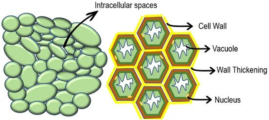
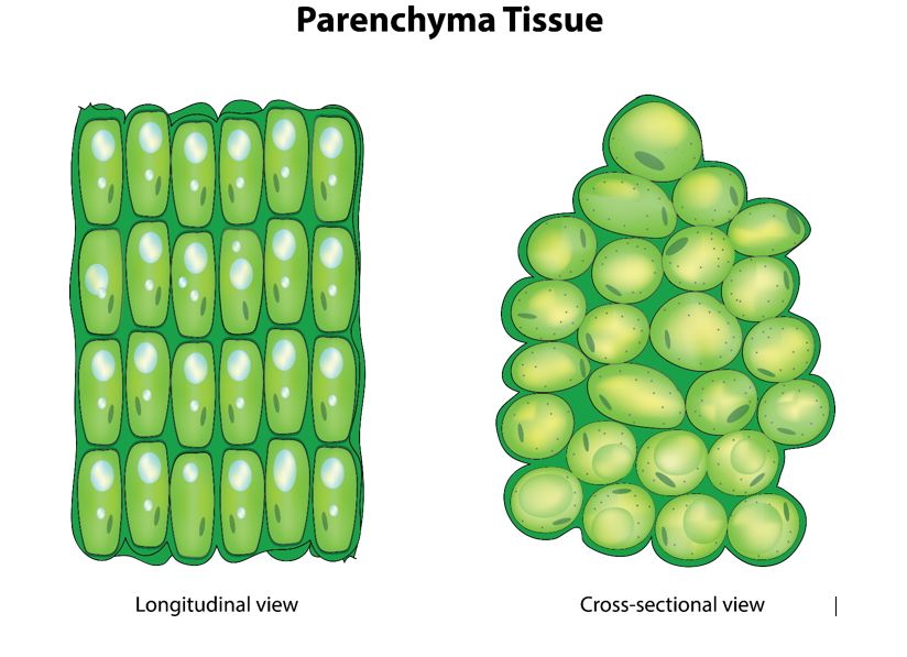
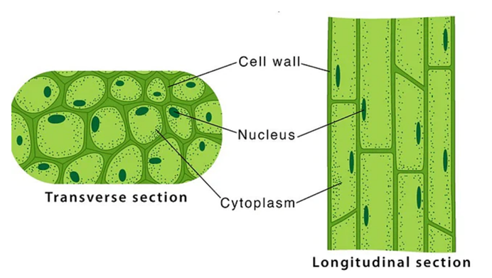
(i) Parenchyma
→ It is the fundamental tissue.
→ Tissue first time evolved in bryophyte.
→ Thin walled cells, oval or spherical in structure.
→ Cell wall mainly composed of cellulose & pectin.
→ Large central vacuole for food & water storage.
→ Primary function is food storage.
→ Some parenchyma involved in excretory substance storage are so called as idioblast, storing
such as resin, tannin, gums & oils.
→ In typical parenchyma chlorophyll is absent.
→ Chloroplast containing parenchyma tissue are chlorenchyma which perform
photosynthesis such as mesophyll of leaves.
→ In hydrophytic plants aerenchyma (a type of parenchyma containing air spaces) provides
buoyancy.
→ Parenchyma provides turgidity to cells.
(ii) Collenchyma
→ It is the living mechanical tissue.
→ Elongated cells with thick corners.
→ Localized cellulose & pectin thickening.
→ Provides flexibility to plant parts & easy bending of various parts of plant.
→ Present only in herbaceous dicot stem.
→ Present at thin margin of leaves.
→ Few chloroplasts may be present.
→ Gives mechanical strength & elasticity to the growing stems.
(iii) Sclerenchyma (Scleras–hard) Strengthening tissue.
→ Composed of extremely thick walled cells with little or no protoplasm.
→ Cells are dead & possess very thick lignified walls.
→ Lignin is water-proof material.
→ Intercellular spaces are absent.
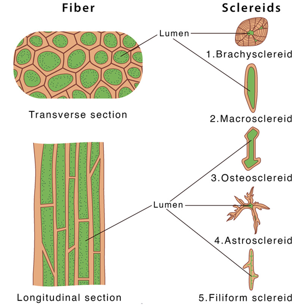
• Cells of sclerenchyma are of two types Sclereids and Fibres.
• Sclereids
→ These are also called grit cells or stone cells.
→ These are small cells, where lumen is so small due to higher thickening of cell wall, as present in
drup fruit (mango, coconut, walnut) in legume seeds (Macrosclereid).
• Fibers
→ They are very long, narrow, thick, lignified cells. Lumen is large as compared to sclereids.
They are generally 1-3 mm long.
→ In the thick walls of both the fibres and sclereids are present thin areas called as pits.
→ Sclrenchyma Fibres are used in the manufacture of ropes, mats & certain textile fibres.
→ Jute and coir are obtained from the thick bundle of fibres.
Difference between Parenchyma, Collenchyma and Sclerenchyma

Complex Permanent Tissues
→ It consists of more than one type of cells which work together as a unit.
→ It helps in transportation of organic materials, water & minerals.
→ It is also known as conducting or vascular tissue.
→ Xylem & phloem together form vascular bundles.
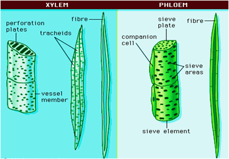
Xylem
→ It is also known as wood and is a vascular and mechanical tissue.
→ Thick walled cells are found in the form of tubular passages.
→ Xylem consists of four types of cells called as elements Tracheids, Vessels, xylem parenchyma
and xylem sclerenchyma.
(i) Tracheids
→ They are elongated angular dead cells (primitive elements) mainly involved in conduction of
water and minerals in gymnosperms.
(ii) Vessles
→ They are advance element (generally found in angiosperms).
→ Vessels are cylindrical tube like structures placed one above the other end to end which form a
continuous channel for efficient conduction of water.
(iii) Xylem parenchyma
→ They are small & thick walled parenchymatous cells subjected for storage of starch (food).
(iv) Xylem sclerenchyma
→ Thy are non-living fibres with thick walls and narrow cavities provide mechanical support.
→ Except xylem parenchyma all other xylem elements are dead.
→ The annual rings present in the trunk of a tree are xylem rings.
→ By counting the number of annual rings, we can determine the age of a tree.
Phloem
→ They also consist of both parenchymatous and schlerenchymatous cells.
→ Phloem consists of four types of element which are Sieve tubes, Companion cells, Phloem fibre
and Phloem parenchyma.
(i) Sieve tubes
→ Sieve tubes are slender tube like structures made up of elongated, thin walled cells placed end to
end.
→ The end walls of sieve tube cells are perforated by numerous pores, called as sieve plates.
→ Nucleus of sieve cell degenerates at maturity. However, cytoplasm persists, because of
protoplasmic continuation of sieve tube with companion cell through plasmodesmata.
→ Sieve cells possess slime protein or protein which is concerned with growth and repair of sieve
cells.
(ii) Companion cells
→ Companion cells have dense cytoplasm and prominent nuclei.
→ Sieve cells & companion cells are so called sister cells because they originate from single mother
cell.
(iii) Phloem fibre
→ They give mechanical support to sieve tubes.
(iv) Phloem parenchyma
→They store food and help in radial conduction of food.
(v) Leptome
→ Main part of phloem involved in conduction of food, which is sieve tube.
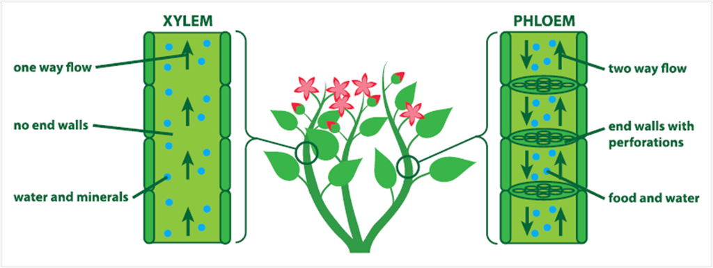
→ In xylem, only unidirectional movement is possible while in phloem bidirectional movement can
occur.
→ In phloem, except phloem sclerenchyma all elements are living.
1. Introduction and Plant Tissues
Tissues
Introduction
• Living organisms in this world comprise of cells.
• There are unicellular as well as multicellular organisms present in this world.
• In unicellular organisms, the only single cell is capable of performing several functions such as Respiration, Digestion and Clearing of the cell.
• In multicellular organisms, there is a division of labour. There are different types as well as groups of cells that perform different functions in a multicellular organism.
• Cells form groups cells that need to perform a single task often group together.
• This grouping of cells together to perform a function efficiently is called a Tissue. For Example, Muscles and Blood.
• The tissue cells have the same structure and they perform the same function.
Plant Tissues
Types of Plant Tissues
Meristematic Tissue
• Only certain parts of a plant tend to grow. The tissues located in such parts are called meristematic tissues.
• They have the capability to divide themselves and form new tissues. They have thin cell wall made of cellulose. Also have dense nucleus and cytoplasm but lack vacuoles.
• They can further we classify differently based on the areas of the plants where they are located -
o Apical
o Lateral
o Intercalary
Location of meristematic tissue
|
Apical Meristem |
Lateral Meristem |
Intercalary Meristem |
|
|
|
Why there are no vacuoles in the intercalary meristem?
- Vacuoles are responsible for storage of food in water. The intercalary tissues do not store them. They are rather responsible for manufacturing them.
- Moreover, vacuoles contain sap which provides rigidity to a cell. This property of vacuoles may not allow the intercalary tissues to divide and manufacture new cells. Hence vacuoles are not present in them.
1. Introduction and Plant Tissues
- Books Name
- Yash Tyagi Coaching Science Book
- Publication
- ACERISE INDIA
- Course
- CBSE Class 9
- Subject
- Science
Chapter-2
Tissues
Introduction and Plant Tissues
Introduction
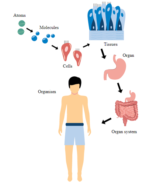
As you all know that we have a complex body organization starting from cellular level to a well-developed individual. If we look at the hierarchy of organization we see that:
Cells → Tissue→ organ→ organ system→ organism
As far as cells are concerned, we have studied that cells are structural and functional units of life. The cells, when formed, undergo the process of differentiation according to the functions that they will perform. The cells that have the same structure, features and perform the same functions form a particular type of tissue. Let us start with the chapter where we will learn all about it.
The cells which are specialized in a function are grouped together and form a particular type of tissue. We have different tissues in our body that performs different functions like:
1. They play the role of protection.
2. They help in control and coordination.
3. They help in transportation.
4. They act as insulators.
5. Many more functions are performed.
Let us learn about Plant tissue
Types of plant tissues
They are classified into mainly two types as given below
- Meristematic tissue
- Permanent tissue
Meristematic tissue and its types
You all have seen that plants have a tendency to grow and keep growing throughout their life. This is because of certain cells in it that keep on dividing and they are called meristematic cells. Let us learn about them. They are formed of cells that have the ability to divide continuously throughout their life and help in increasing the length and girth of the plant.
Characteristics of meristematic cells
- They have thin cellulose cell walls.
- The meristematic cells may be spherical, oval, polygonal or rectangular in shape. The meristermatic cells are compactly arranged.
- Each meristematic cell contains dense or abundant cytoplasm and a single large nucleus.
- The meristematic cells contain few vacuoles or no vacuoles at all.
Occurrence
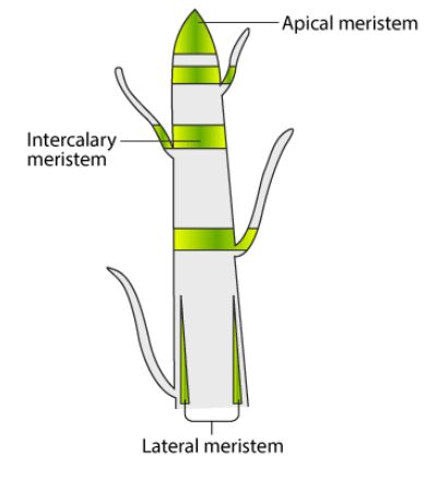
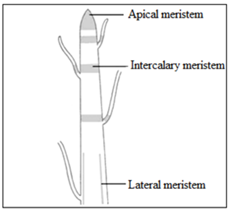
According to their position in the plant, meristems are of three types:
- Apical meristem
- Lateral meristem
- Intercalary meristem
Apical meristems
- It is present at the growing tip of stems and roots. It is at shoot apex and root apex.
- Its function is to increase the height of the plant.
Lateral meristems
- These are found beneath the bark (called cork cambium).
- The function is to help increase in diameter.
Intercalary meristems
- It is located at the base of leaves or at internodes.
- Its function is to increase the number of branches.
2. Permanent Tissues
Permanent Tissue
The cells that are formed by the meristematic tissues often have to take a certain role in the plant and thus, they lose their ability to divide and form more cells. They then become the permanent tissues of the plants.
- Differentiation - The process by which cells of the meristematic tissues convert themselves into a permanent tissue by taking a fixed shape, size and function is called differentiation.
- Types of Permanent Tissues:
- Simple Permanent Tissues
- Complex Permanent Tissues
- Parenchyma
- Chlorenchyma
- Aerenchyma
- Collenchyma
- Sclerenchyma
|
Parenchyma |
Collenchyma |
Sclerenchyma |
|
These tissues are responsible for photosynthesis, storage of food, gaseous exchange and floating of plants. |
These tissues are responsible for providing flexibility to the plants so that they can bend easily. |
These tissues are responsible for making plants hard and rigid. |
|
They are a group of living cells with cell wall made of cellulose. |
They are a group of living cells with cell wall made of cellulose and pectin. |
They are made up of dead cells having cell wall made of lignin. |
|
The parenchyma cells have large intercellular spaces between them. |
They have a little intercellular space in between them. |
The cells do not have any intercellular spaces. |
|
There are thin walls that surround each cell. |
The cells present in these tissues are broad and irregularly thick at corners. |
The cells have a long structure with thick walls. |
|
They are found in leaves and newly formed branches. |
They are present in leaves and stems of a plant. |
They are found in stems, veins of the leaves and coverings of nuts and seeds. |
Chlorenchyma
- These tissues are similar to that of parenchyma but they also contain chlorophyll in them.
- Due to the presence of chlorophyll, they are capable of performing the process of photosynthesis in plants.
Aerenchyma
- They are found in aquatic plants.
- They are also similar in structure to that of the parenchyma but they have large air cavities in them.
- These cavities allow the aquatic plants to float in water.
What is Lignin?
The cell walls of dead cells have a substance called lignin in them which provides rigidity to the cells. Lignin acts as the cement for the cells.
Epidermis
- The outermost layer of the cell is known as the Epidermis.
- It covers the entire plant.
- It is a thin layer of single cells but in places with less water, the epidermis of the plants can become thick in order to avoid frequent water loss.
- The cells are flat and they have no intercellular spaces between them.
- The outer walls of the epidermal cells are thick and the inner walls are thin.
- The epidermal cells often have long hair-like structures in roots which facilitate the absorption of water.
- The main function of the epidermis is to protect the plants from fungi, water loss and any injuries by secrets a wax-like water-resistant substance called as Cuticle on the surface of the plants which protects the plants.
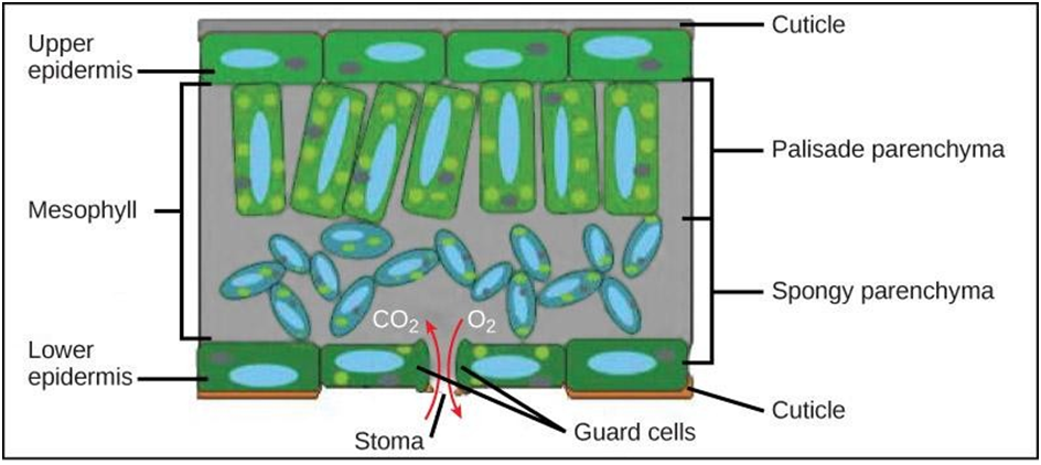
Stomata
- Stomata are pore-like structures that are present in the epidermis of the leaves.
- These pores are enclosed by two cells that have a similar shape as a kidney. These are called Guard Cells of Stomata. Guard cells are modified epidermal cells.
- Guard cells are responsible for the exchange of gases and transpiration.
Why do plants in desert areas have a waxy coating of cutin over them?
The epidermis cells of plants that are found in deserts have a waxy coating of cutin over them because it prevents water loss from the plants surface since water is already scarce in such areas.
Why do branches of old trees are different than the stems of a new plant?
- As a plant grows older the meristematic cells start covering the upper layer of the plants instead of the epidermis.
- These are the dead cells that have no special function in the plants but to provide them rigidity. They make the branches of the plants thick.
- This is often called the Bark or the thick cork of the tree.
- The bark of the trees contains a substance called Suberin which makes it waterproof and does not allow gaseous exchanges.
Complex Permanent Tissues
Complex Permanent Tissues comprise of different kinds of cells. These different types of cells coordinate with each other and perform a common function in these tissues. Two Complex Permanent Tissues are
Xylem and Phloem.
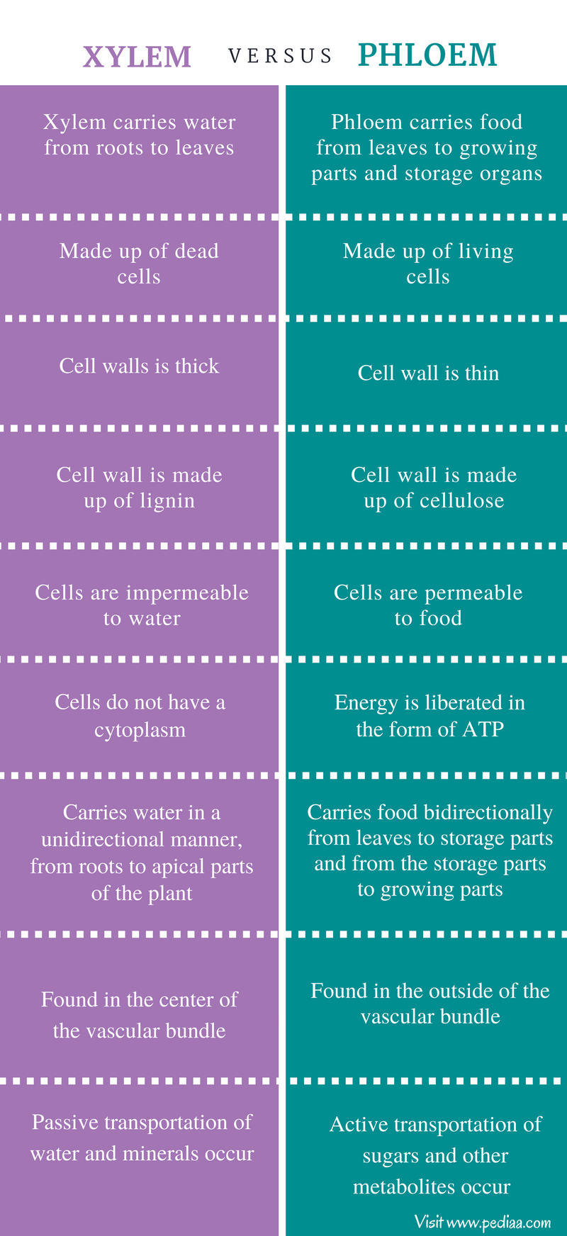
Xylem and Phloem
Similarities between Xylem & Phloem
- Their main function is to carry food and water in the plant.
- Both have a vascular bundle which is a conductive tissue in plants that helps them survive in different environmental conditions.
Xylem
Xylem is made up of dead cells having a thick cell lining. It consists of following elements-
- Tracheids and Vessels – They have broad tubular structure so that we can allow transportation of food and water in the plants vertically.
- Xylem Parenchyma – It stores food and helps in transportation of water horizontally in the plants.
- Xylem Fibers – They support transportation
Phloem
Phloem is made up of living cells and it allows the movement of food from leaves to other parts of the plant. It has the following elements –
Sieve Tubes – Broad shaped cells with porous walls
- Companion Cells – They facilitate the functions of the sieve tubes
- Phloem Fibers – Provide flexibility to the phloem
- Phloem Parenchyma – Stores starch and proteins
|
|
Xylem |
Phloem |
|
Made of |
Dead Cells |
Living Cells |
|
Cell wall thickness |
Thick |
Thin |
|
Cell wall material |
Lignin (rigid) |
Celluloses |
|
Permeability |
Impermeable |
Permeable |
|
Cytoplasm |
None |
Cytoplasm lining |
|
Transports… |
Water & minerals |
Food |
|
Carried to…. |
Leaves |
Growing parts & storage organs |
|
Direction of flow |
Upwards |
Up & down |
|
Tissue alos has … |
Fibres |
Companion cells |
2. Permanent Tissues
- Books Name
- Yash Tyagi Coaching Science Book
- Publication
- ACERISE INDIA
- Course
- CBSE Class 9
- Subject
- Science
Permanent Tissues
Permanent Tissues
There are certain meristematic cells that can not divide throughout their life. They actually lose their tendency to form new cells. As a result, they form another tissue that is Permanent. They have particular functions except forming new cells. These tissues are derived from the meristematic tissue but their cells have lost the power of division. They are of two types:
- Simple permanent tissue
- Complex permanent tissue
Simple permanent tissues
These tissues are composed of cells which are structurally and functionally similar. Thus, these tissues are all made of one type of cells. Depending upon the composition and function they are further classified into three types:
- Parenchyma
- Collenchyma
- Sclerenchyma
Let us learn about the difference between the three:
Parenchyma
This tissue is widely distributed in plant body such as stem, roots, leaves, flowers and fruits. It performs various functions and also has distinct features in xerophytes and hydrophytes. Let us see the features it possesses.
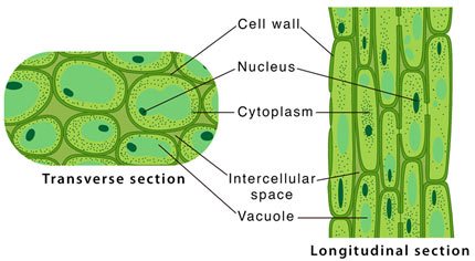
Characteristics
- Parenchyma cells are living and possess the power of division.
- The cells are rounded or isodiametric that isequally expanded in all sides.
- The parenchymatous cells are oval, round, polygonal or elongated in shape.
- The cell wall is thin and enclosed but dense cytoplasm which contains a small nucleus.
- Intercellular spaces are present
Functions
1. Parenchyma serves as a packing tissue.
2. Parenchyma acts as the main support to the stem.
3. Parenchyma serves as food storage tissue.
4. It helps in transporting materials.
5. It allows gaseous exchange.
6. It stores waste products of plants.
7. If chloroplast is present, the parenchyma tissue is called chlorenchyma and it helps in performing photosynthesis.
8. In hydrophytes large air cavities present called arenchyma that provide buoyancy to plants.
Collenchyma
You must have seen that some plant parts can bend without breaking. This feature is due to the presence of a mechanical tissue called collenchymas. They are generally found below epidermis of dicot stem and petiole. They also occur in midribs of dicot leaves but are absent in monocots.
Characteristics
- They are living cells.
- They are characterized by the deposition of extra cellulose at the corners of the cells.
- The intercellular spaces are generally absent.
Functions
- It is a mechanical tissue so, it provides mechanical support to plant and its parts.
- It also provides strength and flexibility.
Sclerenchyma
You all have used and seen coconut for various purposes like performing some rituals, for eating, drinking coconut water, etc. The husk that is found outside the coconut looks like thin fibres and is hard too. This is made up of cells that is Sclerenchyma. Let us study about it.
Characteristics
- They are actually dead cells.
- The walls of these cells are thickened with deposition of lignin.
- There are no intercellular spaces.
Cells of sclerenchyma are basically of two types:
- Fibres
- Sclereids
Fibres: They consist of very long, narrow, thick and lignified cells. They are usually pointed at both ends.
Sclereids: They are irregular in shape. They are also dead and are found under different parts like cortex, pith, phloem, etc.
Both fibres and sclereids have thin areas on them that are called pits.
Functions
- They provide mechanical support andare protective in nature.
Protective tissue
Plants and plant parts also need protection from external factors. So, there are certain tissues that perform this action. Let us learn about them. It includes epidermis and cork (or phellem).
Epidermis
Epidermis is one cell thick layer and is covered with cuticle. It is a waterproof layer as it has a waxy substance called cutin. The cells of the epidermis are elongated and flattened and do not contain any intracellular space. Function of the epidermis is to protect the plant from desiccation and infection.
Cork
As plants grow older, epidermis undergoes certain changes and transforms into phellogen or cork. The cambium cells of cork are rectangular and are dead. The walls of cork cells are heavily thickened by the deposition of suberin which makes these cells impermeable to water and gases. Cork cells prevent desiccation, infection and mechanical injury.
Stomata
Epidermis of a leaf has small pores, called stomata. Each stoma is bounded by specialized epidermal cells called guard cells. These Guard cells are the epidermal cells only and contain chloroplasts. The stoma allows gaseous exchange to occur during photosynthesis and respiration.
Complex permanent tissues
You all know that green plants can carry out photosynthesis and also absorb water through roots. But it may make you curious to know that how these substances are transported to whole plant. As we know, they don’t have blood, lymph or so. It is due to this complex tissue that water is transported and food is translocated. Let us learn about it. They consist of more than one type of cells. They are of the following two types:
- Xylem
- Phloem
Xylem and phloem are popularly known as vascular tissues
Xylem
Xylem is composed of cells with four different parts:
- Tracheids
- Vessels
- Xylem parenchyma
- Xylem sclerenchyma.
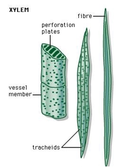
Except xylem parenchyma, all other xylem elements are dead and bounded by thick lignified walls. Vessels are shorter and wider than tracheid. Vessels are very long tube-like structures. Tracheids are elongated cells with tapering ends. They also conduct water. Since, tracheids do not have open ends like vessel, so the water has to pass from cell to cell via the pits.
Functions
- The main function of xylem is to carry water and mineral salts upward from the root to different parts of shoots.
- Since walls of tracheids vessels and sclerenchyma of xylem are lignified they give mechanical strength to the plant body.
Phloem
It consists of four components:
- Sieve tubes
- Companion cells
- Phloem parenchyma
- Phloem fibres.
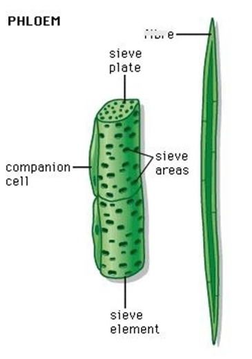
Sieve tubes
Sieve tubes are slenderical tube –like structures composed of elongated thin-walled cells, placed end to end. Their end walls are perforated by numerous pores and are called sieve plates.
Companion Cells
They are small thin-walled cells containing dense and very active cytoplasm and large elongated nucleus.
Functions
- Photo-synthetically prepared food materials are transported from the leaves to the storage organs.
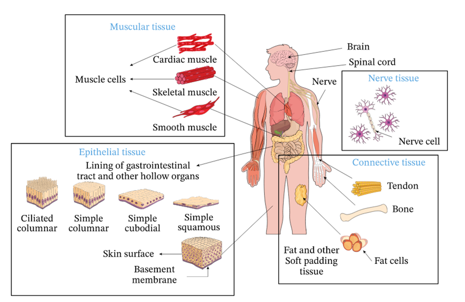
3. Animal Tissues
Animal Tissues
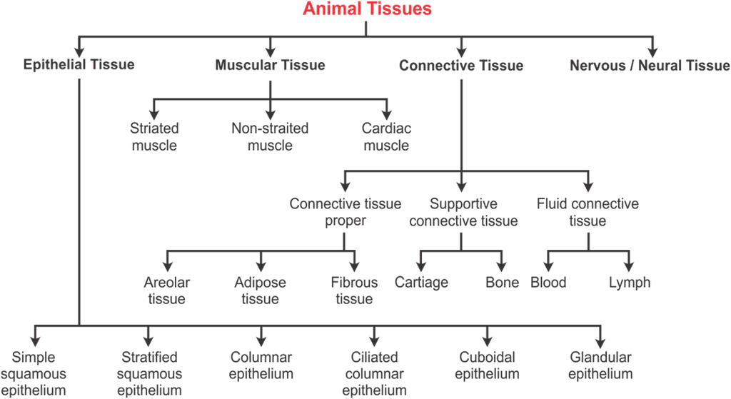
Types of Animal Tissues
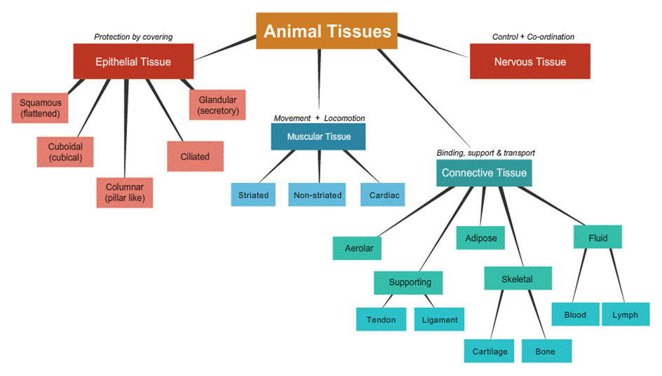
Types of Animal Tissues
-
- Epithelial Tissue
- They are the protective tissues of the human body. They cover many organs and cavities that are present inside the body.
- Where are the epithelial tissues found in the human body?
-
- The lining of the blood vessels
- The lining of the mouth
- Kidney tubules
- Skin
- Lung alveoli
Structure and functions of the epithelial tissues -
-
- The main function of the epithelial tissues is to act as a barrier and separate different organs and systems from each other.
- There is no space between the cells of epithelial tissues
- The cells are permeable. This makes it possible for them to exchange materials between different parts of the body and also between the body and the external environment.
- The epithelial tissues remain separated from the tissues beneath them because of a thin membrane over them.
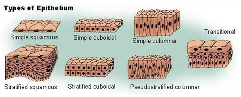
Types of Epithelium
|
Different types of epithelium tissues |
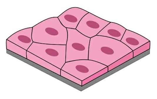
Simple Squamous |
Stratified Squamous |
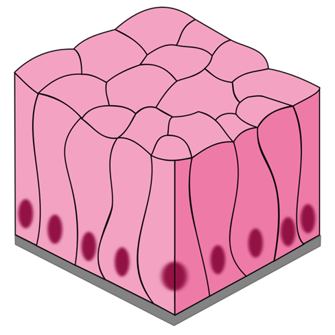
Columnar |
Ciliated Columnar |
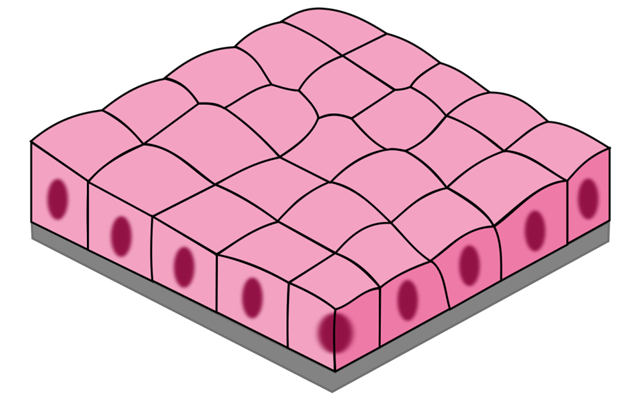
Cuboidal |
Glandular |
|
Structure |
They have delicate cell lining and possess a flat thin structure |
The epithelium Squamous cells are arranged in several layers |
They are the column-like shape tissues |
Columnar epithelial tissues which have Cilia present on them |
They are cube-shaped cells which are involved in absorption and secretion. |
These are special gland cells that can secrete substances |
|
Found in |
Alveoli and bowman’s capsule- nephron in kidney |
Skin |
Intestine |
Respiratory system |
Kidney tubules |
Sweat glands in the skin |
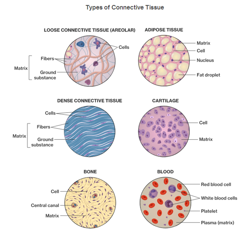
Connective Tissue
Structure and function of connective tissues
-
- They are loosely bound cells present in an intercellular Matrix.
- This matrix can be of different types – Dense, Rigid, Fluid or Jelly-like.
- Depending upon the functionality of the connective tissue, the nature of the matrix varies in them.
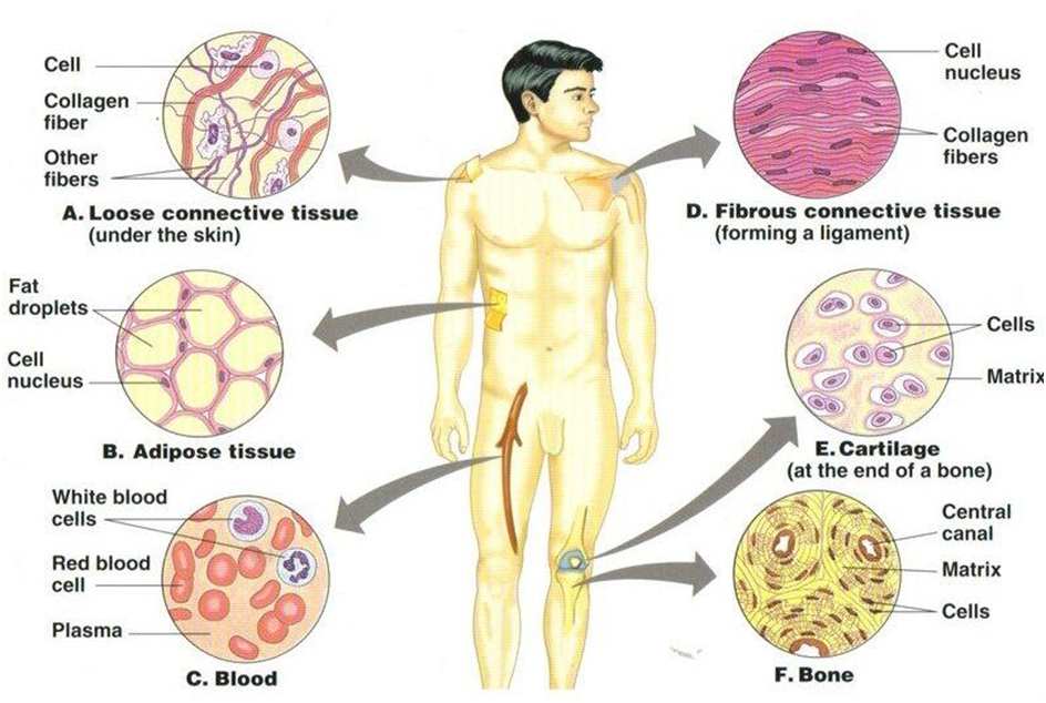
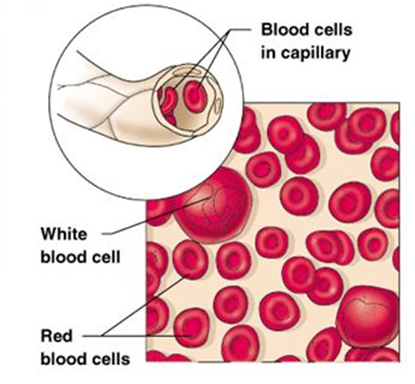
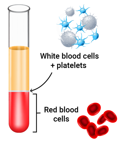
Blood
- The main function of blood is to transport gases, food, waste materials and hormones in the body.
- Therefore, blood has a fluid Matrix present in it which is called Plasma.
- The plasma contains the red blood cells, the white blood cells and blood platelets.
- The RBC have hemoglobin pigment which carries oxygen to tissues.
- White blood cells fight diseases and platelets are involved in clotting of blood when injured.
- The plasma also contains proteins and hormones in it.
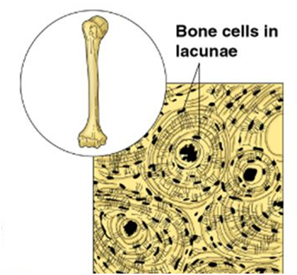
Bones
- Bones form a framework of the body over which the muscles are wrapped together.
- The bone tissue is strong and inflexible in nature.
- Therefore, the bone cells are present in a rigid matrix which is formed from calcium and phosphorus.
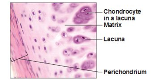
Cartilage
- Cartilage is present over the joints of the bones and provides them with a smooth structure.
- For Example, in the nose tip and ear pinna, trachea, larynx.
- They contain solid matrix made of protein and sugar. They have homogenous matrix.
- It provides support and flexibility to various parts of our body.
Ligaments
- A ligament connects two bones together.
- It has an elasticity which facilitates the connection.
- The cells of ligaments have a little matrix.
Tendons
- The tendons tissues are responsible for connecting bones and muscles together.
- They have limited flexibility but very great strength.
Areolar
- This tissue acts as a filter in between the spaces present inside the organs of the body.
- It helps in repairing other tissues as well.
- It is found in the skin and bone marrow.
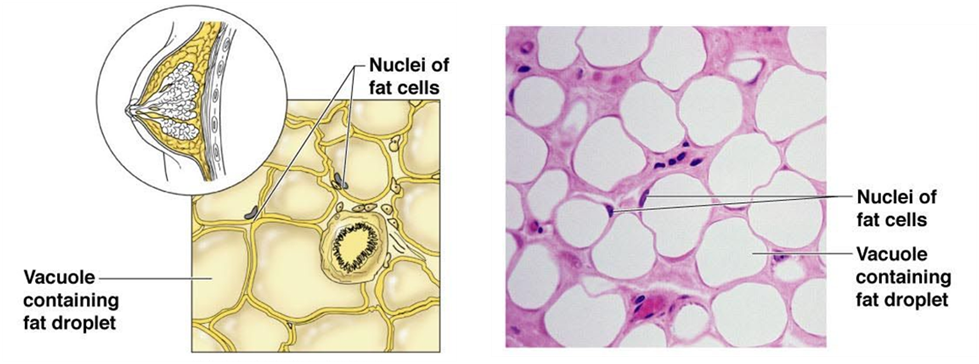
Adipose
- Fats are stored in our body in the adipose tissues.
- They are found below the skin and between the organs of the body.
- Provides cushioning to the organs.
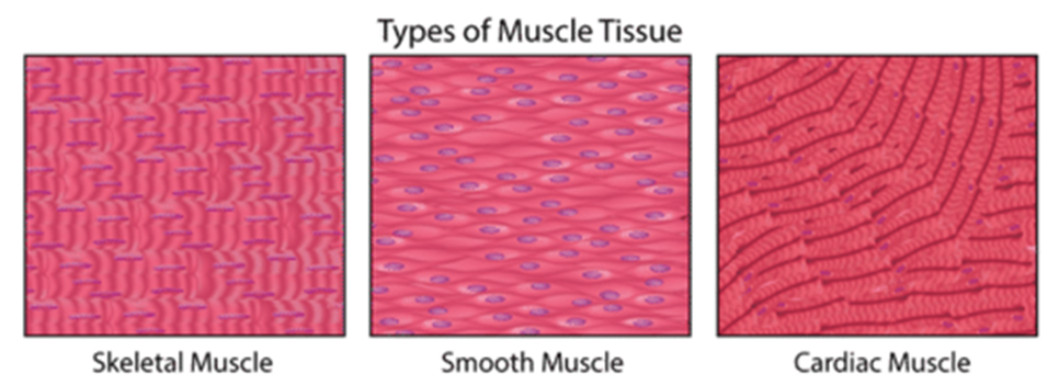
Muscular Tissue
- It is made up of muscle fibers which are long cells.
- It allows movements in our body.
- How muscles can cause movement?
They contain special proteins called Contractile Proteins. These proteins cause contraction and relaxation of the muscles.
- There are two kinds of muscles found in our body - Voluntary Muscles and Involuntary Muscles.
|
Striated/ Skeletal/ Voluntary muscles |
Smooth/ Unstriated/Involuntary muscles |
|
We can move them according to our own will |
We cannot start or stop the movement of involuntary muscles. |
|
They are also called Skeletal Muscles as they are attached to the bones. |
They also called Smooth Muscles. |
|
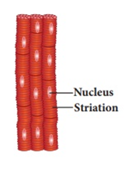
They are also called Striated Muscles because of the presence of dark and light bands over them |
They are also called Unstriated Muscles because they do not have any light or dark bands on them. |
|
The cells of voluntary muscles have more than one nucleus, they do not have any branches, and have a long cylindrical structure. |
The cells of the involuntary muscles are long and have pointed ends. |
|
For Example, Muscles of our hands and legs. |
For Example, The muscles in the alimentary canal and the Iris of our eyes. |
Cardiac Muscles
- These are special kinds of involuntary muscles.
- The muscles of the heart are called Cardiac Muscles they perform rhythmic contraction and relaxation throughout our life.
- They are cylindrical in shape; they have branches and there is a single nucleus.
- Cardiac muscle consists of individual heart muscle cells connected by intercalated discs to work as a single functional organ
3. Animal Tissues
- Books Name
- Yash Tyagi Coaching Science Book
- Publication
- ACERISE INDIA
- Course
- CBSE Class 9
- Subject
- Science
Animal Tissues
Animal Tissue
Man is multicellular, containing many organs and many systems operating in the body which help to carry out different activities like Breathing, Transportation of substances, Movement and many more. All these systems need different types of tissues that help perform these functions.
Epithelial tissue
This tissue forms the outer layer of all body parts. It is protective in nature as it acts a barrier to keep different organs separate. It is present almost everywhere like in skin, lining of buccal cavity, blood vessels, etc. The cells of this tissue are tightly packed and form a continuous sheet. Cells of epithelium contain very little or no intercellular matrix. Epithelial cells lie on a delicate non-cellular basement membrane which contains a special form of matrix that contains a protein called collagen.

Functions
- These cells protect the underlying cells.
- Epithelial cells form the lining of the mouth and alimentary canal and protect these organs.
- It helps in absorption of water and nutrients.
- It helps in the elimination of waste products.
- It also acts as a secretor.
Types of epithelial tissue
- Squamous epithelium
- Cuboidal epithelium
- Columnar epithelium
- Ciliated epithelium
Squamous Epithelium
You all are familiar with cavities of our body parts like mouth, esophagus, nose pericardium, alveoli, etc. and blood vessels. This tissue forms the lining of cavities of all these parts and also the covering of the tongue and skin. It is made up of thin, flat, irregular shaped cells which fit together like floor tiles to form a compact tissue.
Function
- It protects the underlying parts of body from mechanical injury, entry of germs, chemicals and drying.
Cuboidal Epithelium
We all know that we have kidneys for purifying blood and also, vessels that run into or through it. This tissue forms those tubules and also the glands in our body that perform functions of secretion. It consists of cube-like (cubical) cells.
Functions
- It helps in absorption, excretion and secretion.
Columnar Epithelium
We all know that we have a slimy substance called mucus in our body that performs function in respiration activities and also in digestion activities. The mucus membrane is formed by it. It also forms lining of gall bladder and oviducts. It consists of cells which are taller in comparison to being broad. Their nuclei is towards the base and the free ends of cells contain thread – like structures called microvilli.
Ciliated Epithelium
In our body, substances are made to move like the ovum, air, etc. So, in our body, parts where propelling of substances takes place, there we have thread – like structures that act as propellers. They are villi. The villi are formed of these cells. They also line the trachea (wind-pipe), bronchi (lungs), kidney tubules and oviducts (Fallopian tubes). They are certain cubical or columnar cells that have a free border which bears thread-like cytoplasmic outgrowths called cilia. Such cells form the ciliated epithelium.
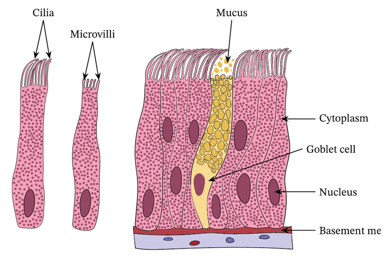
Function
- The rhythmic beating of the cilia moves solid particles in one direction through the ducts.
Muscle Tissue
We have 600 muscles in our body and they are contractile in nature. They help in bringing about movements in body parts. Like it helps us to move, it helps our heart to beat, it helps in bringing about all movements through our limbs. On the basis of their location, structure and functions there are following three types of muscle fibres:
- Striated muscles
- Smooth muscles
- Cardiac muscles
Striated Muscles
(Also called as Striped, skeletal or voluntary muscles) It is seen in muscles of limbs, body wall, neck, etc. Striated muscles are present in tongue, pharynx, diaphragm and upper part of the esophagus. They are called visceral striated muscles. It is cylindrical un-branched. These cells have a number of nuclei and each muscle cell is multinucleated. Each muscle cell is enclosed in a thin but distinct plasma membrane called sarcolemma and contain a fluid called sarcoplasm.
- The entire muscle fibres show alternate dark and light stripes. They are called striped muscles.
- They are attached to bones and are responsible for body movements they are called skeletal muscles.
- These muscles work according to our will. They are also called voluntary muscles.
- They are long or elongated with non-tapering ends.
Functions
- Striated muscles are powerful and undergo rapid contraction. These muscles can be tired and need rest.
- Striated muscles provide the force for locomotion and all other voluntary movements of the body.
Smooth Muscles
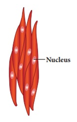
They are seen in the walls of the hollow (tubular) visceral organs except that of the heart that is why they are called visceral muscles.
- There is a single centrally located cigar-shaped nucleus in the centre of cytoplasm or sarcoplasm.
- These fibrils do not bear any bands , stripes or striations across the muscle hence, called smooth or unstrained muscles.
Functions
- Smooth muscles do not work and contract according to our will. So they are also called involuntary muscles. The movement of food in the alimentary canal, opening and closing of tubes are involuntary movements.
- Smooth muscles contract slowly but can remain contracted for a long period of time.
Cardiac Muscles
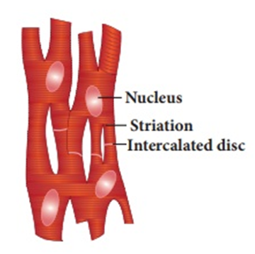
As you all know that we have an important organ that is heart and it keeps on pumping blood to all body parts without getting tired. It is because of a special muscle in it that is cardiac muscle. These muscles show the characteristics of both smooth and striated muscles. Cardiac muscles are composed of branched fibres, the branches join to form a network. Each fibre or cell is surrounded by sarcolemma and has cytoplasm (sarcoplasm) with longitudinal myofibrils and a centrally located nucleus. Cardiac muscles have stripes or light and dark bands. They show densely stained cross-bands called Intercalated discs.
Functions
- Cardiac muscles contract and relax rapidly.
- The contraction and relaxation of the heart muscles help to pump and distribute blood to various parts of the body.
Connective Tissue
The connective tissue is specialized to connect and anchor various body organs. It binds the tissue and gives support to various parts of the body by forming packing around organs so that they do not get displaced by body movements. They act as binding, supporting and packing tissue.
Its cells are living and separated from each other and are few in number. They have homogeneous gel-like intercellular substance called medium or matrix which forms the main bulk of the connective tissue. Thus, the space between cells is filled with the non-living matrix which may be solid like in bones and cartilages and fluids as in the blood. Matrix is fibrous in nature and binds other tissues in fact the nature of matrix decides the function of connective tissue.
Types of connective tissue In animals there are of following five types as given below:
- Areolar (loose) connective tissue
- Dense regular connective tissue
- Adipose tissue
- Skeletal tissue
- Fluid connective tissue
Areolar loose connective tissue
The tissue that joins skin to muscles and fills space inside organs is none other than the areolar connective tissue. It is a loose and cellular connective tissue. It has a matrix that consists of two kinds of fibres that is white collagen fibres and yellow elastic fibres or elastin.
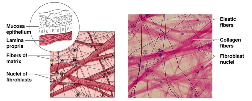
Functions
- It acts as a supporting and packing tissue between organs lying in the body cavity. Matrix of this tissue is important in diffusion of oxygen and nutrients from small blood vessels.
- It helps in repair of tissues after an injury.
- It also helps in combating foreign toxins.
- It fixes skin to underlying muscles.
Dense Regular Connective Tissue
It is a fibrous connective tissue. It is characterized by ordered and densely packed matrix composed of fibres and cells. It is of two types:
- Tendons
- Ligaments
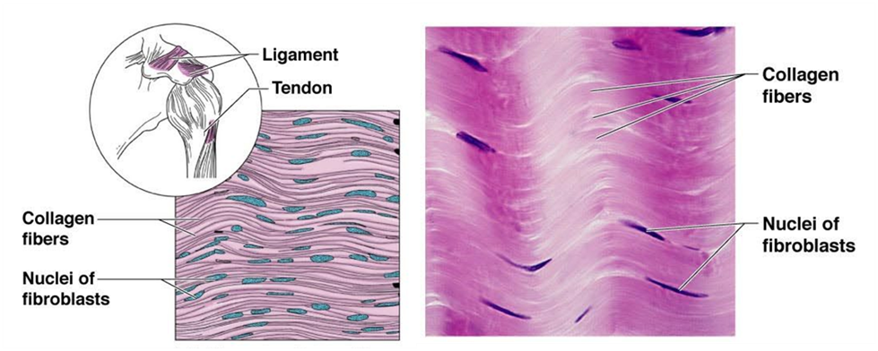
Tendons are cord-like, strong inelastic structures that join skeletal muscles to bones. They have great strength but their flexibility is limited. The cells of tendons are called tendinocytes.
Ligaments are elastic structures which connect bones to bones and have great strength but contain very little matrix. Sprain is caused by excessive pulling (stretching) of ligaments.
Adipose Tissue
This adipose tissue is abundant below the skin. It is the tissue that gives shape to our body and is a fat reservoir. Adipose tissue is basically an aggregation of fat cells or adipocytes. Each fat cell is rounded or oval and contains a large droplet of fat that almost fills it. It has collagen and elastin fibres. The partitions carry blood vessels.
Function
- It serves as a fat reservoir.
- It provides shape to the limbs and the body.
- It keeps visceral organs in position.
- It acts as an insulator.
Skeletal Tissue
The skeletal or supporting tissue includes cartilage and bone which forms the endoskeleton of vertebrate body. It mainly consists of:
- Bone
- Cartilage
Bone
It is very strong and non-flexible tissue. Its matrix is made up of protein that is heavily coated with salts of calcium and magnesium that are responsible for the hardness of the bone. Bone cells called osteoblasts or osteocytes, are present lamellae with fluid-filled spaces is called lacunae.
Functions
Bones form endoskeleton of human beings and other vertebrates except the sharks. It serves the following functions.
- It provides shape to the body.
- It provides skeletal support to body,
- It protects vital body organs such as brain, lungs, etc.
- It serves as a storage site of calcium and phosphate.
- It anchors the muscles.
Cartilage-
It has less vascular matrix which is composed of calcium salts. Its cells are called chondrocytes. The matrix of cartilage consists of collagen fibres. Chondrocytes are present in fluid-filled space known as lacunae, blood vessels are absent in matrix. It forms the ear pinna , nose tip, epiglottis, etc.
Functions
- It provides support and flexibility to the body parts.
Fluid connective tissue
This tissue helps in creating link between different parts of the body and maintains a continuity of life. They are of two types: Blood and Lymph
Blood It is formed of two components:
- Plasma
- Blood cells
Plasma
It is a yellow colored fluid which is 90% water and 10% organic and inorganic substances. Due to the presence of these substances, it acts as a nutrient medium for blood cells.
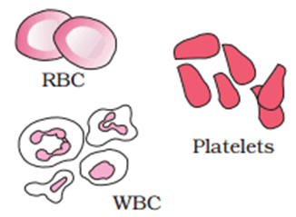
The blood cells present are:
- Red blood cells
- White blood cells
- Platelets
Red blood cells
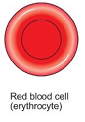
- These are incomplete cells with no nucleus, mitochondria and endoplasmic reticulum
- These cells are the smallest cells of our body.
- They are cells that have disc – like shape.
- These cells have red colour as they contain a pigment made up of iron called haemoglobin.
- The function of this hemoglobin is to transport gases by forming complexes i.e oxyHb and carbinoHb.
- These cells are synthesized in bone marrow of adults and in infants in spleen from stem cells.
- The life span of these cells is 120 days.
- These cells are also called erythrocytes.
White blood cells
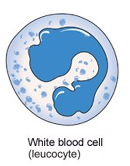
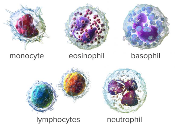
- These cells are irregular in shape and are complete cells enclosing all organelles.
- These cells do not have any pigment in them that is the reason that they have no colour.
- These cells have a life span of 2 to 4 weeks.
- They are also called leucocytes.
- They are also called soldiers of our body as they protect our body from infection causing agents – either they engulf infection causing agent or they secrete certain proteins that make infection causing agent harmless.
Platelets
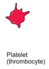
- Platelets are tiny fragments that may or may not have a nucleus.
- Their function is to secrete certain proteins that help in blood clotting.
- Their life span is 60 to 120 days.
- They are also called thrombocytes.
Lymph
It is a colorless fluid that gets filtered out of the blood capillaries.

Functions
- Lymph transports the nutrients (oxygen, glucose) that may have filtered out of the blood capillaries back into the heart to be re-circulated in the body.
- It brings CO2 and nitrogenous wastes from tissue fluid to blood.
- Being loaded with WBCs such as lymphocytes, the lymph protects the body against infection.
- It forms the defense or immune system of the body.
4. Nervous Tissue
The Nervous Tissue
- How do we react to stimuli?
This is becaus e of the nervous tissues present in our body. They are capable of transmitting information quickly from the brain to different parts of the body and vice-versa.
- Therefore, nervous tissues are found in nerves, brain, and spinal cord.
- The Nervous tissue is made up of cells called the Nerve Cells or Neurons.
- These neurons connect together to form the nerves of our body.
Structure of a Neuron
-
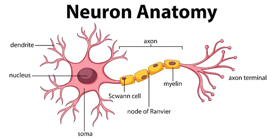
- It is an elongated cell with a Cell Body that consists of some branch-like structure called Dendrites.
- There is a Nucleus present in the centre of the cell body.
- The Nerve Endings of the cell are connected with the cell body via Axon.
- A nerve cell can be up to 1 m long.
Structure of Neuron
1.Dendrites
- They are tree-like extensions (highly-branched) at the beginning of a neuron.
- They increase the surface area of the neuron.
- They receive chemical signals from different neurons of the body.
- They then convert these chemical signals into electrical signals and pass them to the neuron cell body.
- A neuron can have a single dendrite or multiple dendrites
2. Cell Body
- Also called Soma.
- The main function of the cell body and nucleus of the neuron is to maintain the functionality of the cell.
- It does not play an active role in the transmission of the signal.
- It produces proteins that are required by different parts of the neuron to work properly.
- It contains different cell organelles such as mitochondria, Golgi apparatus etc that perform various functions of the cell.
3. Axon
- Neurons have one axon in general.
- It is a long structure that connects the cell body to the terminals and it also connects with other neurons, cells and organs of the body through nerve terminals.
- It allows in fast transmission of signals. The larger the diameter of the axon the faster it will transmit signals.
- It is covered with a special insulating substance called myelin. It helps in rapid transmission of signals.
4. Nervous Tissue
- Books Name
- Yash Tyagi Coaching Science Book
- Publication
- ACERISE INDIA
- Course
- CBSE Class 9
- Subject
- Science
Nervous Tissue
Nervous tissue
This tissue helps us in thinking, listening, conveying messages, etc. On the whole, we can say that it helps in control and coordination in the body. It is a specialized tissue that helps in transmitting messages within our body. It contains highly specialized cells called neurons. The neurons have the ability to receive stimuli from within or outside the body and to conduct impulses to different parts. Each neuron consists of:
- Cyton
- Dendrite
- Axon
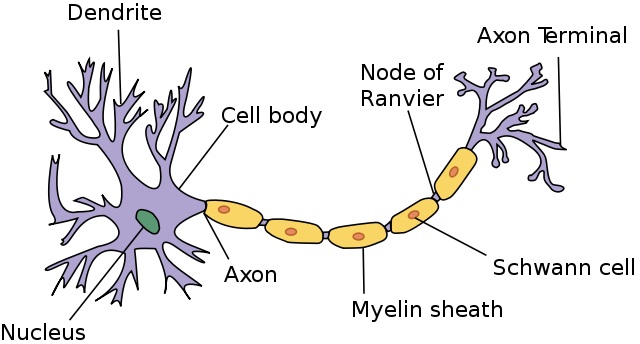
The irregular structure called cell body encloses a nucleus in neuroplasm. From cell body, small branches arise on upper side called dendrite. On the lower side, it gives out only one branch that is elongated called axon. The whole neuron is made up of neurolemma. It has a fatty layer on it as a modulated sheath that creates nodes of ranvier that help in saltatory conduction. Axon ends into nerve endings.
Function of neuron
- It is to form nerves that further helps in control and coordination of body.
