 ACME SMART PUBLICATION
Animal Tissues
- Books Name
- ACME SMART COACHING Biology Book
- Publication
- ACME SMART PUBLICATION
- Course
- CBSE Class 11
- Subject
- Biology
Tissue : A group of cells similar in structure, function and origin.
In a tissue cells may be dissimilar in structure and function but they are always similar in origin.
– Word animal tissue was coined by – Bichat
– N. Grew coined the term for Plant Anatomy.
– Study of tissue – Histology
– Histology word was given by – Mayr
– Father of Histology – Bichat
– Study of tissue is also called Microscopic anatomy.
– Founder of microscopic anatomy – Marcello Malpighi
TYPES OF TISSUES
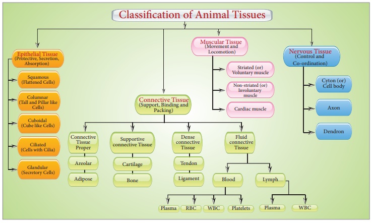
Based on the location and function the animal tissues are classified into four types :

Animal Tissues
Chapter 7
STRUCTURAL ORGANISATION IN ANIMALS
In unicellular organisms, a single cell performs all tasks such as digesting, respiration, and reproduction. The same basic duties are carried out by different groups of cells in a well-organized manner in the complex body of multicellular organisms. The body of a basic organism like Hydra is made up of many distinct types of cells, each with thousands of them. The human body is made up of billions of cells that each perform a different purpose. A set of related cells, as well as intercellular chemicals, execute a defined purpose in multicellular creatures. Tissue is the name for this type of arrangement.
There are just four fundamental types of tissues in all complex creatures. To form an organ such as the stomach, lung, heart, or kidney, these tissues are organized in a certain proportion and pattern. When two or more organs interact physically and/or chemically to fulfill a shared function, they create an organ system, such as the digestive system or the respiratory system. Cells, tissues, organs, and organ systems divide up the work and contribute to the body's overall survival through the division of labor.
Animal Tissues
Cells have different structures depending on their purpose. As a result, the tissues differ and can be divided into four categories:
Epithelial Tissue
- Books Name
- ACME SMART COACHING Biology Book
- Publication
- ACME SMART PUBLICATION
- Course
- CBSE Class 11
- Subject
- Biology
EPITHELIAL TISSUE
Cells of the epithelium are set very close to each other, separated by very thin films of extracellular material.
Neighbouring cells are held together by cell junctions.
The epithelial tissue rests on a noncellular basement membrane, which separates it from the underlying connecting tissue.
The basement membrane is a non-cellular membrane made of two layers:
(i) Upper thin layer called basal lamina, made up of glycoproteins and mucopolysaccharides and secreted by epithelial cells.
(ii) Lower thick fibrous layer called reticular lamina made of reticular fibres and collagen fibres which are a part of underlying connective tissue.
Blood vessels are absent in the epithelial tissue. Materials are exchanged between epithelial cells and vessels of the connective tissues by diffusion across the basement membrane.
The epithelial tissue is classified into simple and compound epithelia.
Specialized junctions between epithelial cells:
To provide mechanical support for the tissue plasma membrane of adjacent epithelial cells modified to form following structures called as Intercellular Junctions.
Tight junctions (Zonula occludens) : help to prevent substances from leaking across the tissue. Plasma membranes in the apical parts become tightly packed together or are even fused.
Interdigitations : These are interfitting, finger like processes of the cell membranes of the adjacent cells.
Intercellular Bridges : These are minute projections that arise from adjacent cell membrances.
They make contact with one anther.
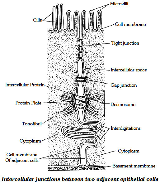
Gap Junctions : Facilitate the cells to communicate with each other by connecting the cytoplasm of adjoining cells, for rapid transfer of ions, small molecules and sometimes big molecules.
Intermediate Junctions (= Zonula adherens) : These usually occur just below tight junctions. The intercellular space at these places contains a clear, low electron density fluid. There is a dense plaque like structure on cytoplasmic side of each plasma membrane from which fine microfilaments of actin (protein) extend into the cytoplasm. There is no intercellular filaments between the adjacent cell membranes. There is an adhesive material at this point. They probably serve anchoring functions.
Desmosomes ( =Macula adherens) : Perform cementing to keep the neighbouring cells together. These are like zonula adherens but are thicker and stronger and are disc like junctions. They have intercellular protein. The plaque-like structures (= protein plate) are much thicker. The microfilaments which extend from protien plat are called tonofibrils. Desmosomes serve anchoring function. Hemidesmosomes (single sided desmosomes) are similar to desmosomes, but the thickening of cell membrane is seen only on one side. Hemidesmosomes join epithelial cells to basal lamina (outer layer of basement membrane).
Classification of Epithelial Tissues
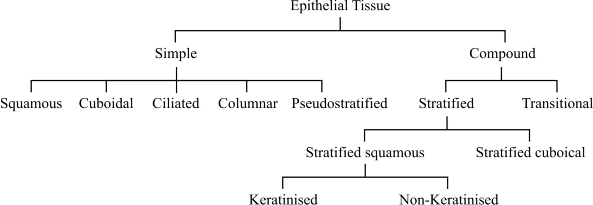
1. Simple Epithelium
It is formed of a single layer of cells.
The adjacent cells are held together by means of desmosome, resting on the basement membrane.
Simple epithelium occurs mainly on secretory and absorptive surfaces.
It helps in nutrition, excretion and secretion but not for protecting the underlying tissue.
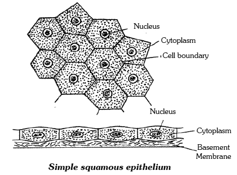
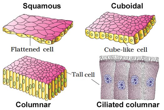
(i) Squamous Epithelium :
It consists of a layer of thin, flat, scale-like cells with prominent nuclei.
The cells have irregular boundaries that fit closely into those of neighbouring cells.
It forms the inner lining of lung alveoli and blood vessels (Endothelium).
It is also known as pavement or tesselated epithelium.
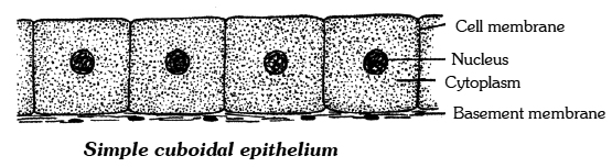
(ii) Cuboidal Epithelium
It has cells which are polygonal in outline, but appear cuboidal in vertical section.
It lines small salivary and pancreatic ducts and thyroid vesicles.
The cells participate in secretion, excretion and absorption.
The cells of cubical epithelium in absorptive surfaces often bear microvilli on their free ends. This gives a brush-like appearance to their free border.
They are, therefore, called brush-bordered cubical epithelial cells e.g., in proximal tubules of kidneys.
Microvilli greatly increase the area of the free surface of the cell and thereby enhance absorption.
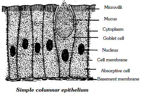
(iii) Columnar Epithelium :
It is characterised by the presence of tall cells shaped like polygonal columns.
The nucleus is usually located at the base of the cell.
Columnar epithelium covers the inner surface of the intestine, stomach and gall bladder.
It also occurs lin gastric and intestinal glands.
Its function is secretion or absorption.
The intestinal mucosa is lined by Brush Bordered Columnar Epithelium which is highly absorptive.
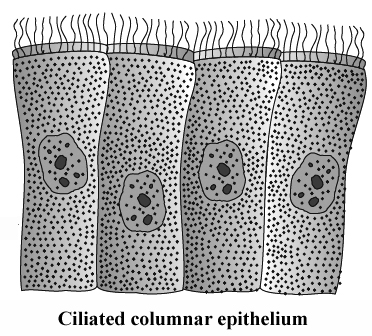
(iv) Ciliated Epithelium :
It consists of columnar or cubical cells bearing cilia on their free surfaces.
The function of the cilia is to move particles, free cells or mucus in a specific direction over the epithelial surface.
Ciliated columnar epithelium lines the inner surfaces of some hollow organs such as fallopian tubes, bronchioles and small bronchi.
Ciliated columnar epithelium lining the ventricles of brain and spinal canal is called as ependyma.
Cilia is of two types
(a) Kinocilia are motile cilia with 9 + 2 organisation,
(b) Stereocilia - Basal granule absent, non-motile, Ciliated columnar epithelium 9 + 2 organisation is absent. Stereocilia are found in some parts of the male reproductive tracts such as the epididymis and vas deferens.
(v) Pseudostratified Epithelium :
It covers the inner linings of trachea and large bronchi.
Although made up of a single layer of columnar cells, it appears two-layered, because some cells are shorter than the others and have their nuclei at different levels.
The shorter cells lack cilia and secrete mucus which traps particles on the epithelial surface.
The longer cells are ciliated.
The ciliary movements propel the mucus and the particles towards the larynx.
Pseudostratified non ciliated columnar epithelium tissue is found in urethra of male and parotid salivary gland.
Squamous showing some keratinisation
Concept Builder
Special Types of Epithelium
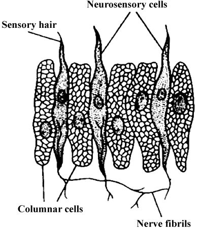
(a) Neuro sensory epithelium :
In between piller shaped supporting cells modified sensory cells are present. On the free end sensory hair is present. Base of these cells is attached with sensory nerve.
e.g. – Gustatory Epithelium – Cover taste bud of tongue and receive taste sensation.
– Olfactory epithelium – Schneidarian membrane receive smell sensation.
– Stato – acoustic – Lining of internal ear.
– In retina of eye receive optic sensation.
(b) Myoepithelium : Around mammary and sweat gland
(c) Pigmented epithelium (Cuboidal) : In Retina of eye
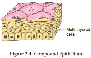
2. Compound Epithelium
It consists of more than one layer of cells.
Only the cells of the deepest layer rest on the basement membrane.
Being multilayered, compound epithelia have little role in secretion or absorption, but they provide protection to underlying tissues against mechanical, chemical, thermal or osmotic stresses.
Compound epithelia may be stratified or transitional.
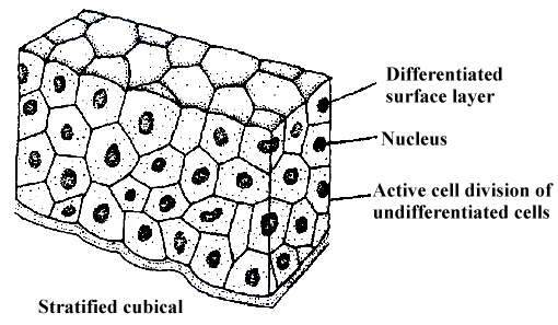
(i) Stratified Epithelium :
It has many layers of epithelial cells.
The deepest layer is formed by cuboidal cells.
But the morphology of the superficial layers varies in the different kinds of stratified epithelia.
In stratified cuboidal epithelium, the superficial cells are cuboidal.
It lines the inner surfaces of larger salivary and pancreatic ducts.
Stratified non-keratinised Squamous Epithelium covers moist surfaces such as those of buccal cavity, pharynx and oesophagus.
It has several superficial layers of living squamous cells and deeper layers of interlinlked polygonal cells.
Stratified Keratinised Squamous Epithelium covers the dry surface of skin.
It has many superficial layers of horny, scale-like remains of dead squamous cells and several deeper layers of living polygonal cells.
Heavy deposits of the insoluble protein keratin in the dead superficial cells make the epithelium impervious to water and highly resistant to mechanical abrasions.
In contrast, nonkeratinised stratified epithelia cannot prevent water loss and afford only moderate protection against abrasions.
(ii) Transitional Epithelium :
It is much thinner and more stretchable than the stratified epithelium.
It has a single layer of cuboidal cells at the base, 2-3 middle layers of large polygonal or pear-shaped cells and a superficial layer of large, broad, rectangular or oval cells.
It lines the inner surface of the urinary bladder and ureters.
It allows considerable expansion of these organs to accommodate urine, because stretching considerablly flattens and broadens the cells of superficial and middle layers.
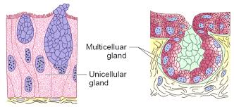
3. Glandular Epithelia
The cells of glandular epithelia are generally columnar or cuboidal.
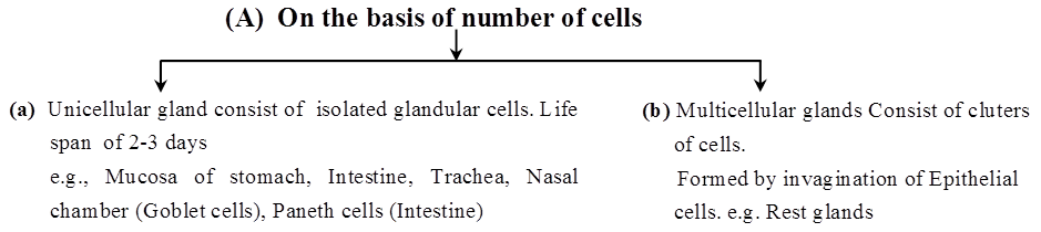
The glandular epithelium can be classified into two types : unicellular, consisting of isolated glandular cells (e.g., goblet cell of alimentary canal), and multicellular (e.g. salivary glands), consisting of cluster of cells.
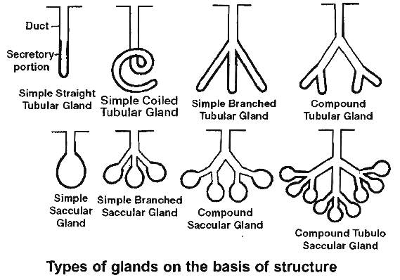
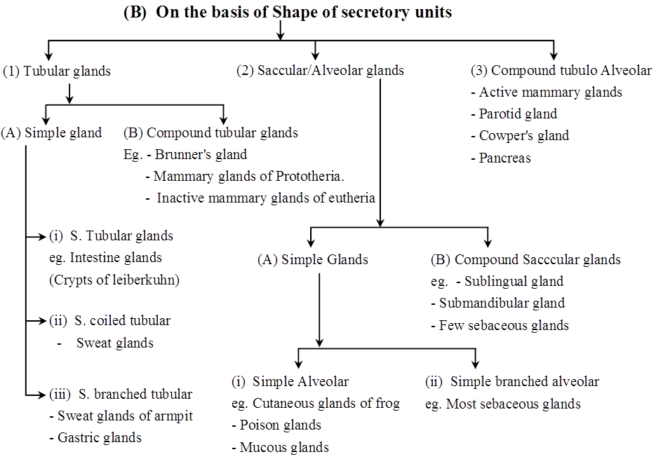
A gland with a single unbranched duct is called a simple gland.
The secretory part of the gland consists of epithelial cells arranged in the form of tubes (tubules) or sacs (acini, alveoli) or a combination of both.
The duct is also made up of epithelial cells.
A gland with a branched system of ducts is called a compound gland.
In these glands, the secretory tubule or acinus may be coiled or branched, opens into the single duct of the gland.
Compound glands are present in the pancreas and sub-mandibular salivary glands.
Concept Builder
Types of simple gland
(a) Simple tubular: Simple tubular glands are present in the intestine (e.g., Crypts of Leiberkuhn).
(b) Simple alveolar: Terminal part forms alveolus e.g., Mucous glands in skin of frog, poison glands in toad.
(c) Simple coiled tubular: Terminal part is coiled e.g., sweat glands.
(d) Branched tubular: Gastric glands in stomach.
(e) Branched alveolar e.g., Sebaceous g.land .
Types of compound gland
(a) Compound tubular gland: e.g., mammary glands of prototherians.
(b) Compound saccular or alveolar gland: e.g., Salivary glands, (sub-maxillary and sub-lingual).
(c) Compound tubulo alveolar or tubulo saccular: They are tubular as well as alveolar and are found in mammary glands, pancreas, parotid salivary gland, Cowper's glands and Bartholin glands.
(ii) Exocrine glands have a secretory portion which contains the cells for secretion of milk, digestive enzymes, mucus, saliva, ear wax, oil and ducts which transport their secretions to the respective sites of action, for example, salivary gland, tear gland, gastric gland and intestinal glands. When a gland performs both exocrine and endocrine functions, it is called a mixed gland or Heterocrine gland (e.g., the pancreas, testis, ovaries).
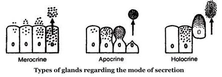
(iii) On the basis of mode of secretion, glands can be :
(a) Holocrine glands: In holocrine glands (e.g., sebaceous gland), the product of secretion is shed with the whole cell leading to its destruction.
(b) Merocrine glands: When the secretory granules leave the cell by exocytosis (simple diffusion) with no loss of other cellular material , the glands are called merocrine glands (e .g. , the pancreas, salivary glands, intestinal glands and sweat glands).
(c) Apocrine glands: In apocrine glands (e.g., mammary gland and axillary sweat glands), only the apical portion of the cytoplasm is discharged along with the secretory product.
Epithelial Tissue
Epithelial Tissue
Epithelium or epithelia refers to epithelial tissue. This tissue has a free surface that is exposed to either a body fluid or the outside environment and serves as a covering or lining for a body part. The cells are densely packed and there is a little intercellular matrix. Simple epithelium and compound epithelium are two forms of epithelial tissues.
(A) SIMPLE EPITHELIUM: A single layer of cells makes up simple epithelium, which serves as a lining for bodily cavities, ducts, and tubes. Basedon structural modification of the cells, the simple epithelium is further divided into three types. These are
(i) Squamous epithelium: A single thin layer of flattened cells with uneven borders makes up the squamous epithelium. They're present in the walls of blood arteries and the air sacs of the lungs, and they help build diffusion boundaries.
(ii) Cuboidal epithelium: A single layer of cube-like cells makes up the cuboidal epithelium. Its main roles are secretion and absorption, and it is usually found in gland ducts and tubular sections of nephrons in the kidneys. Microvilli are found in the epithelium of the proximal convoluted tubule (PCT) of the nephron in the kidney.
(iii) Columnar epithelium: A single layer of tall and thin cells makes up the columnar epithelium. Their nuclei are near the bottom of their bodies. Microvilli may exist on a free surface. They help with secretion and absorption and are located in the lining of the stomach and intestine.
(iv) Ciliated epithelium: Ciliated epithelium is defined as columnar or cuboidal cells with cilia on their free surfaces. Their job is to transfer particles or mucus over the epithelium in a precise direction. They're mostly found on the inside of hollow organs like the bronchioles and fallopian tubes.
(v) Glandular epithelium: The glandular epithelium is formed when some columnar or cuboidal cells become specialized for secretion.They are divided into two types: unicellular glandular cells (goblet cells of the alimentary canal) and multicellular glandular cells (clusters of cells) (salivary gland). Exocrine and endocrine glands are split into two types based on how they secrete their secretions. Mucus, saliva, earwax, oil, milk, digestive enzymes, and other cell products are secreted by exocrine glands. Ducts or tubes are used to release these products. Endocrine glands, on the other hand, lack ducts. Hormones, which are their products, are secreted straight into the gland's fluid bath.
(B) COMPOUND EPITHELIUM: Compound epithelium is made up of multiple layers of cells, therefore it has little function in secretion and absorption. The compound epithelium, like our skin, is made up of two or more cell layers and serves a protective role.Their primary purpose is to protect the body against chemical and mechanical stress. They protect the skin's dry surface, the moist surface of the buccal cavity, the pharynx, the inner lining of the salivary gland, and pancreatic ducts.
The epithelium's cells are bound together by a thin layer of intercellular substance. Specified junctions offer structural and functional linkages between individual cells in practically all animal tissues. In the epithelium and other tissues, there are three types of cell junctions. Tight, adherent, and gap junctions are the three types of junctions. Tight connections prevent chemicals from leaking through a cell's surface.Adhering junctions bind cells by gluing them together. Gap junctions allow cells to communicate with one another by connecting the cytoplasm of neighboring cells, allowing for the rapid movement of ions, small molecules, and occasionally large molecules.Adhering junctions bind cells together by gluing them together.
Connective Tissue
- Books Name
- ACME SMART COACHING Biology Book
- Publication
- ACME SMART PUBLICATION
- Course
- CBSE Class 11
- Subject
- Biology
CONNECTIVE TISSUE
All connective Tissue in the body are developed from Mesoderm.
O. Hartwig called them Mesenchyme because they originated from embryonic mesoderm.
Only connective Tissue constitute 30% of total body weight.
(Muscle – 50%, Epithelium – 10% Nervous – 10%)
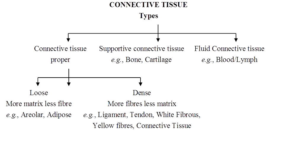
On the basis of matrix connective tissue is of 3 types -
1. Connective Tissue Proper – Matrix soft and fibrous
2. Connective Tissue Skeleton – Dense and mineralized matrix. Due to deposition of minerals it becomes hard.
3. Connective tissue Vascular – Liquid and fibres free matrix
Concept Builder
1. Connective Tissue Proper
Connective Tissue Proper is composed of three components (A) Different types of cells, (B) Fibres, (C) Matrix.
(A) Cells of connective tissue proper
Fibroblast cells :
Largest cell of connective tissue proper.
Maximum in number.
Cell body and nucleus both are oval shaped.
Branched cytoplasmic process arise from these cells so they appear irregular in shape.
Rich in rough ER because main or primary function is to produces fibres. Fibres are composed of protein.
Chief matrix producing cells.
Old fibroblast cells (fibrocyte) are inactive cells.
These are also considered as undifferentiated cells of conn. Tissue because they can be modified into Osteoblast & Chondrioblast cells to produce bone & cartilage.
Function : (1) To produce fibres (2) To secrete matrix.
Plasma Cell - Cart Wheel Cell
Less in number
Amoeboid in shape
Chromatin material is arranged like spokes in wheel so they are also called as Cart wheel cells.
According to research these cells are formed by the division of lymphocytes. So they are also called as clone of lymphocytes.
Function : Produce, Secrete & transport antibody.
Mast cells/Mastocytes
Numerous , amoeboid and small in size.
Structurally and functionally similar to basophils.
2-3 lobed S-shaped nucleus
Cytoplasm contains basophilic granules which can be stained with basic dye Methylene Blue.
It is important cell of connective tissue proper as they perform important functions.
(a) Histamine – Histamine is a protein, a vasodilator
Increase permeability of blood capillaries.
Take part in allergy and inflammatory reactions.
(b) Serotonin -
Also called as 5-Hydroxy tryptamine
It is a protein, a vasoconstrictor & decrease blood circulation but increases blood pressure.
At the site of cut or injury serotonin decrease blood loss.
(c) Heparin –
A mucopolysaccharide, a natural anti-coagulant, prevents clotting of blood in blood vessels by preventing the conversion of prothrombin into thrombin.
Adipose Cells/Fat Cells
Oval shaped stores fat.
Fat is collected in the form of fat globule formed by the fusion of small oil droplets.
On the basis of number of fat globules adipocytes are of two types.
(a) Monolocular adipocytes/ White fat tissue-cell
In these cells single large and central fat globule is present.
nucleus & Cytoplasm is peripheral and Cytoplasm is less in amount.
Due to compression of fat globule, nucleus become flattened in shape . These adipocytes form White Fat.
(b) Multilocular adipocytes/Brown fat tissue cell
In these cell 2-3 fat globules are distributed in the cytoplasm around nucleus
Cytoplasm is more in quantity.
Nucleus is rounded & found in the centre
These adipocytes form Brown Fat.
Mesenchymal Cells
Less in numbers. Small sized with cytoplasmic process having irregular shape.
Oval shaped nucleus
These are undifferentiated cells of connective tissue because they can transform into any cell of connective Tissue proper. (Totipotent in nature)
Function : To form other cells of connective tissue.
Macrophages/Histeocyte/Clasmatocytes.
– It is 2nd largest in size and in number.
– Amoeboid in shape with bean or kidney shaped nucleus.
– Cytoplasm quantity is more agranular but due to presence of a greater number of lysosomes it appears granular.
– Phagocytic in nature, destroy bacteria & viruses by phagocytosis. They arise by the fusion of monocytes
– Also called as scavenger cells of connective tissue because they destroy dead or damaged cells to clean connective tissue.
- Macrophages are named differently in different organs.
Lung – Dust cells
Liver – Kupffer cells
Blood – Monocytes
Brain – Microglial cells
Thymus gland – Hessels granules
Spleen – Reticular cells
Lymphocytes
Less in number and small in size having amoeboid shape.
A large nucleus is present cytoplasm is present as peripheral layer. Cytoplasm quantity is less.
Produce, transport & secretes antibodies.
They divide to form plasma cells of connective tissue proper.
(B) FIBRES
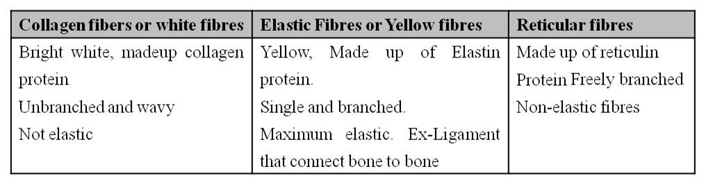
Collagen fibres (White fibres)
They are shining white fibres composed of collagen protein (Tropocollagen).
It is present in maximum quantity in vertebrates, (only collagen fibres constituted one third part of connective tissue fibres in human beings.)
They are wavy & tough fibres always arranged in bundle called fascia.
On boiling they convert into gelatin.
They can be digested by Pepsin enzyme.
Elastic fibres – (Yellow fibres)
Precursor in colour and composed of elastin protein.
They are branched fibres but always arranged singly. Branches of these form network.
In these fibres maximum elasticity is present.
They are highly resistant to chemicals.
When boiled they do not dissolve.
They can be digested by trypsin enzyme.
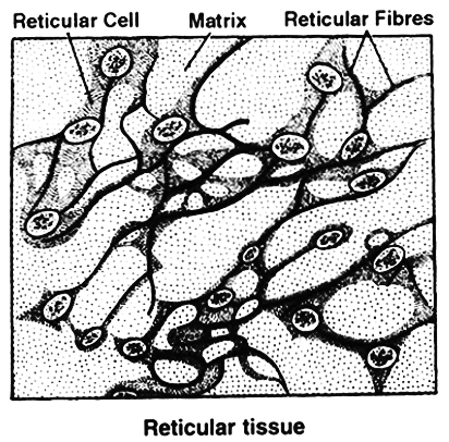
Reticular Fibres: -
Precursor of Collagen fibres, delicate with no elasticity
Also known as Arzyrophil fibre since they can be stained with silver salts.
They are composed of recticulin protein highly branched fibres which always form dense network.
These are mainly distributed in lymphoid organs like spleen or lymph nodes.
 (C) MATRIX
Matrix is composed of Mucopolysaccharide which is present in the form of Hyaluronic acid.
(1) Connective Tissue Proper
(I) Loose Connective Tissue:
It consists of cells scattered within an amorphous mass of proteins that forms a ground substance.
The gelatinous material is strengthened by a loose scattering of protein fibres such as collagen, elastin, which makes tissue elastic and reticulin, which supports the tissue by forming a meshwork.
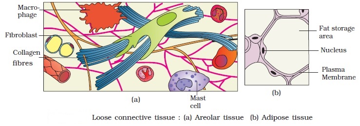
(a) Adipose Tissue:
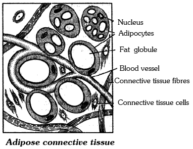
Adipose tissue is a connective tissue rich beneath the skin, around kidneys in mesentery and bone marrow.
Besides fibroblasts, macrophages, collagen fibres and elastic fibres, the adipose tissue also contains large, spherical or oval cells called Fat Cells or Adipocytes.
The cytoplasm and organelles in adipocytes are pressed by fat globule into a narrow annular layer just beneath the plasma membrane.
The adipose tissue synthesises, stores and metabolises fat.
Functions:
(i) Preventing heat loss by forming heat-insulating layer beneath the skin.
(ii) Forming shock-absorbing cushions around kidneys and eyeballs.
(iii) Acting as a food reserve.
White and Brown Tissue:
The cells of adipose tissue are characterized by droplets of fats in the cytoplasm of connective tissue cells.
There are two kinds of fatty tissues. In the white adipose tissue, there is a single large fat droplet in the cells surrounded by a small amount of the cytoplasm.
The brown adipose tissue-cell on the other hand has many small droplets of fat, suspended in a considerably larger amount of cytoplasm.
Whereas brown fat cells contain many mitochondria, the white fat cells have comparatively few.
The colour in the brown fat is due to a high concentration of iron-containing cytochrome pigments.
Brown fat is particularly found in new-born babies and hibernating mammals.
It accounts for 5-6 percent of the body weight of the new-born rabbit and also of man.
Brown fat has a larger capacity for generating heat.
It is because of brown fat that new-born mammals generally do not shiver inspite of lower temperature outside.
Brown fat cannot be a substitute of food. Adipose tissue may be examined from the fat bodies of frog or from the skin of rabbit.
(b) AreolarTissue:
It occurs beneath the epithelia of many hollow visceral organs, skin and in the walls of arteries and veins. The areolar tissue contains different types of cells.
(i) Fibroblasts are the principal cells of this tissue. They are irregularly-shaped flat cells with long protoplasmic processes. Fibroblasts synthesise two kinds of protein-collagen and elastin. Fibroblast secrete the major amount of matrix.
(ii) Macrophage / Histiocytes / Clasmatocytes: They are phagocytic in nature.
(iii) Mast cells / Mastocytes: They are irregular or ovoid cells and contain basophilic granules which are made of:
Histamine - Inflammatory substance produced during allergic reactions.
Heparin - Natural anti-coagulant.
Serotonin - Vasoconstrictor.
(iv) Plasma cells / cart wheel cells synthesise antibodies.
The areolar tissue joins different tissues and forms the packing between them and helps to keep the organs in place and in normal shape.
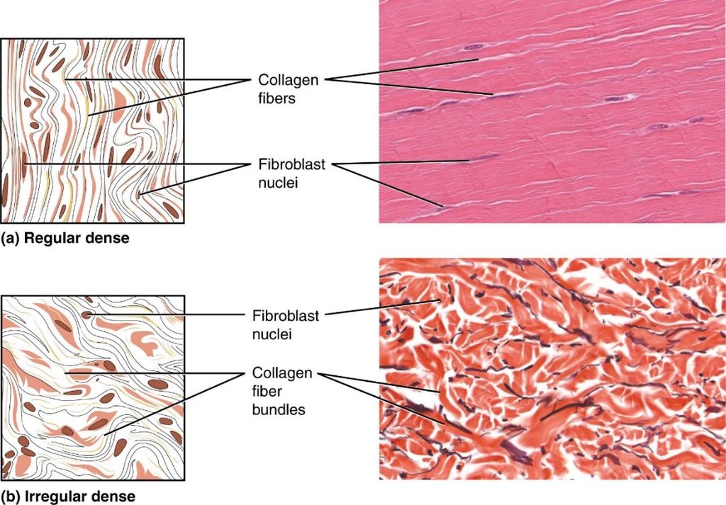
(II) Dense Connective Tissue:
Fibres and fibroblasts are compactly packed in the dense connective tissues.
Orientation of fibres show a regular or irregular pattern and are called dense regular and dense irregular tissues.
In the dense regular connective tissues, the collagen fibres are present in rows between many parallel bundles of fibres e.g. tendons and ligaments.
Dense irregular connective tissue has fibroblasts and many fibres (mostly collagen) that are oriented in different directions.
This tissue is present in the skin, perimysium, perineurium and around bones as periosteum.
(a) White Fibrous Tissue:
It carries only a few fibroblasts scattered amidst the dense network of thick collagen fibre bundles. It has great tensile strength. The presence of white fibrous tissue at the joints between skull bones makes them immovable.
(b) Tendon:
It is a very dense, strong and fibrous connective tissue with thick parallel bundles of collagen fibres. A few flat, elongated tendon cells lie in a single rows between the fibre bundles. Tendon forms the strong inextensible attachment of a skeletal muscle to a bone. Colloidal protein gelatin is obtained by boiling collagen.
(c) Ligament:
Ligaments connect the bones at the joints and hold them in position. Sprain is caused by excessive pulling of ligaments. They are made of bundles of elastic fibres and few collagen fibres. Many year old mummies still have their arteries intact due to well preserved elastic fibres.
Difference between Tendon and Ligament

(d) Reticular tissue:
It consists of star-shaped reticular cells whose protoplasmic processes form a network. These cells are phagocytic in function. Matrix and some other types of cells are also found in the spaces of the network. Reticular tissue is present in the spleen, lymph nodes, bone marrow, etc.
(2) SUPPORTIVE CONNECTIVE TISSUE
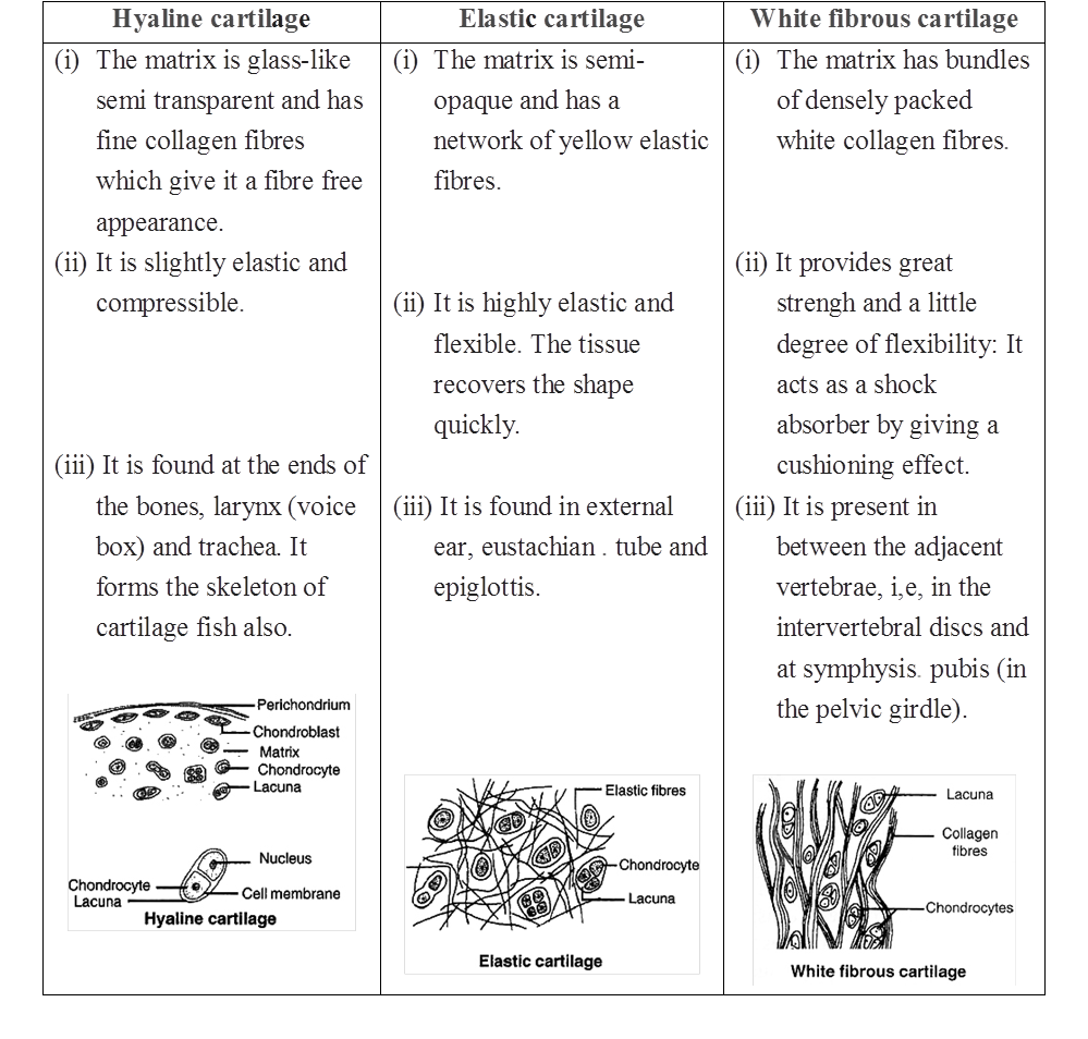
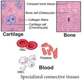
(I) Cartilage:
Cartilage is a solid but semi-rigid and flexible connective tissue. Chondrocytes are large, blunt, angular cartilage cells.
They occur in clusters of 2 or 3 cells in small spaces (lacunae) scattered in the matrix.
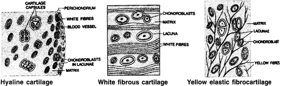
(a) In Hyaline Cartilage, the matrix is apparently fibre-less and glass-like (hyaline) but translucent. It occurs in the larynx, nasal septum, tracheal rings and costal cartilage, It gives those structures a definite but pliable form. White Fibrocartilage carries thick dense bundles of collagen fibres between rows of chondrocytes in lacunae. It occurs in joints between vertebrae. Its collagen fibres make such joints strong but less elastic and only slightly movable.
Nucleus Pulposus - In the centre of the intervertebral disc, a soft area is present called nucleus pulposus which is supposed to be a remnant of notochord.
(b) Elastic Cartilage contains a dense network of elastic fibres between scattered chondrocytes. It forms the eustachian tube, epiglottis and pinna of ear. The elastic fibres make those organs considerably elastic and pliable.
(c) Calcified Cartilage -Initially it is like hyaline cartilage but later on it gets hardened like bone due to deposition of calcium salts, e.g., supra scapula of frog's pectoral girdle, pubis of pelvic girdle of frog.
Table : Types of Cartilage
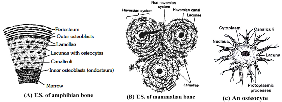
(II) BONE
(i) It is a solid, rigid connective tissue. The matrix of the bone has the deposition of apatite salts of calcium and phosphorus. e.g, hydroxyapatite salts and fluoroapatite salts.
(ii) 60-70% of bone is made up of inorganic matter and 30-40% is made up of organic matter.
(iii) If the bone is put in dil. HCl, the bone becomes decalcified, soft and flexible. Nothing will happen to bone if we put the bone in KOH.
(iv) Osteoblast are bone forming cells which secrete ossein protein.
(v) Osteocytes are bone cells, they are metabolically inactive cells present in lacuna.
(vi) Bone is a solid, rigid and strong connective tissue. Its matrix is heavily deposited with apatite salts of calcium and phosphorus. Flat irregular spaces called Lacunae occur in the solid matrix. Each lacuna lodges a flat bone cell or Osteocyte. A bone cell has an irregular shape and long cytoplasmic processes. These processes extend into minute canals (Canaliculi) radiating from each lacuna.
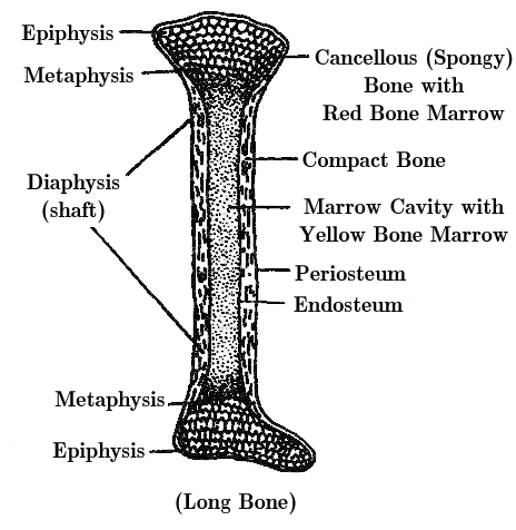
(vii) Compact Bone forms the dense outer layers of all bones. It is composed of many parallel, longitudinal, column-like structures called Haversian Systems, cemented to each other. Haversian canals are connected to each other by Volksman canals. In each Haversian system, several concentric layers (Lamellae) of bony matrix encircle a longitudinal central canal (Haversian Canal). This canal carries blood vessels and nerves. Lacunae containing osteocytes occur in a layer between two lamellae.
(viii) Spongy Bone -The ends of long bones are composed of an open lattice of bone called spongy bone. The spaces within contain marrow, where most blood cells are formed. It carries no concentric organisation like the Haversian system. It consists of a network of many fine irregular bony plates or Trabeculae. Each trabecula consists of many irregularly arranged lamellae with lacunae between them. It has red bone marrow. Spongy bone is also called as cancelfous bone and is found in epiphysis, i.e., the ends of long bones.
Table : Differences between bone and cartilage

Table : Differences between a Dried bone and a Decalcified bone

Concept Builder
Types of Bones :
(i) Cartilage bones / Endochondrial / Replacing bones - They are formed by the replacement of cartilage by the bone e.g. humerus, femur, vertebrae, ribs, girdle bones except clavicle. Chondroclasts are cartilage eater cells.
(ii) Membrane / Investing bone / Dermal - e.g. skull bones, clavicle. The bones are formed in the dermis of the skin and are invested over the already present cartilages.
(iii) Sesamoid bones - They are formed by the ossification of the tendons e.g., Patella.
(iv) Visceral bones - They are those bones which get detached from the skeleton and come to lie in visceral organs e.g.,
(a) os cordis - Present in interventricular septum of heart of deer.
(b) os falciparum - Palm of mole.
(c) os penis - Penis of rat and carnivores.
(d) os palbebrae - In the eyelids of crocodile.
(e) os rostralis - Snout of pig.
1. Bone China: Porcelain was first made in China during tang dynasty. English found a new way of making porcelain with bone ash. Bone china is a form of porcelain made from burned animal bones. Bone ash is mixed with kaolin, a white clay. The bone ash increases the porcelain's translucence.
2. Word Roots and Origins : Periosteum from the Greek "peri" meaning "around" and "osteon" meaning bone.
(3) FLUID CONNECTIVE TISSUE
BLOOD
Blood is a fluid connective tissue.
Its cells are quite distinct from other connective tissue cells both in structure and functions.
The extracellular material in blood is a fluid devoid of fibres.
Fluids outside the cells are generally called Extracellular Fluids (ECF).
Blood is heavier than water.
The extracellular material in blood is a straw-coloured, slightly alkaline (pH =7.4) aqueous fluid called plasma.
Constituents, having characteristic forms, float in the plasma.
They are collectively called the Formed Elements of blood.
They include the blood cells and blood platelets.
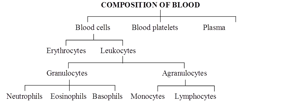
Blood cells are of two types-Erythrocytes and Leukocytes.
Blood circulates within blood vessels in higher animals.
But other extracellular fluids such as cerebrospinal fluid, interstitial fluid, lymph and aqueous humour occur outside blood vessels.
COMPOSITION OF BLOOD
PLASMA
Plasma contains three major classes of plasma proteins viz. serum albumin, serum globulins and fibrinogen.
Plasma proteins serve as a source of proteins for tissue cells.
Tissue cells may utilise plasma proteins for forming their cellular proteins.
Additionally, albumin and globulins retain water in blood plasma by their osmotic effects.
A fall in plasma proteins leads to movement of excessive volumes of water from blood to tissues.
That is why hands and feet get swollen with accumulated fluid (oedema) in persons suffering from dietary deficiency of proteins.
Albumins and globulins also transport many substances such as thyroxine and Fe3+ in combination with them.
One class of globulins, called immunoglobulins, act as Antibodies.
Plasma proteins also maintain the blood pH by neutralising strong acids and bases.
Thus, they act as Acid-Base-Buffers.
It is slightly alkaline non-living inter-cellular substance which constitutes about 60% part of the blood.
It is a pale yellow but transparent and clear fluid.
Composition of Plasma. Plasma forms 55-60% by volume of blood.
1. Water-Water alone forms about 90% to 92% of the plasma. Solids form about 8% of the plasma.
2. Mineral Salts -These are chlorides, bicarbonates, sulphates and phosphates of sodium, potassium, calcium, iron and magnesium. All salts constitute about 0.9% of plasma. Buffer of the blood is sodium bicarbonate.
3. Nutrients - These include glucose, fatty acids, phospholipids, cho'lesterol, fats, aminoacids, nucleosides, etc. Mineral salts have been mentioned above.
4. Plasma proteins -They constitute about 7 to 8% of plasma. These mainly include albumin 4.4% , globulin 1.5 to 2%, prothrombin and fibrinogen both 0.3%.
5. Defence proteins - Immunoglobulins which act as antibodies and some other substances, such as lysozyme and properdin (a large protein) are always found in the plasma. They destroy bacteria, viruses and toxic substances that may enter into the blood from outside.
6. Excretory substances - These include ammonia, urea, uric acid, creatinine, etc.
7. Dissolved gases - Water of blood plasma contains oxygen, carbon dioxide and nitrogen in dissolved form.
8. Anticoagulant - Blood plasma contains a conjugated polysaccharide, heparin which prevents coagulation of blood inside blood vessels.
9. Hormones - These are secreted and released in blood by endocrine glands.
10. Vitamins and Enzymes-Different kinds of vitamins and enzymes are present in the blood plasma.
Functions of Blood plasma-
These can be summarised as under (i) transport, (ii) retention of fluid in blood, (iii) maintenance of blood pH, (iv) body immunity, (v) prevention of blood loss, (vi) conducting heat to skin for dissipation and (vii) uniform distribution of heat all over the body.
Blood Glucose
Glucose is mainly absorbed in the small intestine.
Glucose is also absorbed in the stomach.
After absorption glucose reaches the blood.
Excess of glucose is converted into glycogen by insulin hormone in the liver and muscles.
Whenever it is required, glycogen is changed back into glucose by glucagon hormone.
Usually blood glucose level is about 80-100 mg per 100 ml of blood, 12 hours after a normal meal.
But its concentration rises soon after a carbohydrate rich diet.
If blood glucose level exceeds 180 mg per 100 ml, it starts appearing in urine.
This condition is called glucosuria.
Fasting glucose is 70 -110 mg/dl. Glucose PP[1] is 110 -140 mg/dl.
If it is higher, it causes diabetes mellitus (hyperglycemia).
If it is less, it causes hypoglycemia (less amount of glucose in blood).
Blood Cholesterol
Usually cholesterol is considered a harmful substance. But it is quite useful in limited amount.
Cholesterol is used in the synthesis of biomembranes, vitamin D, bile salts and steroid hormones.
Its normal amount is 80 -180 mg in 100 ml of blood plasma.
Cholesterol comes in the blood plasma either by intestinal absorption of fats or by the synthesis from the liver or by both.
Saturated fats such as ghee and butter increase cholesterol level in the blood.
Increased blood cholesterol may lead to its deposition in the internal wall of the blood vessels like arteries and veins which causes high blood pressure and heart problems.
Functions of Plasma Proteins
1. Prevention of blood loss - Fibrinogen and prothrombin playa role in blood clotting.
2. Retention of fluid in the blood - Albumin helps in osmotic balance.
3. Body immunity - Certain globulins called immunoglobulins (glycoproteins) act as antibodies in blood and tissue fluid. Antibodies belong to a class of proteins called as immunoglobulins.
4. Maintenance of pH - Plasma proteins serve as acid-base buffers. It means they maintain pH of the blood by neutralizing acids and bases.
5. Transport of certain materials - Thyroxine (hormone) is bound to albumin or specific globulin for transport in the plasma.
6. Distribution of heat - Plasma proteins help in uniform distribution of heat all over the body.
7. Enzymes - Some proteins acting as enzymes also occur in the plasma.
BLOOD CELLS
(i) Erythrocytes:
Erythrocytes (red blood corpuscles or RBC) are the most numerous of the formed elements of blood.
Their most important characteristic feature is the presence of hemoglobin, the red oxygen carrying pigment.
The total number of erythrocytes per microlitre (1 µl = 1mm3 =10–6) of blood is known as the Total Count of RBC.
It averages 5 millions and 4.5 millions in adult man and adult woman respectively.
The total count would be low in anaemia and after profuse bleeding.
On the contrary, the abnormal rise in the total count of RBC is called Polycythemia.
Anaemia is caused due to the deficiency of folic acid, vitamin B12 and haemoglobin.
The size and shape of erythrocytes vary in different classes of animals.
In fishes, amphibians, reptiles and birds, erythrocytes are usually nucleated, oval and biconvex. But in mammals they are non nucleated, biconcave and circular.
Only camel and llama possess oval red blood corpuscles.
Human erythrocytes measure 7-8 µm (1µm =10–6 m) in diameter and 2 µm thickness near the rim.
Old and damaged erythrocytes are phagocytosed and destroyed by macrophages.
The pigment part (porphyrin) of hemoglobin is then catabolised to the yellow pigment Bilirubin which is excreted in the bile.
The pale yellow colour of plasma is largely due to bilirubin.
If a sample of blood is rendered non-coagulable by adding potassium or sodium oxalate and then centrifuged at a high speed in a graduated centrifuge tube (hematocrit tube), the centrifugal force rapidly sediments the erythrocytes tothe bottom of the tube.
They become packed into a solid, red, bottom layer while plasma forms a clear, fiuid upper layer.
On the upper surface of the erythrocyte layer, leukocytes form a thin, buff-coloured layer.
From the graduations on the tube, the relative volume of erythrocytes may be read as a percentage of the total blood volume. This is called the Hematocrit Value or Packed Cell Volume.
It normally forms 45 percent of the blood volume.
RBC of mammals are circular, biconcave, non-nucleated except family camelidae. e.g. camel, which has non-nucleated and oval RBC. Largest RBCs are found in amphibia.
Smallest RBCs are found in mammals.
In mammals smallest RBCs are found in 'Musk Deer', Tragulus javanicus (1.5 µm).
In mammals, largest RBCs are found in elephant. (9.4 µJ).
Graveyard of RBC is spleen.
Life Span
Life span of RBC in man =120 days
Life span of RBC in frog = 100 days
Life span of RBC of rabbit = 80 days
Radioactive chromium method (Cr51) is used for estimation of life span of RBC.
Count of RBC
In embryo = 8.5 million/mm3
In Man = 5 to 5.5 million/mm3
In Woman = 4.5 million/mm3
Daily destruction of RBC = 1%
ESR (Erythrocyte sedimentation rate) : It is measured by Wintrobe's method. It is rate of settling down of RBC.
It is also estimated by Westergen's method.
ESR is very useful in diagnosing various diseases including tuberculosis, ESR in men is 0-5 mm/hour and in women it is 0-7 mm/hr in Westergen method.
Haemocytometer : It is instrument for counting the number of both WBCs and RBCs.
Rouleaux: In resting and slow flowing blood, the RBCs aggregate to form rouleaux (the RBCs are piled on top of each other). Fibrinogen favours rouleaux formation.
Bone marrow : It is the main site for formation of RBC. Volume of bone marrow at the time of birth is 70 ml. In adult volume of bone marrow is 4,000 ml.
Structure of RBC of man : Biconcave non-nucleated bounded by Donnan's membrane (plasma membrane of RBC). Haemoglobin is filled in RBC which is respiratory pigment.
Normal Range of Hb
Infants 16.5 ± 3.0 g/dl (dl = deciliter)
Children 3 months 11.0 ± 1.5 g/dl
Children 3 to 6 years 12.0 ± 1.0 g/dl
Children 10 to 12 years 13.0 ± 1.5 g/dl
Men 15.5 ± 2.5 g/dl
Women 14.0 ± 2.5 g/dl
Structure of Haemoglobin :
Each molecule of haemoglobin contains 4 molecules of haem and 1 molecule of globin.
These are attached by co-ordinate bonds.
Haem is protoporphyrin compound and has 4 pyrrole groups jointed together to form ring structure.
In Hb, Fe is present in (Fe++) Ferrous form.
Haem is 5% & Globin is 95%. Globin is made of 4 polypeptide chains.
(ii) Leukocytes:
Leukocytes (white blood corpuscles or WBC) are devoid of hemoglobin and are consequently colourless.
Leukocytes are nucleated blood cells.
They are of two major classes : granulocytes (with cytoplasmic granules) and agranulocytes (without granules).
Granulocytes are of three types, viz. neutrophils, eosinophils and basophils, each with lobed nucleus.
Agranulocytesare of two types, viz. lymphocytes and monocytes.
Neutrophils and monocytes protect the body against microbes by phagocytosing them.
Lymphocytes secrete antibodies in the blood to destroy microbes and their toxins.
The number of leukocytes per microlitre (1 µl = 1 mm3 = 10–6) of blood is called the Total Count of WBC.
It is 6000-8000/mm3 of blood normally.
It may rise abnormally in acute infections (e.g., pneumonia), inflammations (e.g. appendicitis) and malignancies (e.g., leukemia).
In some conditions such as folic acid deficiency, the total count falls abnormally (leukopenia).
The total count of WBC is also of diagnostic value in many diseases.
Monocytes have kidney shaped nucleus.
The process by which monocytes and neutrophils squeeze out through thin capillary walls is Diapedesis.
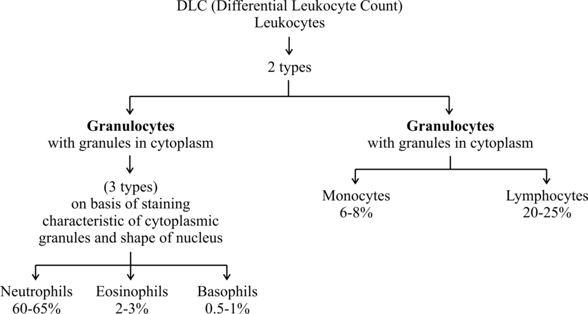
(a) Neutrophils : They are maximum in number, stain equally with both basic and acidic dyes and have many lobed nucleus, granules are in abundance in cytoplasm and help in phagocytosis.
(b) Eosinophils : They have bilobed nucleus, stain with acidic stains. Their number increases during allergic reactions (Eosinophilia).
(c) Basophils: They stain with basic dyes. Their nucleus is 'S' shaped. Coarse granules are few in the cytoplasm. Basophils release heparin and histamine in the blood and have a function similar to the mast cells.
(d) Lymphocytes have large and rounded nucleus. The cytoplasm forms a thin peripheral film. They have their stem cells in the bone marrow and are differentiated in the bone marrow or in the thymus. Lymphocytes are of two types, B-lymphocytes and T-lymphocytes.
B-lymphocytes produce antibodies against antigens and they mature in the bone marrow.
(e) Monocytes are the largest leucocytes (12-15 µm). The nucleus is kidney shaped. They are produced from bone marrow monoblast cells. They help in phagocytosis.
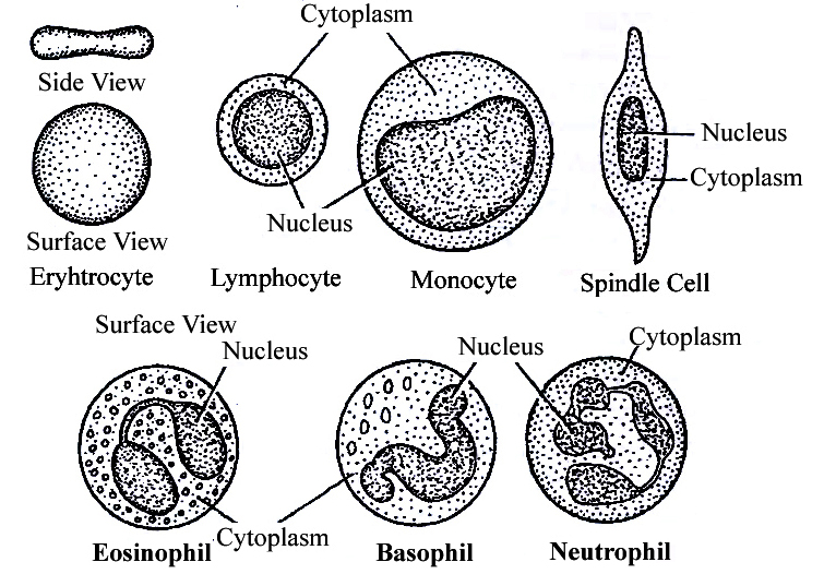
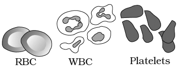
Table : Differences between different types of Leucocytes

(iii) Blood platelets:
Also called thrombocytes, blood platelets are non-nucleated, round or oval, biconvex disc-like bodies.
They are 2-3 micrometres in diameter and their number normally varies from 0.15 to 0.35 million/mm3 or 150000 -350000 platelets/mm3.
They bud off from the cytoplasm of very large Megakaryocytes of the bone marrow.
Their normal life-span is about a week.
When a blood vessel is injured, platelets get clumped at the injured spot and release certain chemicals called Platelet Factors.
These promote blood coagulation.
Thrombocytopenia is decrease in platelet count and Purpura is a group of bleeding diseases due to thrombocytqpenia.
BLOOD COAGULATION
When blood oozes out of a cut, it sets into gel within a few minutes. This is called coagulation.
Coagulation is brought about by hydrolysis of soluble fibrinogen of plasma to insoluble fibrin.
This is catalysed by an enzyme called thrombin.
Fibrin precipitates as a network of fibres.
This network traps many blood cells, particularly RBCs, to form a red solid mass called the Blood Clot.
The clot seals the wound in the vessel to stop the bleeding.
The straw coloured fluid left after clotting of blood, is called Serum.
The serum cannot be coagulated as it lacks fibrinogen.
Thrombin occurs in normal blood as an inactive globulin called Prothrombin.
It must be activated to thrombin before blood coagulation can occur.
In case of injury to a blood vessel, coagulation promoting substances called Thromboplastins are released into the blood from clumped platelets and damaged tissues.
Thromboplastins help in the formation of the enzyme Prothrombinase.
This enzyme hydrolyses prothrombin to thrombin to initiate coagulation.
Ca2+ ions are essential for both activation and action of thrombin.
Blood normally contains an anticoagulant, Heparin which prevents activation of prothrombin, Heparin is released from mast-cell granules.
Blood also contains Antithrombin which inhibits any thrombin formed accidentally.
Blood drawn from a blood vessel can be kept uncoagulated by adding a pinch of oxalate (sodium or potassium oxalate) to it.
Oxalate precipitates Ca2+ and consequently prevents coagulation.
Chilling of blood also delays coagulation because lesser temperature depresses the action of coagulation promoting enzymes.
Concept Builder
ABO Blood cloting factor :
Karl Landsteiner reported for the first time ABO blood groups in human beings.
A. B and O blood groups were discovered by Landsteiner (1900) while AS blood group was found out by de Castello and Steini (1902).
Agglutinogens (antigens) are present on the surface of red blood corpuscles and agglutinins (antibodies) are found in the blood plasma. Both antigens and antibodies are proteins.
When two different type of blood are mixed, the red blood corpuscles form a clump.
The clumping of red blood corpuscles is called agglutination.
Clotting Factors :
13 factors help in blood clotting.
These factors are mainly produced in liver.
Vitamin K is required in the synthesis of these clotting factors.
These factors are represented in Roman number.
I – Fibrinogen
II – Prothrombin
III – Thromboplastin
IV – Ca+2 (cofactor in each step of blood clotting)
V – Proaccelerin
VI – Accelerin (Rejected)
VII – Proconvertein
VIII – AHG Anti Haemophelic Globin (Absent in Haemophilia-A)
IX – Christmas factor
X – Stuart factor
XI – PTA (Plasma Thormboplastin Anticedent)
XII – Hagman factor
XIII – FSF Factors (Fibrin stabilizing factor) (Laki Lor and factor).
Other natural anticoagulants are
Hirudin – found in leech.
Anophelin – found in female Anophelese.
Lampredin – found in Peteromyzon (Lamprey)
Cumerin – obtain from plants
Warfarin – obtain from plants
To collect blood in bottle in blood bank artificial anticoagulants are used like
Sodium citrate
Sodium oxalate
EDTA (Ethylene diamine tetra acetic acid)
These chemicals act as Calcium binding units and remove Ca+2 ions from blood.
Blood group
Agglutination is due to the interaction of antigens and antibodies.
There are two kinds of antigens that are named A and B.
There are also two kinds of antibodies which are called a and b.
The antigen A and antibody a are incompatible (antagonistic) and cause self clumping and cannot exist together.
Similarly, the antigen B and antibody b are incompatible and cause self clumping and cannot exist together.
Thus, A and b can exist together and B and a can exist together.
The corpuscle factors A and B can occur together if their antagonistic plasma factors a and b are not present.
The plasma factors a and b can occur together if their antagonistic corpuscle factors A and B are absent.

Rh Factor
Another-antigen, the Rh antigen similar to one present in Rhesus monkeys (hence Rh), is also observed on the surface of RBCs of majority (nearly 80 per cent) of humans.
In India % ratio of Rh is
Rh+ – 97%
Rh– – 3%
In World
Rh+ – 80%
Rh– – 20%
Such individuals are called Rh Positive (Rh+ve) and those in whom this antigen is absent are called Rh negative (Rh-ve).
An Rh-ve person, if exposed to Rh+ve blood, will form specific antibodies against the Rh antigens.
Therefore, Rh group should also be matched before transfusions.
A special case of Rh incompatibility (mismatching) has been observed between the Rh-ve blood of a pregnant mother with Rh+ve blood of the foetus.
Rh antigens of the foetus do not get exposed to the Rh-ve blood of the mother in the first pregnancy as the two bloods are well separated by the placenta.
However, during the delivery of the first child, there is a possibility of exposure of the maternal blood to small amounts of the Rh+ve blood from the foetus.
In such cases, the mother starts preparing antibodies against Rh antigen in her blood. In case of her subsequent pregnancies, the Rh antibodies from the mother (Rh-ve) can leak into the blood of the foetus (Rh + ve) and destroy the foetal RBCs.
This could be fatal to the foetus or could cause severe anaemia and jaundice to the baby.
This condition is called erythroblastosis foetalis.
This can be avoided by administering anti-Rh antibodies to the mother immediately after the delivery of the first child
Connective Tissue
Connective Tissue
In the bodies of sophisticated animals, connective tissues are the most abundant and widely dispersed. Connective tissues get their name from their unique ability to join and support other body tissues and organs. They include soft connective tissues as well as specialized forms such as cartilage, bone, adipose tissue, and blood. Cells in all connective tissues, except blood, secrete collagen or elastin fibers, which are structural proteins. The fibers provide the tissue its strength, elasticity, and flexibility. These cells also release modified polysaccharides, which form a matrix between cells and fibers (ground substance). There are three forms of connective tissue:
1. Loose connective tissue: Areolar tissue present beneath the skin, is loose connective tissue with cells and fibers loosely distributed in a semi-fluid ground substance. It frequently acts as a support structure for epithelium. Fibroblasts (cells that generate and secrete fibers), macrophages, and mast cells are all present. Another form of loose connective tissue found primarily beneath the skin is adipose tissue. This tissue's cells arespecialized for fat storage. Excess nutrients are converted to lipids and stored in this tissue if they are not utilized right away.
2. Dense connective tissue: The dense connective tissues are densely packed with fibers and fibroblasts. Dense regular and dense irregular tissues have a regular or irregular pattern in their fiber orientation.Collagen fibers are found in rows between several parallel bundles of fibers in dense regular connective tissues. Tendons that connect skeletal muscles to bones and ligaments that connect two bones are examples of this tissue. Fibroblasts and numerous fibers (mainly collagen) are orientated differently in dense irregular connective tissue. The skin contains this tissue.
3. Specialised connective tissue: Specialized connective tissues include cartilage, bones, and blood. The cartilage intercellular substance is firm and malleable, and it resists compression. Chondrocytes (tissue cells) are encased in little cavities within the matrix they secrete. In adults, most cartilages in vertebrate embryos are replaced by bones. In adults, cartilage can be found in the tip of the nose, outer ear joints, and between neighboring spinal column bones, limbs, and hands. Bones have a hard, non-pliable ground substance that is rich in calcium salts and collagen fibers, and this is what gives them their strength. It is the major tissue that gives the body its structural framework. Soft tissues and organs are supported and protected by bones.Lacunae are places where bone cells (osteocytes) can be found. The lengthy bones of the legs, for example, serve as weight-bearing structures. They also interact with the skeletal muscles that are linked to them in order to form blood cells in the marrow of some bones. Plasma, red blood cells (RBC), white blood cells (WBC), and platelets make up blood, which is a fluid connective tissue. It is the primary circulating fluid that aids in the transportation of a variety of chemicals.
Muscular Tissue
- Books Name
- ACME SMART COACHING Biology Book
- Publication
- ACME SMART PUBLICATION
- Course
- CBSE Class 11
- Subject
- Biology
MUSCLE TISSUE
Muscles cause movements of limbs and internal organs and also locomotion of the organism.
Cells of muscle tissue can shorten forcefully and again return to the relaxed state.
This specialised property is called Contractility.
It is based on the organised arrangement of some protein filaments in the cytoplasm of a muscle cell.
The cell shortens or relaxes according to the relative positions of different intracellular filaments.
Whenever adequately stimulated, muscle cells respond by contracting.
This property of the muscle tissue is responsible for various movements in an animal.
Muscle cells are usually called Muscle Fibres because they are thin and elongated.
In higher animals, some muscles remain associated with the skeleton, but many others form walls of visceral organs, blood vessels and heart.
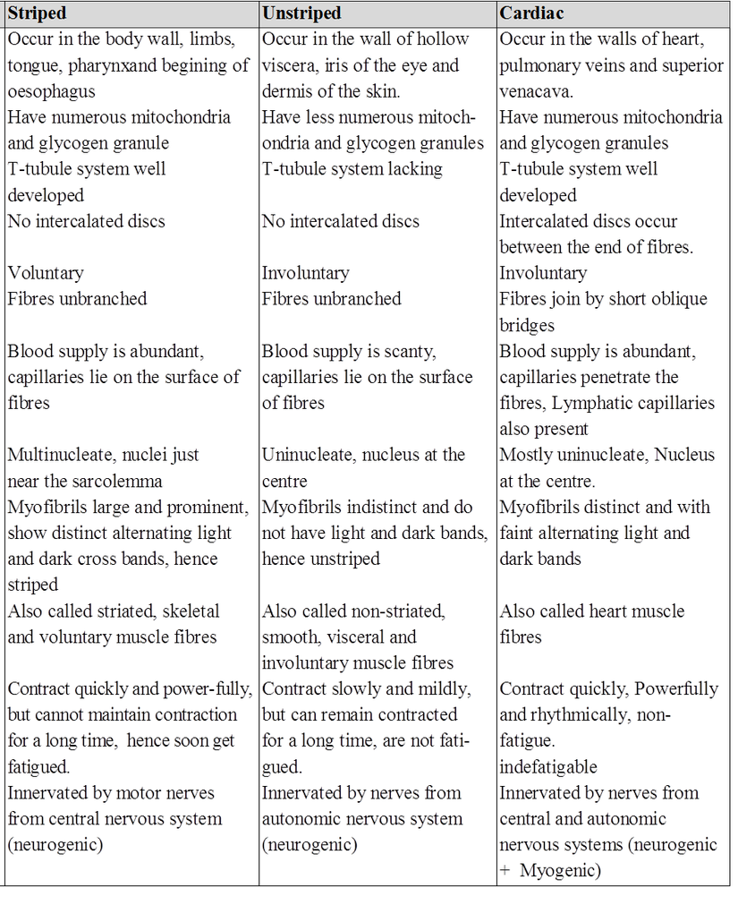
Muscle tissue may be classified into striated, non-striated and cardiac muscles, according to their structure, location and functions.
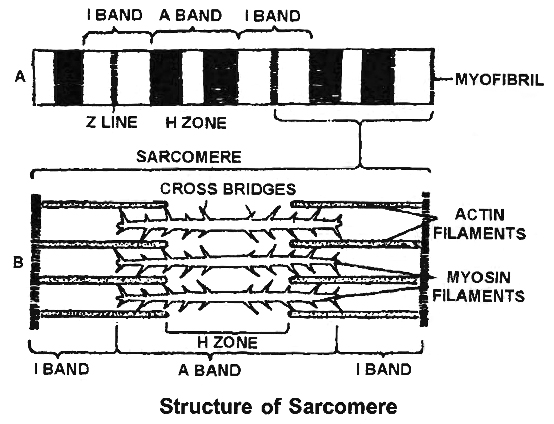
(i) Striated / Skeletal / Voluntary muscles are attached to bones by tendons. A voluntary muscle is composed of long bundles of striated muscle fibres. Each fibre is a long, unbranched, cylindrical cell. It shows transverse striations in the form of regular alternate dark (A) and light (l) bands.
At the centre of the I band is a fine, dense Z band or Z-line (Krause's membrane). The plasma membrane covering the fibre is called Sarcolemma. The cytoplasm inside the fibre is called Sarcoplasm.
The sarcoplasm contains many long, thin, unbranched, cross-striated cylindrical structures called Myofibrils. They are arranged along the long axis ofthe fibre. Dark A bands of neighbouring myofibrils are located side by side, so also are their light I bands. This gives crossstriated appearance to the entire muscle fibre also.
A-band has both actin and myosin filaments. The portion of A-band, where actin filaments are absent is called H-zone. Z-line or Krauze membrane is a dark membrane which bisects I band or isotropic band.
Muscle is rich in proteins. Most of these proteins occur as two types of filaments arranged longitudinally in myofibrils. The thick filaments are made up of the protein Myosin. Myosin filaments are located inside A bands.
Thin filaments are more numerous. They are composed of the protein Actin. From a fine, dense, dark Z band at the centre of each I band, actin filaments extend through the I band and encroach between myosin filaments upto a considerable distance into the A band.
Each segment of the myofibril from one Z band to the next, functions as a contractile unit and is called a Sarcomere.
Various parts of a sarcomere have a specific arrangement of actin and myosin filaments as given below.
I band – Has only actin filaments
A band – Has both actin and myosin filaments
H band – Has only myosin filaments
Z line – A membrane to which actin filaments are attached on both the sides.
(ii) Non-Striated or Smooth muscle fibres do not show cross-striations, instead, they look smooth. Smooth muscles cannot be moved voluntarily. So they are also called Involuntary Muscles. Functionally, smooth muscles are of two types. Single-Unit Smooth Muscles are composed of muscle fibres closely joined together.
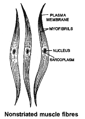
All its fibres contract together as a single unit. They may contract automatically and rhythmically. Such smooth muscles occur on the walls of hollow visceral organs such as the urinary bladder and the gastrointestinal tract. Multi-Unit Smooth Muscles are composed of more independent muscle fibres, not so closely joined together. Individual fibres of such smooth muscles contract as separate unit. These occur at hair roots and in the walls of large blood vessels. e.g., Erector pili muscles.
Smooth muscle fibres are elongated spindle-shaped cells. They are packed parallel to each other in branching bundles. Each fibre contains a single, spindle shaped nucleus at its thick central part. The smooth muscle fibre is generally shorter than a striated muscle fibre. Mitochondria and other organelles are less extensive and protein filaments are not regularly arranged to give rise to striations.
Table : Differences between Single-unit and Multi-unit Smooth Muscles

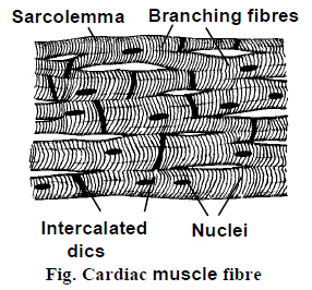
(iii) Cardiac muscle occurs in the heart. It possesses considerable automatic rhythmicity and generates its own wave of excitation. The excitation can also pass directly from one fibre to another in the cardiac muscles. It is not under voluntary control. It shows cross-striations, but striations are much fainter than those of striated muscle.
Between the cardiac muscle fibres, intercalated discs are present. They are specialised regions of cell membrane of two adjacent fibres. The intercalated discs function as boosters of contraction wave and permit the wave of contraction to be transmitted from one cardiac fibre to another.
Cardiac muscle cells are short cylindrical cells joined end to end to form rows. They possess abundant cytoplasm with myofibrils (sarcoplasm) and numerous mitochondria and glycogen granules.
This is because they need a large amount of energy. Faint but regular, alternate dark and light bands give rise to cross-striations in the cardiac muscle fibres and indicate regular and alternate arrangements of thin and thick filaments in the fibre.
Sarcomeres are also present. Cardiac muscle cells frequently branch to form junctions with neighbouring cells. Where two cardiac muscle cells meet end to end, dense zig-zig junction is formed between them. It is called an Intercalated Disc. Longest refractory period is present in cardiac muscles.
Differences between striated, non-striated and cardiac muscles.
Muscular Tissue
Muscular Tissue
Many long, cylindrical fibers are organized in parallel arrays in each muscle. Myofibrils are a type of tiny fibril that makes up these fibers. Muscle fibers contract (shorten) in response to stimulus, then relax (lengthen) and uncontract in a coordinated manner. Their actions cause the body to move in order tochange the environment and maintain the postures of the various body parts. The sarcolemma is the muscle cell membrane, and the sarcoplasm is the muscle cell cytoplasm. The excitability of the sarcolemma allows it to conduct electrical impulses that occur during depolarization. Muscles are involved in all of the body's movements in general. Skeletal, smooth, and cardiac muscles are the three types of muscles. Skeletal muscle tissue is tightly bound to the bones. Striated (striped) skeletal muscle fibers are bundled together in a parallel pattern in a typical muscle like the biceps.Several bundles of muscle fibers are encased in a strong connective tissue sheath.Tendons connect skeletal muscles to bones, and they are responsible for all motions of bodily components in relation to one another. Skeletal muscle, unlike smooth and cardiac muscle, is controlled by the user. Shoulder muscles, hamstring muscles, and abdominal muscles are all examples of skeletal muscles. Smooth muscle fibers have no striations and taper at both ends (fusiform). They are held together by cell connections and bundled in a connective tissue sheath. This type of muscle tissue can be found in the walls of internal organs such as the blood vessels, stomach, and intestine. Smooth muscles are referred to as "involuntary" since they cannot be controlled directly. Cardiac muscle tissue is only found in the heart, where it performs coordinated contractions that allowthe heart to pump blood through the circulatory system.The only contractile tissue found in the heart is cardiac muscle tissue. The plasma membranes of heart muscle cells are fused.At some fusion points, communication junctions (intercalated discs) allow cells to contract as a group, meaning that when one cell receives a signal to contract, its neighbors are also stimulated to contract.
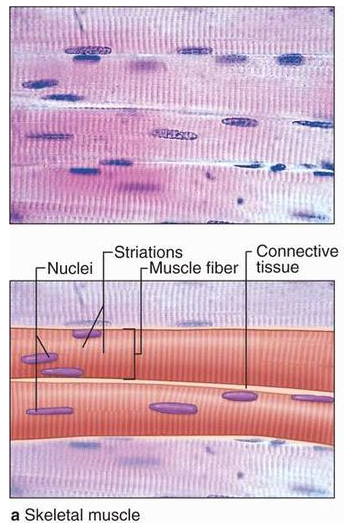
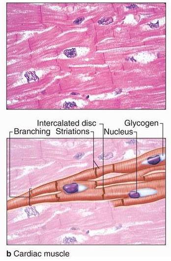
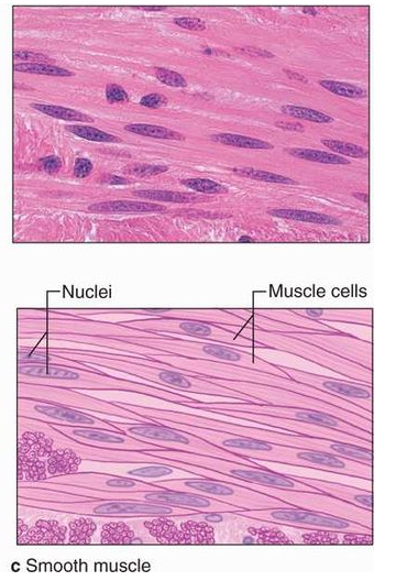
Figure 8: Muscular Tissue
Nervous (Neural) Tissue
- Books Name
- ACME SMART COACHING Biology Book
- Publication
- ACME SMART PUBLICATION
- Course
- CBSE Class 11
- Subject
- Biology
NERVE TISSUE
Ordinary connective tissue is absent inside the central nervous system, the neurons are held together by supportive cells called Neuroglia Cells. Nerve tissue is made of neurons and neuroglia cells.
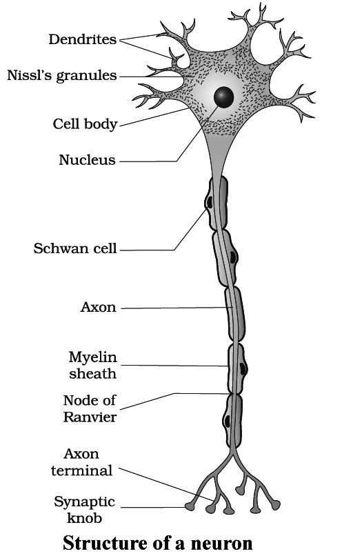
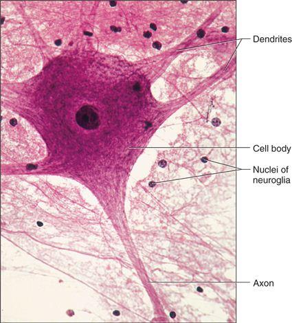
A Neuron has a large cell body with two or more, thin protoplasmic processes extending from it.
One of the processes called the Axon is long and conducts nerve impulses away from the cell body.
It ends in a number of small branches on muscle fibres, gland cells or other neurons.
The remaining one or more processes conduct nerve impulses towards the cell body and are called Dendrites or Dendrons.
The axon terminals may form intercommu- nicating junctions, called Synapses, with dendrite terminals, cell bodies or even axons of other neurons.
Nerve impulses pass between neurons through the synapse with the help of chemicals such as acetylcholine which are termed Neurotransmitters.
The cell body of a neuron is called the Soma.
The soma has various shapes.
Both the soma and the processes are covered by the plasma membrane.
The soma contains abundant granular cytoplasm and a large nucleus.
To serve the high energy needs for impulse conduction, neurons have many mitochondria.
Light microscopy shows many small conical, angular or rhomboidal and highly basophilic structures in the cytoplasm of soma and dendrites, called Nissl Bodies which are absent in the axon and the axon hillock. Nissl's bodies are made of ribosomes, ER, m-RNA.
The processes which arise from neuron are called as neurites. These are of two types-Dendrites, Axon.
(i) Dendrites conduct the nerve impulse towards the nerve cell body and are called as afferent processes.
(ii) Axon is a single, usually long process. The part of cyton from where the axon arises is called as axon hillock. The cell membrane of the axon is called axolemma and its cytoplasm is known as axoplasm. The axon divides to form axon ending ; each with a synaptic knob. The synaptic knobs contain mitochondria and secretory vesicles. The vesicles contain neurotransmitters which are nor-adrenaline, adrenaline or Acetyl choline etc.
SYNAPSE
Nerve signals travels from neuron to neuron all over the body.
These associations are called synapses.
Synapse is a junction between axon endings of one nerve fibre and dendrite of the other.
At a synapse, the membrane of axon and dendrites are not in physical contact with each other but there is a narrow intercellular gap, 10 to 20 nanometres across, separating the axon tip and the target cell.
This gap is Synaptic cleft. The neurotransmitter is always released from axon endings and not by dendrites, so there is only one way transmission of nerve impulse.
Types of Neurons
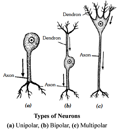
The neurons are of four types based on the number of nerve processes.
(i) Unipol'ar neurons : Which have only axon but no dendron and are found only in early embryos.
(ii) Bipolar neurons : Which have two processes, one axon and another dendron, and are found in olfactory epithelium and retina of eye.
(iii) Multipolar neurons: Which have many processes arising from cell body; out of them one is (longer) acts as an axon and the remaining as dendrites. Multipolar neurons are most common and are found in brain and spinal cord.
(iv) Pseudo-unipolar neurons: They are actually bipolar but appear like unipolar. A single process arises first which divides to form dendrite and axon. This is found in dorsal root ganglion of the spinal cord.
Non-polar Neurons: Each neuron bears several branched processes which are not differentiated into axon of dendrite.
On the basis of function, neurons are of three types:
(i) Sensory (Receptor or Afferent) Neurons: They connect sense organs with the central nervous system (brain and spinal cord).
(ii) Motor (Effector or Efferent) Neurons : They connect tile central nervous system to the effectors (muscles and glands). They carry motor impulses from the central nervous system to the effectors.
(iii) Interneurons (Connector, Relaying or Adjustor Neurons) : They are present in the central nervous system and occur between the sensory and motor neurons for distant transmission of impulses. They are neither sensory nor motor.
Extended axon or dendrite of a neuron is called a nerve fibre. It is generally elongated axon. There are two basic types of nerve fibres :
(i) Myelinated/Medullated nerve fibres are with the myelin sheath. Myelin sheath is formed by the spiral wrapping of Schwann cell membrane around the axon. Outside the myelin sheath, neurilemma is present. Myelin sheath is absent at certain points called Nodes of Ranvier. In myelinated nerve fibres, the impulse jumps from one node of Ranvier to the other, this is called saltatory conduction of the impulse. Node of Ranvier is without myelin but with Neurilemma. Myelinated nerve fibres are found in cranial and spinal nerves.
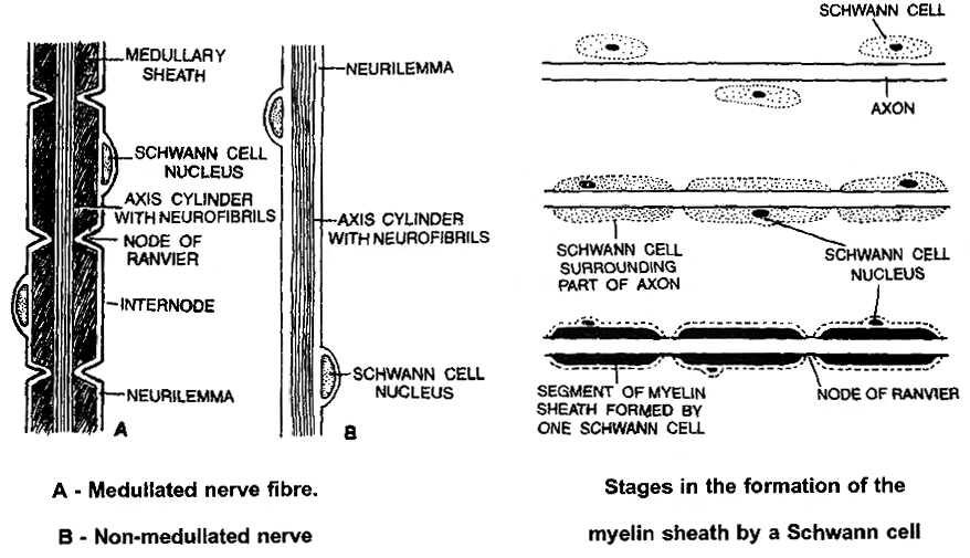
(ii) Non-myelinated/non-medullated nerve fibres are not covered with myelin sheath. They are called non-myelinated or non-medullated nerve fibres. They do not possess nodes of Ranvier, but have neurilemma. Myelinated nerve fibres are generally thicker than non-myelinated ones. These fibres are enclosed by Schwann cell that do not form a myelin sheath around these axons and are commonly found in autonomous and the somatic neural system.
Nerve:
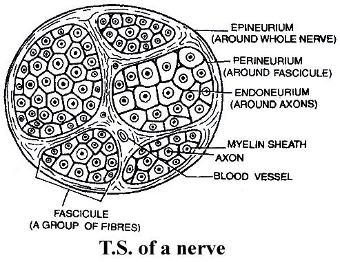
A nerve is a collection of nerve fibres surrounded with connective tissue membranes.
The membrane of the nerve fibre is neurilemma; outside this, each nerve fibres is surrounded by a layer of connective tissue called the Endoneurium.
A nerve consists of several bundles of nerve fibers called fasiculi.
Each fasiculum is surrounded by a layer of connective tissue called the perineurium.
A dense layer of connective tissue that surrounds the entire nerve made of a number of fasiculi is called Epineurium.
A nerve can be :
(i) Sensory Nerve: It is made up of only sensory nerve fibres surrounded by connective tissue membrane. It carries the impulse from the receptor to CNS.
(ii) Motor nerve : It is made up of motor nerve fibres, which carry the impulse from CNS to the effector organs i.e., muscles or glands to bring about their movement.
(iii) Mixed Nerve: It has both the sensory and motor nerve fibres. All the spinal nerves in our body are mixed.
NEUROGLIA CELLS / GLIAL CELLS
They are undifferentiated cells with no Nissl's granules.
(i) Astrocytes / Macrocytes -They are large in size with a number of protoplasmic processes. They form maximum number of glial cells. They help in repair of nerve tissue and form blood brain barrier.
(ii) Oligodendrocytes - They are with few protoplasmic processes and form myelin sheath in eNS.
There is no neurilemma inside the central nervous system. In the absence of Schwann cells, myelin is formed by the spiral wrapping of the nerve fibres by processes of Oligodendrocytes. They are a type of neuroglia cells.
(iii) Microglial cells - They are mesodermal in origin. They are smallest in size with few feathery processes and help in phagocytosis.
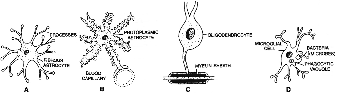
Different kinds of neuroglial cells.
A. Fibrous astrocyte. B. Protoplasmic astrocyte C. Oligodendrocyte. D. Microglial cell.
SUMMARY
Cells, tissues, organs and organ systems split up the work in a way that ensures the survival of the body as a whole and exhibit division of labour.
A tissue is defined as group of cells along with intercellular substances performing one or more functions in the body.
Epithelial tissues are sheet like tissues lining the body's surface and its cavities, ducts and tubes.
Epithelia have one free surface facing a body fluid or the outside environment.
Their cells are structurally and functionally connected at junctions.
Epithelial tissue is classified into different categories on the basis of shape and function of cell.
Diverse types of connective tissues bind together, support, strengthen, protect and insulate other tissue in the body.
Soft connective tissues consists of protein fibres as well as a variety of cells arranged in a ground substance.
Cartilage, bone, blood, and adipose tissue are specialised connective tissues.
Cartilage and bone are both structural materials.
Blood is a fluid tissue with transport functions.
Adipose tissue is a reservoir of stored energy.
Muscle tissue, which can contract (shorten) in response to stimulation, helps in movement of the body and specific body parts.
Skeletal muscle is the muscle tissue attached to bones.
Smooth muscle is a component of internal organs.
Cardiac muscle makes up the contractile walls of the heart.
Connective tissue covers all three types of tissues.
Nervous tissue exerts greatest control over the response of body.
Neurons are the basic units of nervous tissue.
Nervous (Neural) Tissue
Neural Tissue
The brain, spinal cord, and nerves all include nerve tissue or neural tissue. It is in charge of coordinating and controlling various bodily functions. It promotes muscle contraction, raises environmental awareness, and is involved in emotions, memory, and thinking. The body's reactivity to changing conditions is mostly controlled by neural tissue. Nerve cells, often known as neurons, make up nervous tissue. Neurons are specialized cells that respond to stimuli by sending out signals via axons, which are elongated projections that emerge from the cell body.
Nervous tissue contains two types of cells namely neurons and neuroglia. Neurons are excitable cells that make up the neural system. A cell membrane surrounds neuronal cells. The nucleus of neurons includes genes. Cytoplasm, mitochondria, and other organelles can be found in neurons. Neurons don't divide in any manner. Nerves are actually neuron projections.The rest of the neural system is made up of neuroglial cells that protect and maintain neurons. Neuroglia accounts for more than half of all neural tissue in our bodies. Neuroglia is non-neuronal cells that do not produce electrical impulses in the central nervous system and peripheral nervous system. Neuroglia are found in both invertebrates and vertebrates' nervous systems and are distinguishable from neurons by the absence of axons and the presence of only one kind of process. Furthermore, they do not establish synapses and keep their potential to divide throughout their lives.When a neuron is activated sufficiently, an electrical disturbance is produced that travels quickly through its plasma membrane. When a disturbance reaches the neuron's ends or output zone, it sets off a chain of events that can stimulate or inhibit neighbouring cells.
The nervous tissue is the main tissue component of the two major parts of the nervous tissue: the central nervous system (CNS), which is formed by the spinal cord and brain, and the peripheral nervous system (PNS), which controls and regulates the body's functions and activities. Nervous tissue can be found in peripheral nerves throughout the body as well as central nervous system components such as the spinal cord and brain.
organ and organ system
- Books Name
- ACME SMART COACHING Biology Book
- Publication
- ACME SMART PUBLICATION
- Course
- CBSE Class 11
- Subject
- Biology
organ and organ system
The tissues are organized to form organs to perform certain functions. All these organs constitute an organ system. This is very essential for an organism to function.
Cells form tissues form which in turn form organs; and form organ system.
This means that every organ is composed of many tissues.
For example, our heart consists of all the four types of tissues- epithelial, connective, muscular and neural.
organ and organ system
Organ and organ system
In multicellular organisms, the fundamental tissues indicated above arrange to form organs, which then associate to form organ systems. Such organization is required for the millions of cells that make up an organism's activities to be more efficient and coordinated. One or more types of tissues make up each organ in our body. Our heart, for example, is made up of all four types of tissues: epithelial, connective, muscular, and neural. After some thorough examination, we also discover that the complexity of organs and organ systems follows a predictable pattern. The term for this discernible pattern is the evolutionary trend. The morphology and anatomy of three species at various evolutionary levels are reviewed below to demonstrate how they are organized and operate.The study of form or externally observable features is known as morphology. The term morphology only refers to this in the case of plants or microorganisms. This refers to the exterior appearance of organs or sections of the body in animals. The term anatomy is commonly used to describe the morphology of internal organs in animals. The earthworm, cockroach, and frog, which represent invertebrates and vertebrates, will be studied for their morphology and anatomy.
Cockroach
- Books Name
- ACME SMART COACHING Biology Book
- Publication
- ACME SMART PUBLICATION
- Course
- CBSE Class 11
- Subject
- Biology
COCKROACH (Periplaneta americana)
Phylum : Arthropoda
Class : Insecta
Genus : Periplaneta
Species : americana
Morphology
The body is divided into three distinct regions head, thorax, abdomen.
Theirsize ranges from 1/4 inches to 3 inches (0.6-7.6 cm).
Head is hypognathus (facing downwards), and is formed by the fusion of six segments.
Anteriorly, the head bears mouth which is provided with appendages collectively called mouth parts which are used in chewing, cutting and swallowing.
The mouth parts consist of a pair of mandibles and maxillae, labium forming the lower lip and a labrum forming upper lip. Within the cavity enclosed by mouthparts, there is a median flexible lobe called hypopharynx which acts like a tongue.
Thorax consists of 3 segments - the prothorax, mesothorax and metathorax.
A pair ofwings arise from mesothorax which are thick and leathery and are called Elytra or Tegmina.
A pair of membranous wings, used in flying arise from metathorax.
In houseflies and mosquitoes the metathoracic wings are reduced to halteres for balancing.
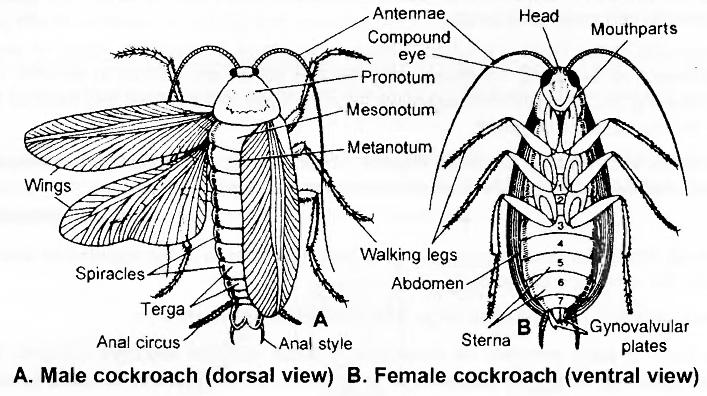
Salient Features of Peripianata americana (Cockroach) Habit and Habitat
Cockroaches are world wide and found in such places where darkness, warmth, dampness and plenty of organic debris is available.
Cockroaches are nocturnal and omnivorous.
Three species of cockroaches are commonly found in India (a) Periplanata americana, (b) Blatta orientalis, and (c) Blatta germaniaca.
P. americana is the largest and most common . It is commonly called, American cockroach, Bombay canary or Ship cockroach. In both the sexes, wings are present which are larger than the body.
Blatta orientalis is a black or dark brown medium sized species. The female has rudimentary wings which are not helpful in flight. Male has wings which are short and are not present up to the end of the body.
Blatta germanica : It is the smallest of the domestic species of the cockroaches. It is pale yell:ow-brown in colour.
External features -(P. americana)
The body is narrow, elongated, bilaterally symmetrical and flattened, measuring about 3 to 4½ cms (34-53 mm) in length and 1½ to 2 cms in breadth.
The colour is reddish brown with a pale yellow area around the edge of pronotum and two dark patches over it.
Exoskeleton
The entire body of the cockroach is covered with a thick, hard, chitinous cuticle, which is secreted by epidermis, forming the exoskeleton.
The exoskeleton of each segment consists of four plate like pieces called sclerites. Dorsal sclerite is called tergum or tergite, ventral is called sternum or sternite and two lateral sclerites are called pleura or pleurites.
The sclerites of each segment are joined with each other and with those of the adjacent segments by means of soft and flexible articular or arthrodial membranes. This gives the sclerites, some freedom of movement upon each other at their edges.
The stiff, immovable bristles or spines covering the body and its appendages are in fact the outgrowths of the cuticle while the movable hair like setae occurring at some places are secreted by special trichogen cells of the hypodermis lying below the cuticle.
Segmentation
Embryologically, the body of cockroach is formed of 20 segments -6 in head, 3 in thorax and 11 in abdomen.
Due to complete or incomplete fusion between some segments during development, the number of distinct segments is reduced in the adult.
All segments of the head are fused. Thorax consists of 3 segments. Only 10 segments are retained in the abdomen of the adult. Of these, only the first seven are distinct in females and first nine in males. The remaining hinder abdominal segments become small and modified into extemal genitalia that are hidden under the last distinct segment.
When wings are removed, the three regions -head, thorax and abdomen become distinctly visible. A small, soft and mobile neck or cervicum connects the head with thorax.
Head
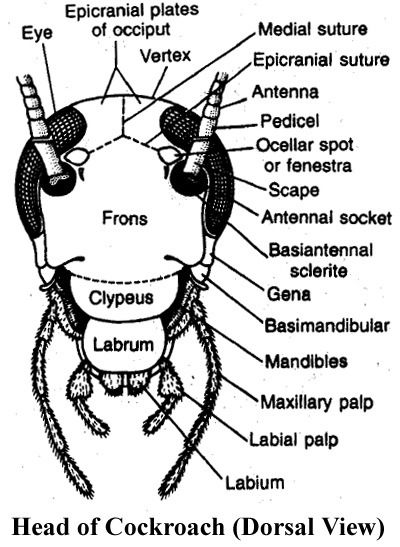
It is small, triangular and its narrow end is bent downwards in hypognathous position i.e., at an angle of 900 with the long axis of the body.
On each lateral side, it bears a large and blackish compound eye.
At the base of each antenna, on inner side, a small rounded and light coloured area called fenestra or ocellar spot representing simple eye is present. Endoskeleton of head is called tentorium.
All sclerites of the head are fused , forming a strong head capsule exhibiting only faint lines of fusion.
Cephalic appendages: The head appendages include 1 pair antennae, 1 pair mandibles, 1 pair first maxillae and 1 pair second maxillae (fused as single labium or lower lip), one labrum or upper lip and one hypopharynx.
Antennae : These are a pair of long, thread like appendages, extending forward from an antennal socket located dorsally upon head capsule near the eye. These are very mobile and act as tactile, thermal and olfactory receptor organs. Each is formed of several small segments called podomeres. The first basal podomere, called scape, is the largest. The second called pedicel, is narrow and elongated. The remaining long, slender and many jOinted part of each antenna is called flagellum.
The Mouth Parts
The remaining cephalic appendages are small and located around the mouth. Hence, together these appendages comprise the mouth parts of the cockroach. These help in 'biting and chewing' its food.
Labrum (upper lip) : It is the broad, flattened terminal sclerite of the dorsal side of head capsule, movably articulated to the clypeus acts as upper lip. It has epipharynx (chemoreceptors) on its inner side.
Mandibles: Thick, hard and triangular appendages beneath the labrum, one on each lateral side of the mouth, which bear pointed, teeth like processes called denticles.
First maxillae: Located on each side of the mouth next to mandibles. These serve to hold food particles in between the mandibles for cutting and chewing. They also bear olfactory receptors.
Labium (lower lip): The second maxillae are fused together forming a single large structure which covers the mouth from ventral side, hence called the 'lower lip' or labium. It bears tactile and gustatory sensory setae.
Hypopharynx: It is a small, cylindrical mouthpart, sandwiched between first maxillae and covered by labrum and labium on dorsal and ventral sides respectively. It bears several sensory setae on its free end, and the opening of common salivary duct upon its basal part.
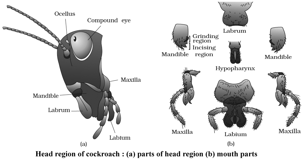
Thorax
(i) It comprises of three segments -prothorax, mesothorax and metathorax.
(ii) The three thoracic segments are covered by relatively thicker and larger tergites called nota.
(iii) The notum of prothorax called pronotum is very large and covers the neck also. Each of the mesonotum and metanotum bears a pair of wings.
Thoracic Appendages
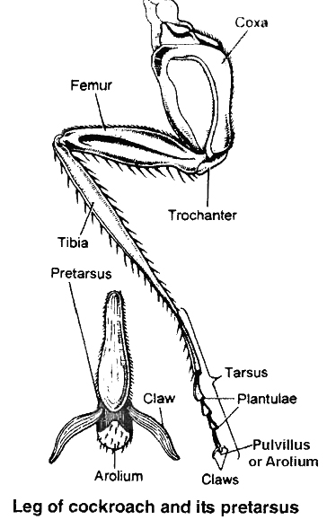
Each thoracic segment bears a pair of walking legs. Each walking leg consists of five segments:
(i) Coxa.
(ii) A triangular short rod like trochanter, articulated with coxa and femur.
(iii) A long, spiny femur.
(iv) A spring like tibia which represents the longest segment.
(v) A long tarsus, divided into five tarsomeres or podomeres. The last tarsomere is called pretarsus forming the claws and bearing an adhesive arolium or pulvillus. Similar but smaller adhesive pads called plantulae are located at each joint of tarsus.
Abdomen in both males and females consists of 10 segments. A typical abdominal segment has a dorsal tergum, ventral sternum and between them a narrow membranous pleuron on each side which bears spiracles. In females, the sclerites of 8th and 9th are overlapped by corresponding sclerites of the 7th segment.
The seventh sternum is boat shaped and together with eighth and ninth sterna forms a brood orgenital pouch. In males only 8th is overlapped by the 7th segment. The tenth segment bears a pair of 15 jointed filamentous structures called as anal cerci. Ventral to these in the males, the 9th segment bears a pair of short, thread like anal styles which are absent in females. Between one sclerite and the other, there is a flexible arthrodial membrane.
Abdomen

It is the largest and the broadest, relatively more flattened and softer part behind the thorax.
There are ten tergites. In both males and females, the 8th and 9th tergites are mostly covered by the 7th. The 10th tergum is somewhat bowl-shaped and posteriorly bifurcated into two lobes.
Ventrally, the abdomen has 9 sternites in male and 7 in females.
In females the last sternite (7th) is larger and boat shaped and together with indistinct 8th and 9th sternites it forms a chamber Anal styles like structure called gynatrium, posterior part of this chamber is MALE called oothecal chamber. Behind this chamber, 7th sternite Opening of oothecal chamber bifurcates into two prominent oval plates called apical lobes. Female gonopore is located between them.
In males, the 9th sternite bears a pair of spine like anal-styles.
In both male and female cockroaches, several small chitinous appendages are located around the gonopore. These help in reproduction and hence called gonapophyses.
At several places, certain processes of exoskeleton extend into the body and form endoskeletal elements which provide attachment to muscles and hence called apodemes.
Abdominal Appendages
Abdominal segments lack locomotory appendages. There are certain small structures associated with gonopore, which are different in male and female cockroaches.
The 10th tergum posteriorly bears a pair of many jointed anal cerci. They bear minute sensory hair sensitive to sound and other vibrations.
The 9th sternum of males bears, in addition, a pair of small and spine-like, unjointed anal styles.
Digestive System of Cockroach
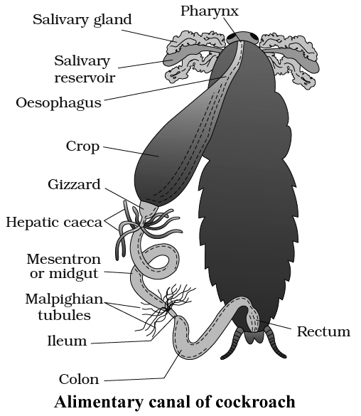
The alimentary canal is long and somewhat coiled, divisible into three main parts namely foregut, midgut and hindgut.
Foregut (stomodaeum) is lined by cuticle and is differentiated into five parts: buccal chamber, pharynx, oesophagus, crop and gizzard. Crop is used for storage of food.
Gizzard is muscular and internally provided with six cuticular teeth which crush the food.
A stomodaeal valve is present between gizzard and mesenteron.
Midgut (mesenteron or ventriculus) is short, tubular and lined with glandular endoderm.
At the anterior end of mesenteron, there are 6-8 blind glandular hepatic or gastric caecae which secrete digestive enzymes.
Internally mesenteron is not lined by cuticle but it is covered by a very thin and transparent peritrophic membrane formed of chitin and proteins.
Peritrophic membrane is secreted by gizzard, it serves to protect the wall of midgut from abrasion due to friction of food particles.
Peritrophic membrane is permeable to digested food and enzymes in the mesenteron.
Hindgut (proctodaeum) is broader than midgut and it comprises ileum, colon and rectum.
The wall of rectum is provided with six rectal papillae. They help in the absorption of water and salts.
Cockroach is omnivorous. It feeds on all sorts of organic debris.
The digestive enzymes of saliva are mainly zymase and amylase.
Most of the nutrients of food are digested in the crop.
Absorption of digested food takes place in mesenteron.
Respiratory System of Cockroach
The blood of cockroach is not responsible for the transportation of gases, it serves as a stationary medium for exchange of gases.
A complicated system of numerous, shiny, transparent and branched air tubes or tracheae are found for gaseous exchange in the haemocoel cavity. There are 6 longitudinal tracheal tubes -2 dorsal, 2 ventral and 2 lateral which are interconnected by transverse commissures. Chitinous rings prevent collapse of trachea.
Atmospheric air enters into and escapes out from this system through ten pairs of slit-like apertures called stigmata or spiracles located on lateral sides of the body. Two pairs of these are thoracic and eight pairs are abdominal. The openings of spiracles are regulated by the sphincters.
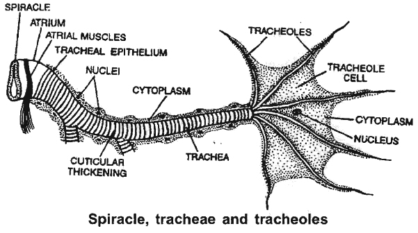
Thoracic spiracles are somewhat larger. One pairofthese is between prothoraxand mesothorax and the other between mesothoraxand metathorax, upon respective pleurites.
The first pair ofabdominal spiracles are dorsolateral upon tergite offirstabdominal segment, butthe remaining seven pairs are present upon the pleurites of second to eighth segments.
Each spiracle is surrounded by a ring-like sclerite called peritreme.
Mechanism
Several tergo-sternal muscles extend vertically between the tergites and sternites of all abdominal segments.
Harmonious contractions and relaxations ofthese at regular intervals cause rhythmic expansion and compression of abdomen leading to inspiration (with relaxation) and expiration (with contraction) of air.
At rest, the oxygen requirement is less, tracheolar ends get filled with tissue fluid.
The movement of O2 is along the pressure gradient as the tracheolar ends are losing oxygen to the cells for performing cellular respiration.
O2 requirement increases during activity.
Tracheolar fluid is withdrawn out of Tracheoles.
Alternate expansion and contraction of abdominal cavity occurs involving tergosternal muscles and abdominal muscles.
High level of CO2 in abdominal cavity make tergo-sternal muscles and abdominal muscles to contract pushing out the air from tracheal system to the outside through spiracles.
With relaxation abdomen expands i.e., tracheal trunks and tracheae expand and as a result, air rushes into tracheae and tracheoles via spiracles, it results in inspirations.
Circulatory System of Cockroach
Blood vascular system is open and lacunar type. Body cavity contains blood (haemolymph) which bathes viscera in it, therefore known as Haemocoel.
Blood vascular system consists of a tubular heart, a blood vessel called anterior aorta and a system of ill defined blood spaces or sinuses.
Heart
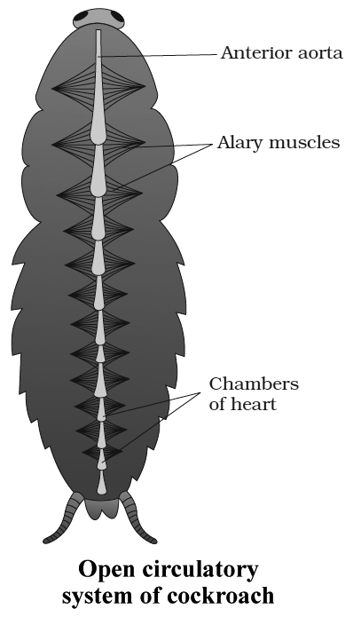
It is a long elongated tube situated in the mid dorsal line of thorax and the abdomen immediately beneath the terga.
Heart consists of thirteen chambers.
The last two posterior chambers are very small.
The chambers are separated from one another by deep constrictions.
The opening of each chamber into another is guarded by valves which allow blood from behind forward.
The Blood Sinuses
The large body cavity or haemocoel is divided by two membranous horizontal partitions, into three wide and flattened sinuses-the dorsal pericardial sinus containing the 'heart', the middle perivisceral sinus containing the gut, and the ventral perineural sinus or sternal sinus containing the nerve cord.
The partition between pericardial and perivisceral sinuses is called dorsal diaphragm and between perivisceral and perineural sinuses is called ventral diaphragm.
The sinuses intercommunicate by pores in the respective diaphragms.
A pair of fan like, triangular alary muscles in the floor of the pericardial sinus in each segment reinforce the dorsal diaphragm by their broad bases and also connect it, by their pointed tips with the tergite of the segment.
Circulation of Haemolymph
The pumping force that propels the haemolymph is provided by the pulsations of the 'heart'. The respiratory movements of abdomen and contraction of alary muscles increase this force.
From the pericardial sinus, the haemolymph enters into heart through ostia. When the heart is filled it contracts from behind forwards. This is its systole phase. Soon the heart becomes relaxed in its diastole phase. Then the next systole follows after a short time interval called diastasis. Thus heart pulsates about 50 times/minute.
During systole, the valve like ostia close, preventing back flow of haemolymph into the pericardial sinus. Therefore, some of its haemolymph is pumped into segmental vessels while most of it is poured into the head sinus through the terminally opening anterior aorta.
From the head sinus, the haemolymph flows backwards into the thorax and abdomen. While flowing backwards from head sinus, the haemolymph remains in the ventral part due to presence of Oesophagus in dorsal part. So, it fills into the perineural sinus.
From the perineural sinus, the haemolymph now flows into the perivisceral sinus through the pores of ventral diaphragm in abdominal region.
Then from perivisceral sinus, it flows into pericardial sinus through the pores of dorsal diaphragm. Then, during heart's diastole, it fills in the heart through the ostia.
Excretion in Cockroach
Cockroach is uricotelic. In cockroach, following structures help in excretion:
(i) Malpighian tubules
(ii) Fat bodies
(iii) Nephrocytes
(iv) Cuticle
(v) Uricose glands in some species.
Malpighian tubules:
Malpighian tubules are attached at the junction of midgut and hindgut. Excretory products, dissolved in haemolymph are absorbed by malpighian tubes are discharged into hindgut.
Fat bodies:
Some fat bodies are also present in haemocoel which have mycetocytes, urate cells, oenocytes and trophocytes.
Nephrocytes :
Nephrocytes present in lateral wall of heart and help in excretion and store nitrogenous waste.
Uricose glands:
In some species, in males, uricose glands are present on the periphery of mushroom glands. These glands synthesize uric acid . Malpighian tubules are analogous to mammalian kidneys. Fat bodies are analogous to vertebrate liver.
Nervous System of Cockroach
It consists of a series of fused, segmentally arranged ganglia joined by paired longitudinal connectives on the ventral side.
Central Nervous System :
It consists of brain or supra oesophageal ganglion. Brain gives off a pair of short, stout cords, the circumoesophageal connectives, that encircle the oesophagus and pass downwards and backwards over the suboesophageal ganglion situated below oesophagus.
From the suboesophageal ganglion passes backwards into the thorax, a double ventral nerve-cord, which bears three ganglia in the thorax and six in the abdomen.
Peripheral Nervous System :
It consists of nerves, which are given off from the ganglia so as to innervate all the parts of the body.
Sympathetic or Somatogastric Visceral Nervous System:
It consists of a frontal ganglion which is situated on the dorsal side of the oesophagus in the head.
From this ganglion, a median unpaired recurrent nerve reaches the visceral ganglion situated on the crop.
Various nerve branches are given off from the visceral ganglion.
The frontal ganglion is joined with the central nervous system by nerves which connect it to circumoesophageal commissures.
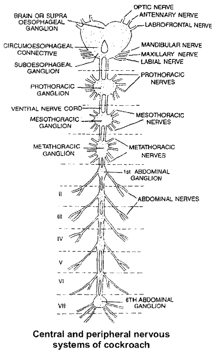
Sense Organs of Cockroach
Receptor cells are present on general body surface.
Proprioreceptors :
They are for hearing or receiving sound vibrations. Auditory receptors are present on antennae and anal cerci.
Thigmoreceptors :
They are receptors for touch. Present on antennae, maxillary palps and legs.
Olfactory receptors: They receive various smells. Present on antennae and palps.
Gustatory receptors: They are for sense of taste. Present on maxillae and labial palps.
Eyes
Cockroach has compound eyes. Each compound eye is formed of about 2000 hexagonal ommatidia.
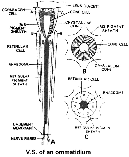
Each ommatidium has a biconvex lens or cornea. Below the lens there are corneagen cells which secrete the lens.
Below the corneagen cells is a transparent crystalline cone surrounded by four vitrellae or cone cell.
The vitrellae secrete the crystalline cone. All this forms the focusing or dioptrical region.
Below the cone there is refractive body the Rhabdome, surrounded by seven retinular cells.
Each ommatidia is isolated from the other by iris pigment sheath and retinal pigment sheath.
The image formed is apposition or mosaic vision, composed of as many separate but adjacent images as there are ommatidia.
In mosaic vision, images are sharp but separate and the eye can use only in bright light.
In cockroach vision is mosaic and apposition image is formed (although cockroach is nocturnal).
If pigmented iris sheath is removed from the compound eye of insects, only superposition image will be formed.
Reproductive System of Cockroach (Male)
In cockroach, sexes are separate, so it is dioecious.
Testes of cockroach are located in the abdominal segments 4, 5 and 6.
Mushroom gland consists of two types of tubules, (a) the long slender tubules the utriculi majores or peripheral tubules and (b) short tubules, the utriculi breviores, making up the major part of the gland. It is present in the 6-7th abdominal segments which functions as an accessory reproductive gland .
Small seminal vesicles are also found associated with mushroom glands.
All sperms of a seminal vesicle are glued together into a large bundle called spermatophore.
Spermatophore has three layered wall: inner layer secreted by utriculi majores; middle layer secreted by ejaculatory duct and outer layer secreted by phallic gland or conglobate gland
There are three asymmetrical chitinous structures called male gonapophyses or phallomeres. Right phallomere, left phaliomere (largest) and ventral phaliomere (smallest).
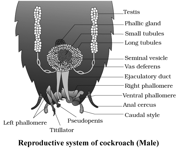
Reproductive System of Cockroach (Female)
Female organs consist of ovaries, oviducts, vagina, genital chamber, spermathecae, collaterial glands and female gonapophysis (ovipositor processes).
Ovaries of cockroach are located in the abdominal segments 2 to 6. Each ovary consists of eight ovarioles.
Two oviducts from each side open into a common oviduct or vagina which opens into genital chamber by female genital pore. A pair of spermathecae (left larger pyriform sac) are present near female genital pore.
A pair of collaterial glands also open in genital chamber.
Genital pouch or gynatrium is divisible into a genital chamber in front and oothecal chamber (vestibulum) behind.
Female genitalia consists of 3 pairs of chitinous processes hanging from the roof of oothecal chamber into its cavity.
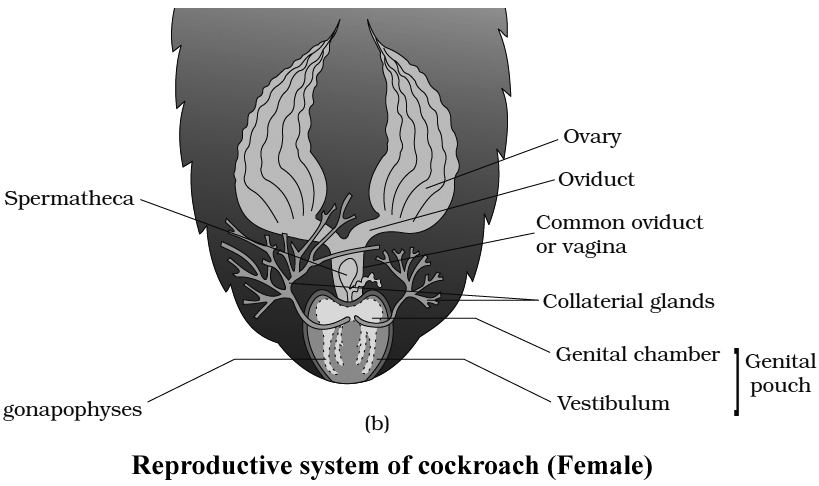
Ootheca of cockroach contains fourteen to sixteen fertilized eggs. Ootheca of cockroach is formed of a protein secreted by collaterial glands. On an average, females produce 9-10 oothecae.
Nymphs of cockroach emerge out from ootheca. A nymph resembles an adult in general structure but lacks wings and mature reproductive organs. The next to last nymphal stage has wing pads but only adult cockroach have wings.
Instar is a stage in the development of insect (larval instar, nymphal instar). Period between two successive moults in insects is termed stadium.
In Periplaneta americana, the nymph grows by moulting about 13 times to reach the adult form and Blatta orientalis, it moults 6 times.
Comparison of Periplaneta and Blatta
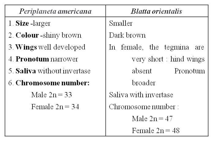
Cockroach
Cockroach
Cockroaches are dark or black-bodied animals belonging to the Phylum Arthropoda's class Insecta. Cockroaches with bright yellow, red, and green colorshave also been documented in tropical areas. Their antennae, legs, and a flat extension of the upper body wall that conceals the head vary in size from 14 inches to 3 inches (0.6-7.6 cm). They are nocturnal omnivores who live in moist environments all over the world. They have become occupants of human homes, making them major pests and disease vectors.
(i)Morphology: The common cockroach, Periplaneta americana, has wings that extend past the tip of the abdomen in males and is around 34-53 mm long inadults. The cockroach's body is divided into three separate regions: the head, the thorax, and the abdomen. A strong chitinous exoskeleton covers the entire body (brown). Hardened plates called sclerites (tergites dorsally and sternites ventrally) are linked to each other by a thin and flexible articular membrane in each segment of the exoskeleton (arthrodial membrane).
The triangular head is positioned anteriorly at right angles to the longitudinal axis of the body. It is made up of the fusion of six segments and has a flexible neck that allows it to move freely in all directions. A pair of compound eyes can be found on the head capsule. Membranous receptacles in front of the eyes sprout a pair of thread-like antennae. Sensory receptors in antennae aid in environmental monitoring. Appendages on the anterior end of the skull produce biting and chewing mouth parts. A labrum (upper lip), a pair of mandibles, a pair of maxillae, and a labium make up the mouthparts (lower lip). The tongue (hypopharynx) is a flexible lobe that resides within the space encompassed by the mouthparts.
Prothorax, mesothorax, and metathorax are the three sections of the thorax. The neck is a short extension of the prothorax that connects the head to the thorax. Walking legs are attached to each thoracic section. The mesothorax produces the first pair of wings, whereas the metathorax produces the second pair. Tegmina, or opaque dark and leathery forewings (mesothoracic), cover the hind wings when at rest. The rear wings are transparent and membranous, and they help the animal fly.
Both males and females have ten segments in their abdomen. The 7th sternum is boat-shaped in females, and it creates a brood or genital pouch with the 8th and 9th sterna, which contains the female gonopore, spermathecal pores, and collateral glands in the front section.The genital pouch or chamber is located at the back end of the abdomen, between the 9th and 10th terga and the 9th sternum. Dorsal anus, ventral male genital hole, and gonapophysis are all found here. Males have a pair of short, threadlike anal styles that females lack. Anal cerci are a pair of jointed filamentous structures found on the 10th segment of both sexes.
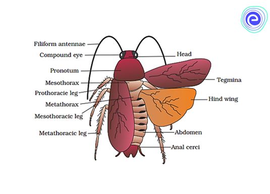
(ii) Anatomy: The alimentary canal is separated into three sections in the body cavity: foregut, midgut, and hindgut. The mouth opens into a short tubular pharynx, which leads to the oesophagus, a tiny tube conduit.
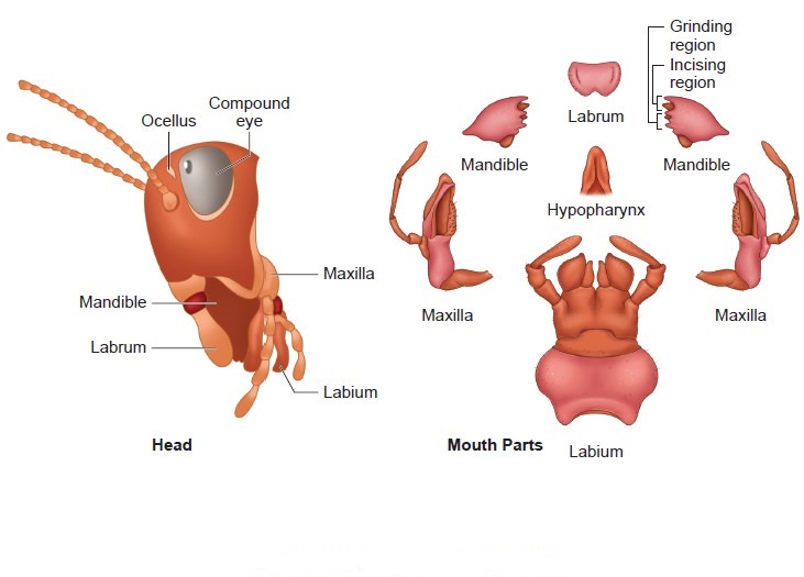
The esophagus expands into a sac-like structure called a crop, which is used to store food. Gizzard or proventriculus follows the crop. It possesses a thick outer layer of circular muscles and a thick inner cuticle that forms six teeth-like chitinous plates. The gizzard assists in the crushing of food particles. The cuticle covers the entire foregut. At the confluence of the foregut and midgut is a ring of 6-8 blind tubules called the hepatic or gastric caeca, which release digestive juice.Another ring of 100-150 yellow-colored thin filamentous Malpighian tubules exists at the midgut-hindgut junction. They aid in excretory product clearance from the hemolymph. The ileum, colon, and rectum make up the hindgut, which is larger than the midgut. Through the anus, the rectum opens up.
Cockroaches have an open blood circulatory system. The blood vessels are underdeveloped and open into the air (hemocoel). The blood bathes the visceral organs in the haemocoel (hemolymph). Colorless plasma and haemocytes make up the haemolymph. The cockroach's heart is an extended muscular tube that runs along the mid-dorsal line of the thorax and abdomen. It is divided into funnel-shaped chambers on both sides by ostia.Through the ostia, blood from the sinuses enters the heart and is pumped anteriorly to the sinuses. The respiratory system is made up of a network of trachea that open through ten pairs of small openings on the body's lateral side called spiracles. Air is carried to all parts by thin branching tubes (tracheal tubes separated into tracheoles). The sphincters control the opening of the spiracles.
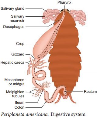
Diffusion occurs at the tracheoles to exchange gases. Malpighian tubules are responsible for excretion. Glandular and ciliated cells line each tubule. They take in nitrogenous waste and convert it to uric acid, which is then expelled through the hindgut. As a result, It is known as uricotelic.
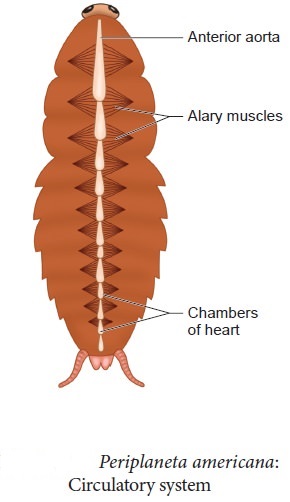
The fat body, nephrocytes, and urecose glands all contribute to excretion. On the ventral side, the cockroach's nervous system is made up of a series of fused, segmentally arranged ganglia connected by paired longitudinal connectives. The thorax has three ganglia and the abdomen has six. Cockroaches have a neurological system that runs throughout their bodies. A portion of the nervous system is located in the skull, while the rest is located on the ventral (belly) side of the animal. Because of this, even if a cockroach's head is removed, it can survive for up to one week. The brain is represented in the head area by the supra-oesophageal ganglion, which sends nerves to the antennae and compound eyes.
Antennae, eyes, maxillary palps, labial palps, anal cerci, and other sensory organs are found in cockroaches. The complex eyes are located on the head's dorsal surface. About 2000 hexagonal ommatidia make up each eye. A cockroach can obtain several photos of an object by using many ommatidia. Mosaic vision is a type of vision that has a higher sensitivity but lower resolution and is common at night (hence called nocturnal vision).
Cockroaches are dioecious, with well-developed reproductive organs in both sexes. A pair of testes, one on each lateral side in the 4th -6th abdominal segments, make up the male reproductive system.
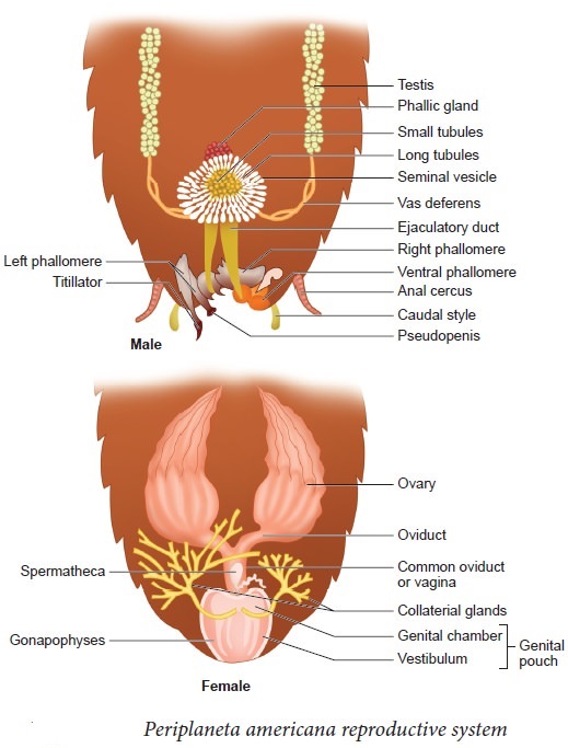
A narrow vas deferens develops from each testis and opens into the ejaculatory duct via the seminal vesicle.The male gonopore is located ventral to the anus and opens into the ejaculatory duct. In the 6th and 7th abdominal segments, there is a mushroom-shaped gland that serves as an accessory reproductive gland. Male gonapophysis or phallomeres depict the external genitalia (chitinous asymmetrical structures, surrounding the male gonopore). The sperm are retained in the seminal vesicles and cemented together in spermatophores, which are expelled during copulation. Two enormous ovaries lie laterally in the 2nd – 6th abdominal segments, forming the female reproductive system. Each ovary is made up of eight ovarian tubules, also known as ovarioles, which contain a chain of growing ova. Each ovary's oviducts join to form a single median oviduct (also known as the vagina), which exits into the genital chamber.The 6th segment, which enters the vaginal chamber, contains a pair of spermatheca. Spermatophores are used to transport sperm. The fertilized eggs are enclosed in oothecae capsules. The ootheca is a 3/8" (8 mm) length dark reddish to a blackish-brown capsule. They're dropped or bonded to a suitable surface, usually near a food source in a crack or crevice with high relative humidity. Females produce 9-10 oothecae on average, each holding 14-16 eggs. P. americana has a paurometabolous development, which means it develops through the nymphal stage. The nymphs have the appearance of adults. To attain adulthood, the nymph must molt 13 times.Wing pads are present in the next to last nymphal stage, however, only adult cockroaches have wings.
Many cockroach species are found in the wild and have yet to be identified as economically important. In and around the human habitat, a few species flourish. They are pests because their stinky excreta spoils food and contaminates it. By polluting food, they can spread a range of bacterial infections.
earthworm
- Books Name
- ACME SMART COACHING Biology Book
- Publication
- ACME SMART PUBLICATION
- Course
- CBSE Class 11
- Subject
- Biology
Morphology, Anatomy & Function of Different Systems of
an Annelid, an Insect & an Amphibian
EARTHWORM
Indian Earthworms (Pheretima posthuma)
Phylum : Annelida
Class : Oligochaeta
Genus : Pheretima
Species : posthuma
There are several types of Earthworms. The most common genus of Earthworm is Pheretima in India and Lumbricus in Europe. Pheretima has 500 species, 13 of them are found in India.
Habitat :
Earthworm is reddish brown terrestrial animal which inhabits upper layer of moist soil where it lives inside burrows during day time.
Earthworm inhabits those soils which have abundant organic matter.
An acre of good moist soil can have upto 50,000 animals. Burrow is made by boring and swallowing the soil.
The burrows are vertical or oblique.
They are 30-45 cm deep during moist season but may go as deep as 2 m in summer.
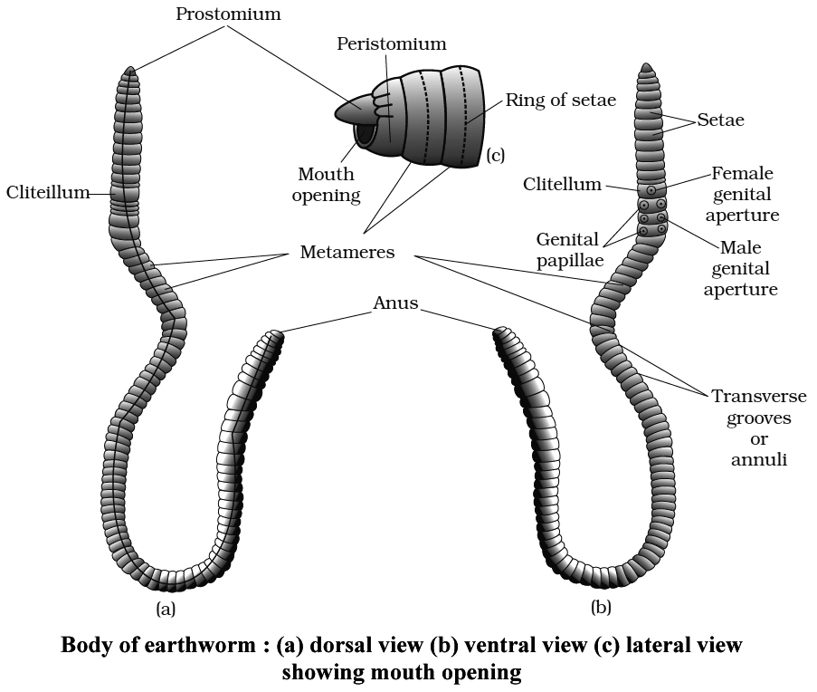
The burrows are lined by debris or mucus secreted by the animals.
The burrow is wider at the base.
During winter the animal drags organic debris into its burrow and plugs the mouth of the burrow.
This keeps the burrow warm.
Even the mouth of the burrow is hidden from view by leaves and small stones.
The area of the burrow can be recognised by faecal pellets called worm castings.
Habit :
Earthworm is nocturnal because it is sensitive to higher light intensities.
It partly creeps out of burrows during night for search of food.
It is only during rainy season that the earthworm comes out of the burrow even during daytime.
After heavy rainfall, they can be seen crawling on the ground in large number.
If the burrow is left, the animal does not re-enter the same.
It digs a new burrow by pushing the body through the soft soil as well as by eating its way through the soil.
The worm keep its skin moist through mucus, coelomic oozings and from moisture of the soil. The animal respires through skin.
The body of earthworm is long cylindrical has about 100-120 segments (metameres). The first segment is called as 'buccal segment' or peristomium which bears a very small terminal opening the mouth.
A small projection is also present which hangs over the cresent shaped mouth and is called prostomium. It serve as a wedge to force open cracks in the soil into which earthworm may crawl. It is sensory in nature.
The skin of earthworm is brown due to the presence of porphyrin pigment and protects the earthworm from UV radiations.
In all the body segments, except the first, last and clitellum, there is a ring of S-shaped setae, embedded in the epidermal pit at the middle of each segment (perichaetine).
Setae are chitinous structures and are not dissolved in KOH.
In the intersegmental grooves of 5/6, 6/7, 7/8, 8/9 segments, 4 pairs of spermathecal pores are present which are the opening of spermathecae.
A thick band of glandular tissue clitellum (cingulum) surrounds segments 14 to 16, forming a thick girdle, its glands secrete mucus and albumin, they also form the cocoon.
On the ventral surface of 18th segment, a pair of male genital apertures are present and on the ventral surface of 14th segment, a median female genital aperture is present.
On the ventral side of each of the 17th and 19th segments, circular raised pairs of genital papillae are present which help in reproduction.
The dorsal surface of body is marked by a dark median mid dorsal line (representing dorsal blood vessel) along the longitudinal axis of the body. The ventral surface is distinguished by the presence of genital openings (pores).
Internal Morphollogy
The body wall of earthworms is thin, soft and slimy. From the surface inwards, it consists of cuticle, epidermis, muscular layers and coelomic epithelium
(1) Cuticle: It is thin and elastic non-cellular protective membrane. It is formed of collagen fibres secreted by underlying epidermis.
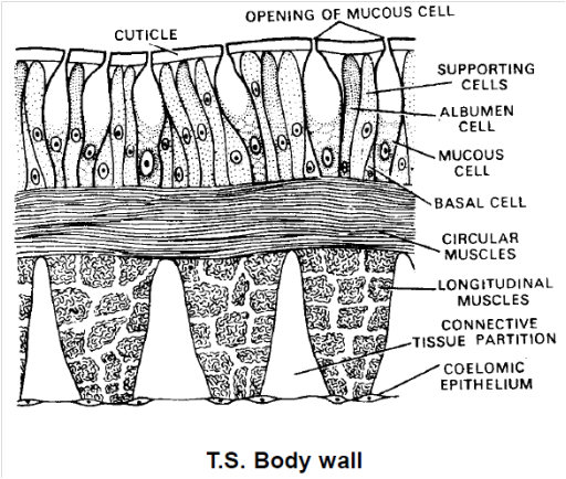
(2) Epidermis: This is a single layer of epithelium of tall, columnar cells which are distinguished into four types as follows :
Supporting cells : Unspecialized epithelial cells that form the major part of the epidermis.
Glandu1lar cells: These are of two types-the more numerous and club-shaped mucus-secreting goblet cells and fewer, narrower albumen cells. The mucus, secreted by these cells, keeps the body wall moist and slimy. It is also used to lubricate and smoothen the walls of burrows.
Basal cells: These are shorter, conical cells wedged in between narrower basal parts of other cells.
Sensory cells: These are narrow, columnar cells occuring, here and there, in small groups. Each sensory cell has small sensory hairs at its free end.
(3) Muscular layer: Beneath the epidermis is the muscullature of body wall. It consists of a thin outer layer of circular and about twice thicker, inner layer of longitudinal muscle fibres. The circular muscle layer is a continuous sheet around the body, but the longitudinal layer is broken into several longitudinal strips or bands, separated from each other by thin connective tissue partitions. These bands appear elliptical or club-shaped in transverse section. Numerous granules of porphyrin pigment are found scattered in circular muscle layer.
(4) Coelomic epithelium: Next to the longitudinal muscle layer is a thin, membrane-like, mesodermal epithelium of flattened, squamous cells. It is the outer envelope of coelomic cavity and hence called parietal or somatic layer of coelomic epithelium or peritonium.
Coelom or Body Cavity
Earthworm has schizocoel type of body cavity.
It lies between the body wall and alimentary canal.
A layer of peritoneum lines both the surfaces, outer parietal in contact with body wall and inner visceral in contact with alimentary canal.
The coelom of first four segments is continuous or undivided.
The coelom is divided by septa from fourth and fifth segment onwards.
The septa lying between segments 5/6, 6/7, 7/8, 8/9 or 9/10 and 10/11 are thick and muscular.
One of the two septa, either between the 8th and 9 th or between 9 th and 10th segments are absent.
The first six septa are cone like and run obliquely backwards from the body wall to the gut wall.
The first nine septa i.e. upto septum 13/14 are without perforations.
The remaining septa beginning from septum 14/15 are perforated by numerous apertures.
Coelomic fluid is milky white and alkaline. It has fluid matrix of watery plasma containing proteins, salts and numerous minute nucleated corpuscles. Corpuscles are of following four types.
(a) Phagocytes: They move like Amoeba and engulf harmful germs.
(b) Leucocytes : These are smaller and fewer. Their function is not fully understood.
(c) Mucocytes: These are elongated, vase like corpuscles, one end forms an expanded fan like structure and the other narrow end contains nucleus. Function of these cells is not known.
(d) Eleocytes : They are formed by mitosis of the yellow cells of the visceral peritoneum. They contain glycogen and fat and distribute their food to various tissues.
Coelomic fluid serves as a hydrostatic skeleton to assist the musculature of body wall in bringing about the locomotion of body. It oozes out upon body surface through dorsal pores, keeping the body wall moist to facilitate respiration and to destroy bacteria and other harmful micro-organism.
Locomotion
The earthworm does not have specialized locomotory organs.
The locomotion is brought about by the circular and longitudinal muscles of the body wall, aided by the chitinous curved setae embedded in the skin.
Due to the contraction of the circular muscles of the anterior end the latter becomes thin, elongated and extends forwards.
At the same time, the setae of the anterior end hold the ground firmly and prevent the animal from slipping backward.
Now the circular muscles of the anterior end relax and the longitudinal muscles contract.
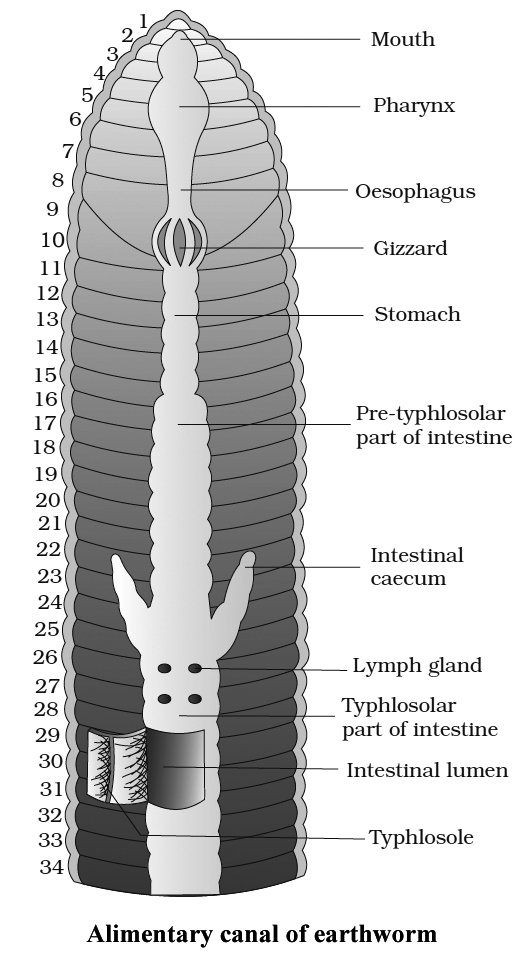
It causes the shortening and thickening of the anterior segments, and thus, the posterior part of the body is pulled ahead.
The process is repeated and the worm is able to move forward with speed. Earthworms move at the rate of about 15 cm per minute.
Alimentary Canal
It is a straight tube and runs between first and last segment of the body.
In 1-3 segments buccal cavity is present, pharynx in 4th segment.
From 5-7 segments oesophagus is present.
Gizzard is present in 8th segment.
From 9-14th segments is a tubular stomach. Calciferous glands are present in the stomach, which produce CaCO3 to neutralise the humic acid.
Intestinestarts from the 15th segment on wards and continues till the last segment.
A pair of shortand conical Intestinal caecae projects from the intestine on the 26th segment. They secrete amylolytic enzyme which digests starch. Other enzymes are lipase, cellulase, invertase etc.
Surrounding the pharynx there are pharyngeal or salivary glands, made of masses of chromophil cells, they produce mucin for the lubrication of the food, and also a proteolytic enzyme which can digest some proteins.
Associated with the intestine are chloragogen cells which are supposed to be excretory in function.
Intestine is divided into
(a) Pre-typhlosolar region from 15 to 26th segment, it has small villi
(b) Typhlosolar region which starts from 27th segment and extends upto 23-25 segments in front of anus. Typhlosole is a large villus as an internal median fold of dorsal wall of intestine. This enhances effective area of absorption after digestion.
(c) Post-typhosolar region: Also known as rectum. It is present in last 23-25 segments and opens to outside through a terminal anus.
Lymph glands: These are white fluffy bodies which are found arranged on either side of the dorsal vessel from 26th segment and extend to the successive segments. These glands are believed to produce phagocytes of the coelomic fluid.
Circulatory system
Earthworm is first to evolve a closed circulatory system in the evolution of animals.
The respiratory pigment is haemoglobin which remains dissolved in the plasma, RBC are absent.
Blood glands are present on 4, 5 and 6th segments, they produce blood cells and haemoglobin.
There are 2 main blood vessels.
Dorsal vessel is the largest vessel and blood flows forwards from postrior end to anterior end. They have contractile wall, and valves are present. Before the 13th segment, it is distributing vessel and behind 13th segment, it is a collecting vessel.
Ventral vessel is the main distributing vessel, blood flows backwards from anterior to posterior end, valves are absent.
Latero-oesophageal vessels are paired vessels which extend from first to thirteenth segment.
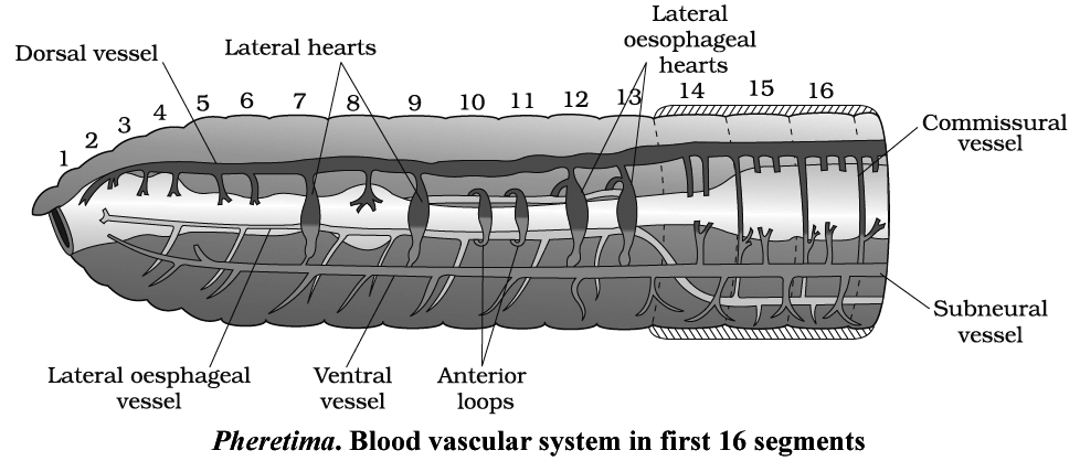
Supra-oesophageal vessel is unpaired and extends between 9th and 13th segments.
Sub-neural vessel
Hearts
There are 4 pairs of hearts with valves.
(a) Two pairs of lateral hearts, one pair in 7th and one pair in 9th segment
(b) Two pairs of lateral oesophageal hearts in 12 and 13th segments.
(c) There are 2 pairs of lateral loops in which valves are absent. One pair is in 10th and one pair is in 11th segment. Blood flows in upward direction in them.
EXCRETION
Concept Builder
Earthworm mainly removes the nitrogenous waste in the form of urea in soil. But when plenty of water is available earthworm is ammonotelic. So earthworm is both Ureotelic and Ammonotelic.
Excretory System
Excretory organs occur as segmentally arranged coiled tubules called nephridia. They deliver the wastes through a pore to the surface of body wall or into digestive tube.
(i) Three main types of nephridia are:
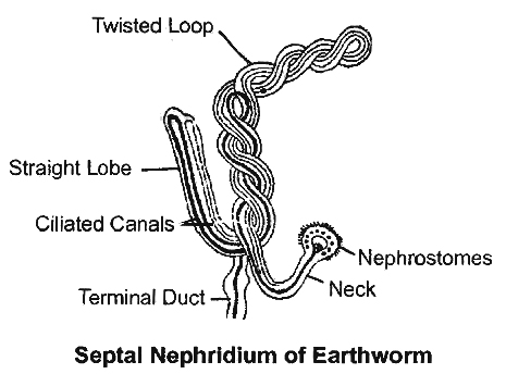
(a) Pharyngeal nephridia are situated in the segments 4,5 and 6. They open in the anterior part of alimentary canal, i.e., buccal cavity and pharynx. They are without nephrostome and are enteronephric type.
(b) Integumentary nephridia are scattered in the body wall. They are smallest, v-shaped, without nephrostome and are exonephric type. In clitellar segments they form forests of nephridia.
(c) Septal nephridia are the largest, attached to both faces of each intersegmental septum behind 15th segment.
(ii) Septal nephridia are the only nephridia with nephrostome or funnel. The terminal duct opens into septal excretory canal. These canals, in turn, open into two supraintestinal excretory canals. Septal nephridia are enteronephric, finally excretory products are poured into intestine. Enteronephric condition is an adaptation for the conservation of water or osmoregulation.
(iii) Excretory products of earthworm are urea (about 50%), ammonia (about 40%) and traces of creatinine. Earthworms are mainly ureotelic.
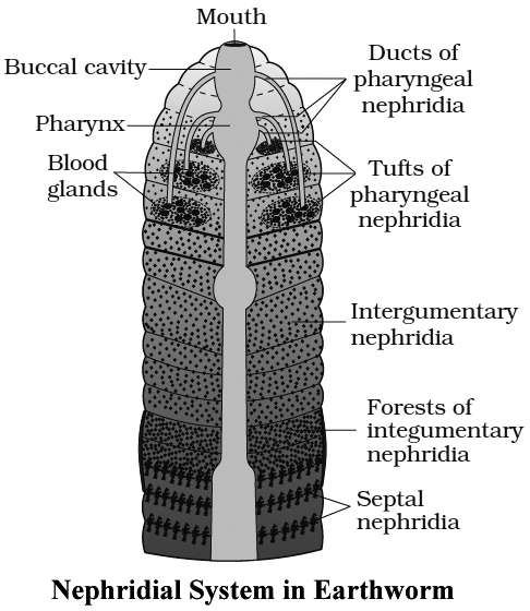
NERVOUS SYSTEM
It consists of central, peripheral and sympathetic nervous system.
CNS
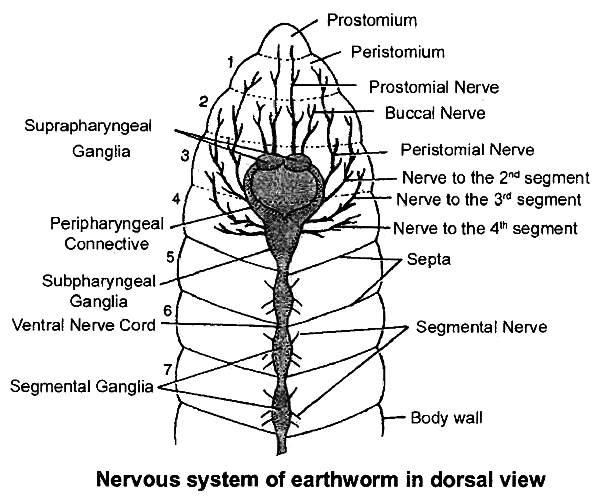
It consists of brain ring / circumpharyngeal ring and a ganglionated double ventral nerve cord.
Circumpharyngeal ring occurs in 3rd and 4th segment. It has a brain consisting of two suprapharyngeal ganglia, two circumpharyngeal connectives and a pair of subpharyngeal ganglia.
Ventral nerve cord arises from subpharyngeal ganglia, which is double, solid and bears paired ganglia in each segment.
PNS
It comprises nerves that extend from CNS to supply various parts. The nerves are mixed in nature.
Two pairs of nerves arise from brain and innervate prostomium and buccal cavity.
Nerves from circumpharyngeal connectives supply segments one and two.
Subpharyngeal ganglia send nerves to second, third and fourth segments.
Each segmental ganglion (actually paired) sends out 3 pairs of segmental peripheral nerves, one pair from anterior part and two pairs from posterior part.
They supply various structure in each segment. All the segmental nerves are mixed in nature, i.e. containing both sensory (afferent) and motor (efferent) nerve fibres.
Sympathetic Nervous System
Consists of various nerve plexuses present in the wall of alimentary canal.
SENSE ORGANS
Group of specialised cells found in skin and lining of buccal cavity.
Photoreceptors located in prostomium and dorsal epidermis. Perceive light intenSity with the help of phaosome (Optic organelle).
Thigmoreceptors located in ventral and lateral epidermis.
Olfactoreceptors located in lining of buccal cavity.
Gustatory receptors located in lining of buccal cavity.
RESPIRATION
Earthworm has no special respiratory organs.
Gaseous exchange takes place simply through the skin, which is thin and highly vascular.
Effective gaseous exchange takes place only when the skin is moist.
The skin is kept moist due to the damp earth, secretion of the mucus by the epidermal gland cell and oozing of coelomic fluid through the dorsal pores.
REPRODUCTION
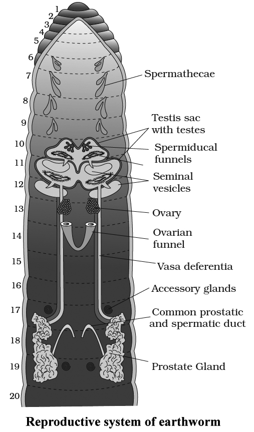
Earthwonn is hermaphrodite.
There are two pairs of testes present in 10th and 11th segments. They are surrounded by 2 testes sacs lying ventrally, one in the 10th and the other in 11th segment.
There are 2 pairs of seminal vesicles, one pair in 11th and the other pair in 12th segment.
Maturation of sperms occurs in seminal vesicles.
The testis sac of 10th segment communicates with the seminal vesicles of 11th segment and testis sac of 11th segment with seminal vesicles of 12th. From each testis sac, vas deferens carries the sperms up to 18th segment where they join the prostatic duct from prostate gland.
Four pairs of spermathecae are present, one pair in each ofthe6, 7, 8, and 9 segments. They receive and store the spermatozoa of another earthworm during copulation.
One pair of ovaries lies in 13th segment, which open through median aperture on 14th segment.
Accessory glands are present on the ventral surface of 17th and 19th segments, which open through the genital papillae. These are a part of male reproductive system.
Testes mature earlier (Protandrous).
Development is direct and there is no larval form.
earthworm
Earthworm
The earthworm is a reddish-brown terrestrial invertebrate that lives in the moist soil's upper layer. They spend the day in tunnels created by burrowing and swallowing earth. Worm castings, or faecal deposits, are used to track them down in the gardens. Pheretima and Lumbricus are the most prevalent Indian earthworms.
(i) Morphology: Earthworms' bodies are long and cylindrical. The body is divided into over a hundred short, identical pieces (metameres about 100-120 in number). Along the longitudinal axis of the body, a dark median mid dorsal line (dorsal blood vessel) marks the dorsal surface. The presence of genital apertures distinguishes the ventral surface (pores).
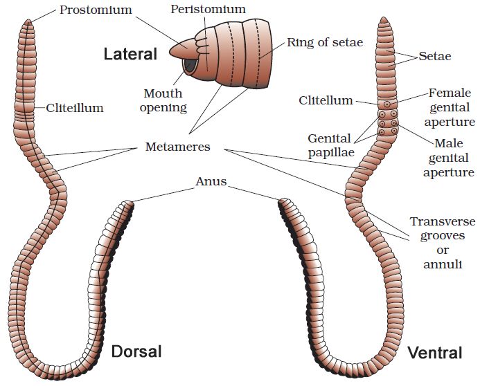
The mouth and the prostomium, a lobe that serves as a mouth cover and a wedge to force open soil crevices into which the earthworm may crawl, make up the anterior end. The role of the prostomium is sensory. The peristomium (buccal segment) is the first body segment and houses the mouth. A conspicuous dark band of glandular tissue termed clitellum covers segments 14-16 in a mature worm. As a result, the body is divided into three distinct regions: preclitellar, clitellar, and postclitellar. The ventrolateral sides of the intersegmental grooves, i.e. the 5th -9th segments, have four pairs of spermathecal openings. In the mid-ventral line of the 14th segment, there is a solitary female genital pore.On the ventrolateral sides of the 18th segment, there are two male genital pores. Nephridiopores are tiny pores that open on the surface of the body. Except for the first, last, and clitellum, each body segment has rows of S-shaped setae inserted in the epidermal pits in the middle. Setae can be retracted or expanded. Their primary function is movement.
(ii) Anatomy: A thin non-cellular cuticle covers the earthworm's body wall, which is followed by the epidermis, two muscle layers (circular and longitudinal), and an innermost coelomic epithelium. The epidermis is a single layer of columnar epithelial cells with secretory gland cells.
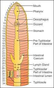
The alimentary canal is a straight tube that connects the first and last body segments. The buccal cavity (1-3 segments) leads into the muscular pharynx through a terminal mouth. The esophagus (5-7 segments) is a tiny, narrow tube that leads to the muscular gizzard (8-9 segments). It aids in the grinding of soil particles and rotting leaves, among other things. The stomach is divided into 9-14 parts. Earthworms eat decaying leaves and organic debris mixed with soil as nourishment.The humic acid in the humus is neutralized by calciferous glands in the stomach. The stomach begins at the 15th section and continues until the last part. On the 26th segment, a pair of short and conical intestinal caecae protrude from the gut. Except for the last 23rd-25th segments, the existence of an internal median fold of the dorsal wall called typhlosole is a distinguishing feature of the intestine after the 26th segment. This expands the absorption surface area in the gut. A little spherical opening called the anus connects the alimentary canal to the outside world. Digestive enzymes break down complicated meals into tiny absorbable components when the organic-rich soil moves through the digestive tract. These simpler molecules are absorbed and used through intestinal walls.
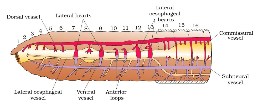
The blood vascular system of Pheretima is closed, consisting of blood arteries, capillaries, and the heart. Blood is restricted to the heart and blood vessels due to the closed circulatory system. Blood circulates in one direction due to contractions. The gut, nerve cord, and body wall are all supplied by smaller blood vessels. The 4th, 5th, and 6th segments all have blood glands. They manufacture blood cells as well as haemoglobin, which is dissolved in blood plasma. Blood cells are naturally phagocytic.
Earthworms do not have specific breathing apparatus. The wet body surface allows for respiratory exchange into the bloodstream.
Nephridia are segmentally arranged coiled tubules that make up the excretory organs (sing.: nephridium). There are three types of nephridia:
(i) Septal nephridia, which are present on both sides of intersegmental septa from segment 15 to the last that open into the intestine,
(ii) Integumentary nephridia, which are attached to the lining of the body wall from segment 3 to the last that open on the body surface, and
(iii) Pharyngeal nephridia, present as a three-paired outfit in 4th, 5th and 6th segment. The structure of these many nephridia is remarkably similar. The volume and composition of the bodily fluids are regulated by nephridia. A nephridium begins as a funnel, collecting surplus fluid from the coelomic chamber.The funnel connects to a tubular component of the nephridium, which transports wastes to the digestive tube through a pore in the body wall.
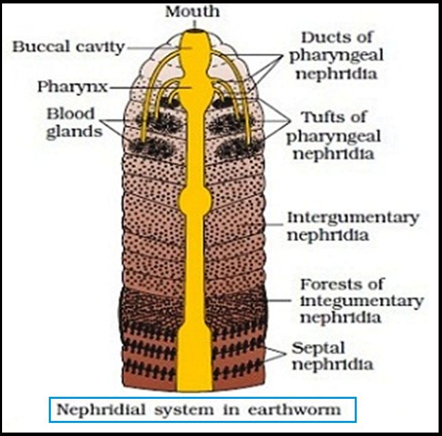
Ganglia are positioned segment-wise on the ventral paired nerve cord to represent the nervous system. In the front region (3rd and 4th segments), the nerve cord bifurcates, laterally enclosing the pharynx and dorsally joining the brain ganglia to form a nerve ring. The cerebral ganglia, along with other nerves in the ring, integrate sensory input and control bodily muscle responses.Although the sensory system lacks eyes, it does have light and touch-sensitive organs (receptor cells) that allow it to discern between light intensities and feel ground vibrations. Chemoreceptors (taste receptors) in worms are specialized to respond to chemical inputs. These sensory organs are found on the worm's front end.
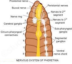
Earthworms are hermaphrodites (bisexual), which means they have both testes and ovaries. In the 10th and 11th segments, there are two pairs of testes. Their vasa deferentia connect to the prostatic duct at the 18th segment. In the 17th and 19th segments, there are two pairs of auxiliary glands, one pair in each.A pair of male genital holes on the ventrolateral side of the 18th segment access the outside the shared prostate and spermatic duct (vasa deferentia). In the 6th-9th segments, there are four pairs of spermathecae (one pair in each segment). During copulation, they receive and store spermatozoa. At the inter-segmental septum of the 12th and 13th segments, one pair of ovaries is attached. Underneath the ovaries are ovarian funnels that continue into the oviduct, unite, and open on the ventral side as a single median female genital hole on the 14th segment.
During the mating process, two worms exchange sperm. One worm must discover another worm, and they mate by exchanging spermatophores (packets of sperm) at opposite gonadal apertures.Cocoons made by clitellum gland cells contain mature sperm and egg cells, as well as nourishing fluid. Fertilization and development take place within the soil-deposited cocoons. The sperm cells within the cocoon fertilize the ova (eggs), which subsequently fall off the worm
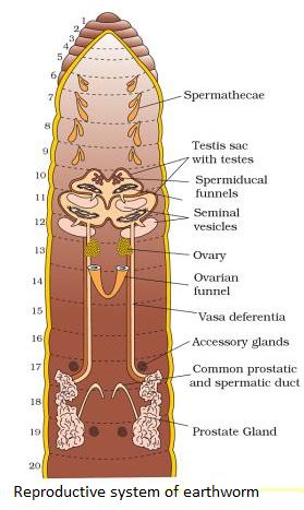
and land in or on the earth. The worm embryos are kept in the cocoon. Each cocoon generates two to twenty baby worms on average after about three weeks. Earthworms develop directly, without the formation of a larva. Earthworms are known as "farmer's friends" because they dig burrows in the soil and make it porous, which aids in plant root penetration and respiration.Vermicomposting is the method of using earthworms to increase soil fertility. They're also utilized as game fishing bait.
frog
- Books Name
- ACME SMART COACHING Biology Book
- Publication
- ACME SMART PUBLICATION
- Course
- CBSE Class 11
- Subject
- Biology
FROG (Rana tigrina)
Phylum : Chordata
Class : Amphibia
Order : Anura
Genus : Rana
Species : tigrina
The most common frog found in India is the Indian bullfrog.
It is the largest frog and is named as bullfrog because of its large size and loud call.
Indian bullfrog is found in fresh water marshes, ditches, ponds, and shallow lakes.
They undergo aestivation (summer sleep) in summer and hibernation (winter sleep) in winter.
They are carnivorous (feeding upon other animals, insects, etc.), poikilothermic i.e. the body temperature changes with environment.
They develop protective coloration to camouflage, i.e., to hide in surroundings.
Frogs belong to the Phylum Chordata, Subphylum Vertebrata or Craniata, Superclass Gnathostomata, Class Amphibia and Genus Rana.
The most common species is known as Rana tigrina (Indian bull frog).
The scientific name of common toad is Bufo melanostictus. Frogs exhibit sexual dimorphism.
Male and female are distinguishable externally only during breeding season when the males develop nuptial pad in the first digit of forelimbs.
Vocal sacs are well developed in males so they produce louder sound as compared to the females which are devoid of vocal sacs.
The vocal sacs help to produce mating calls.
Total number of bones is 153.
External Morphology
Skin is made up epidermis and dermis. Mucous glands are present in the dermis and their ducts open at the surface.
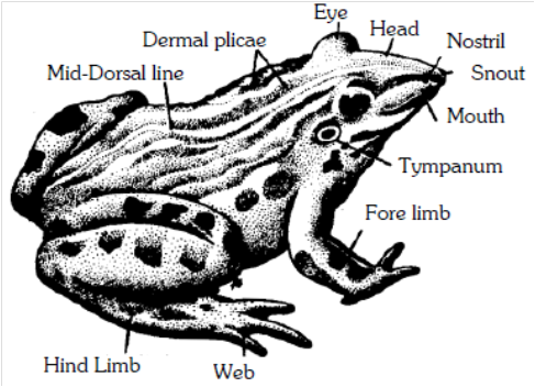
Blood capillaries and pigment cells (chromatophores) are present in the dermis.
Skin is without scales or any other cover or exoskeleton.
Body is divisible into head and trunk, neck is absent. The trunk is provided with a pair of fore and hind-limbs. The hind-limbs are much larger and muscular than the fore-limbs. Fore-limbs end in four digits and the hind limbs end in five digits. The digital formula of fore-limbs is 02233. The digital formula of hind limbs is 22343.
Shank or crus is associated with hind limbs.
Sexual dimorphism :
The male () and female () frogs exhibit certain differences in their external features, which become more pronounced during breeding season.
Generally, male frogs are larger than females.
During breeding season, however, the females become bloated with large ovaries and numerous ova, and appear considerably larger.
Only the males possess a pair of ventro-Iateral, wrinkled pouch-like vocal sacs located a little behind the mouth.
These sacs become specially large and distensible, in breeding season.
By inflating these repeatedly with air from the lungs, the males produce a loud croaking sound meant to call the females for copulation (amplexus).
The sound is actually produced by a pair of vocal cords in the larynx; the sacs only increase its pitch, like resonators.
The females produce a low pitch sound by their vocal cords alone.
The forelimbs in both male and female frogs bear small articular pads dorsally at the joints of digits, but the males also possess a special nuptial, copulatory or amplexusary pad on ventral side of the first finger of each forelimb.
Normally, these pads appear merely as rough patches but during breeding season, these become thick and sticky.
In amplexus, the male strongly grips a female under her armpits by means of these pads.
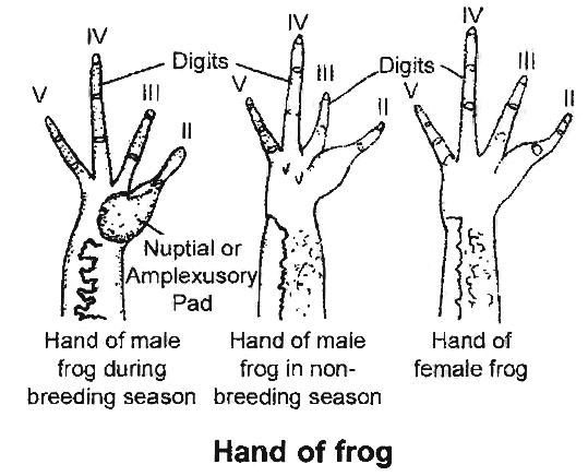
Internal Morphology
Digestive System
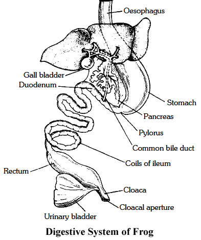
Since the frogs are carnivorous, their alimentary canal is short in length.
Tadpole larva is herbivorous so alimentary canal is very long and coiled in the form of spring.
The mouth is present as a terminal, wide opening.
It opens into bucco-pharyngeal cavity, which contains numerous maxillary teeth arranged along the margin of the upper jaw and vomerine teeth are present in the roof of the buccopharyngeal cavity.
The lower jaw is toothless. Salivary glands are absent.
Opening of eustachian tube, vocal sacs (only in male) gullet and glottis can be seen clearly in the bucco-pharyngeal cavity.
The muscular tongue is bilobed at the tip and free from behind. It is used for capturing the prey.
The gullet opens into a narrow and short tube-like oesophagus, which continues in large and distended stomach.
It contains a thick muscular layer, which helps in converting food into chyme.
It secretes gastric juice containing HCl and proteolytic enzymes. Stomach is followed by a coiled small intestine.
Intestinal wall has numerous finger-like folds called villi and microvilli, projecting into its lumen to enhance surface area for absorption of digested food.
The first part of the small intestine lying parallel to stomach is called duodenum. Intestine continues into a wider rectum, opening into cloaca.
The urinary bladder opens into cloacal chamber through the ureter.
The gastric and intestinal glands occur in the walls of stomach and intestine respectively.
The other important digestive glands associated with the alimentary canal are liver and pancreas.
Liver secretes bile which is temporarily stored in gall bladder before being released into the duodenum.
Bile helps in digestion of food by changing its pH from acidic to alkaline and by emulsifying the fats.
Liver does not secrete any digestive enzymes. Pancreas is an irregular, elongated gland, situated in a thin mesentery and lies parallel to the stomach.
It produces pancreatic juice containing digestive enzymes like trypsin, amylopsin, etc.
Respiratory System
Three types of respiration: cutaneous, buccopharyngeal and pulmonary occur.
Cutaneous respiration on land is through the body surface. During hibernation and aestivation, frog respires only through this method.
Buccopharyngeal respiration occurs through the lining of buccal cavity. It occurs only when frog is out of water. The mucus membrane of the buccal cavity is moist which dissolves oxygen whose diffusion occurs into the blood capillaries.
Pulmonary respiration: Lungs in frog are not efficient respiratory organs because only mixed air enters into them and they mainly function as hydrostatic organs.
Lungs are a pair of thin walled, translucent sacs with inner surface divided into alveoli by septa. Pulmonary respiration has a maximum frequency of 20/minute.
It occurs when more energy is required. Mouth and gullet are kept closed during pulmonary respiration.
Respiratory movements in pulmonary respiration are because of buccopharyngeal cavity which acts as a force pump.
These movements are carried out by set of paired muscles -sternohyal and pterohyal muscles.
Sternohyal muscles are attached with hyoid and coracoid processes, clavicles of the pectoral girdle and on contraction, depress the buccal floor enlarging the buccopharyngeal cavity.
Pterohyals are attached in between hyoid and pro-otics of the skull and on contraction lift the floor of buccal cavity.
With the depression of buccal floor, air enters buccal cavity through the nares.
External nares are then closed by pushing tuberculum prelinguale and the movable premaxillae.
It is followed by raiSing of the buccal floor by pterohyal muscles which reduces the volume and the air is pushed into the lungs where exchange of gases takes place.
Buccal floor is again lowered enlarging its volume which draws air into the buccal cavity.
External nares are opened fol'lowed by raising the buccal floor, pushing the air out through external nares.
Sound producing organ of frog is laryngo-tracheal chamber.
It is supported by one cricoid, two arytenoids and two prearytenoid cartilages.
It has a pair of muscle strands (vocal cords) which actually prod uce sound. Male frog has vocal sacs which act as resonating chambers.
Circulatory System
Circulatory system is closed type.
The heart lies enclosed by a thin, transparent, two layered sac, pericardium.
Frog's heart is a three chambered structure made of two upper auricles and a single lower ventricle.
The two additional chambers connected to the heart of frog are sinus venosus and truncus arteriosus.
Frogs also possess two well developed portal systems: Renal portal system and Hepatic portal system. Frog also has two pairs of lymph hearts.
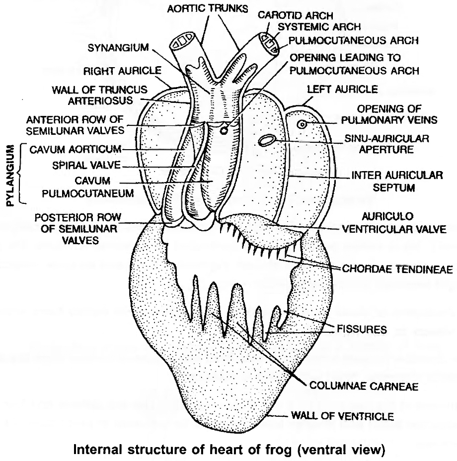
Nervous System
The nervous system is organised into a central nervous system (brain and spinal cord), a peripheral nervous system (cranial and spinal nerves) and an autonomic nervous system (sympathetic and parasympathetic chains ofganglia).
There are ten pairs ofcranial nerves.
Brain is enclosed in a bony structure or brain box (cranium) which has two occipital condyles for attachment with the first vertebra (atlas).
The brain is divided into fore-brain, mid-brain and hind-brain.
Forebrain includes olfactory lobes, paired cerebral hemispheres and unpaired diencephalon.
The mid-brain is characterised by a pair of optic lobes.
Hind-brain consists of cerebellum and medulla oblongata.
The medulla oblongata passes out through the foramen magnum and continues into spinal cord which is contained in the vertebral column.
Jacobson's organ, also called Vomero-nasalorgan opens into nasal chamber and acts as additional olfactory organ.
Eye
Eye is guarded by immovable upper eyelid, movable lower eyelid and transparent nictitating membrane.
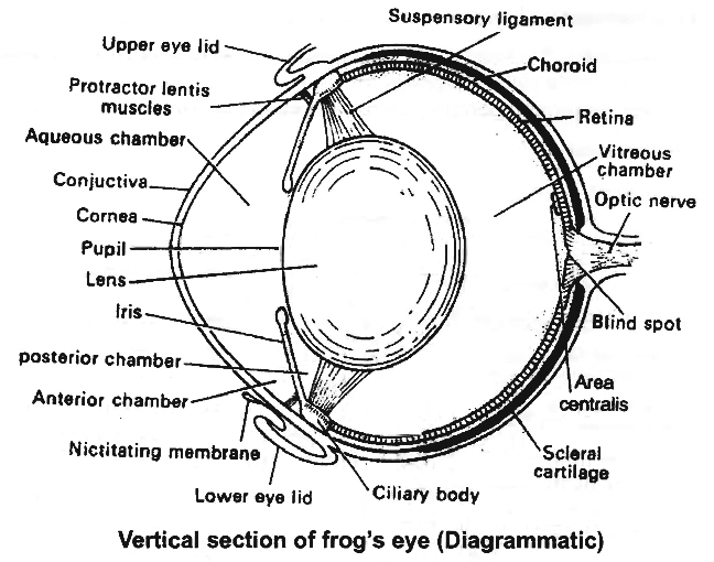
Vertical section of frog's eye (Diagrammatic)
Outersclerotic ring is cartilaginous and cornea is the part exposed out. Middle, highly vascular, pigmented layer is choroid.
Iris is yellow pigmented and perforated by a central aperture, the pupil.
Retina is the innermost coat of eyeball and consists of inner pigmented layer and an outer receptor layer.
Rods and cones are light sensitive structures found in retina.
Rods have rhodopsin or visual purple meant for night vision while cones have iodopsin responsible for colour vision in day light.
The anterior chamber present in front of lens is filled with aqueous humour while the chamber behind the lens is posterior chamber, filled with vitreous humour.
Eye ball is moved in the eye orbit by a set of six muscles -two are oblique and four are recti.
Besides, there are retractor bulbi and levator bulbi muscles for intrusion or protrusion of eye ball into the eye orbit, respectively.
Ear
Ear of frog has only middle and internal ear.
Tympanic membrane is present at the body surface.
Middle ear has single bone called columella auris.
Its outer end is attached to the ear drum while inner end to the stapedial plate.
Pressure of air in middle ear is controlled by eustachian tubes.
Membranous labyrinth or internal ear consists of utriculus, sacculus and semicircular canals. Endolymph fills the membranous labyrinth .
Excretory System
The main organ of excretion is a pair of kidneys.
These are compact, dark red and bean like structures situated little posteriorly in the body cavity on both sides of vertebral column.
The frog excretes urea, thus is a ureotelic animal.
Urea is carried by blood into the kidney where it is separated and excreted.
Each kidney is composed of several structural and functional units called uriniferous tubules or nephrons.
Ureter emerges from the kidney as urinogenital ducts in the males.
A common ureter opens into the cloaca.
A thin-walled urinary bladder is present ventral to rectum which also opens in the cloaca.
Reproductive System
Male reproductive organs consist of a pair of yellowish ovoid testes, which are found adhered to the upper part of kidneys by a double fold of peritoneum called mesorchium.
Vasa efferentia are 10-12 in number and after arising from testes, run through the mesorchium and enter the kidneys of their side.
In kidneys, these open into Bidder's canal which finally communicates with the urinogenital duct.
This duct emerges from the kidneys and finally opens into the cloaca.
The cloaca is a small, median chamber that is used to pass faecal matter, urine and sperms to the exterior.
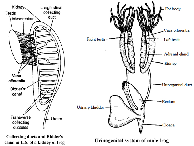
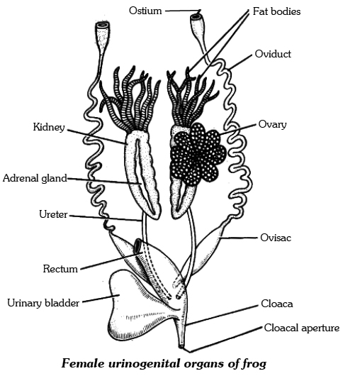
A pair of ovaries, situated near kidneys, comprises the female reproductive organs.
However, these have no functional connection with kidneys.
A pair of oviduct opens into the cloaca, separately.
The release of ovum in female is termed as spawning.
A mature female can lay 2,500 to 3,000 ova at a time.
Fertilisation is external and takes place in water.
Development involves a larval stage called tadpole.
Tadpole undergoes metamorphosis to form the adult.
SUMMARY
Earthworm, Cockroach and Frog show characteristic features in body organisation.
In Pheretima posthuma (earthworm), the body is covered by cuticle.
All segments of its body are alike except the 14th, 15th & 16th segment, which are thick and dark and glandular, forming clitellum.
A ring of S-shaped chitinous setae is found in each segment.
These setae help in locomotion.
On the ventral side spermathecal openings are present in between the grooves of 5 & 6, 6 &7, 7 & 8 and 8 & 9 segments.
Female genital pores are present on 14th segment and male genital pores on 18th segment.
The alimentary canal is a narrow tube made of mouth, buccal cavity, pharynx, gizzard, stomach, intestine and anus.
The blood vascular system is of closed type with heart and valves.
Nervous system is represented by ventral nerve cord.
Earthworm is hermaphrodite.
Two pairs of testes occur In the 10th and 11th segment, respectively.
A pair of ovaries are present on 12th and 13th intersegmental septum.
It is a protandrous animal with crossfertilisation.
Fertilisation and development takes place in cocoon secreted by the glands of clitellum.
The body of Cockroach (Periplaneta americana) is covered by chitinous exoskeleton.
It is divided into head, thorax and abdomen.
Segments bear jointed appendages.
There are three segments of thorax, each bearing a pair of walking legs.
Two pairs of wings are present, one pair each on 2nd and 3rd segment.
There are ten segments in abdomen.
Alimentary canal is well developed with a mouth surrounded by mouth parts, a pharynx, oesophagus, crop, gizzard, midgut, hindgut and anus.
Hepatic caecae are present at the junction of foregut and midgut.
Malpighian tubules are present at the junction of midgut and hindgut and help in excretion.
A pair of salivary gland is present near crop.
The blood vascular system is of open type.
Respiration takes place by network of tracheae.
Trachea opens outside with spiracles.
Nervous system is represented by segmentally arranged ganglia and ventral nerve cord.
A pair of testes is present in 4th and 5th segments and ovaries in 4th, 5th and 6th segments.
Fertilisation is internal.
Female produces 10-40 ootheca bearing developing embryos.
After rupturing of single ootheca sixteen young ones, called nymphs come out.
The Indian bullfrog, Rana tigrina, is the common frog found in India.
Body is covered by skin.
Mucous glands are present in the skin which is highly vascularised and helps in respiration in, water and on land.
Body is divisible into head and trunk.
A muscular tongue is present, which is bilobed at the tip and is used in capturing the prey.
The alimentary canal consists of oesophagous, stomach, intestine and rectum, which open into the cloaca.
The main digestive glands are liver and pancreas.
It can respire in water through skin and through lungs on land.
Circulatory system is closed with single circulation.
RBCs are nucleated.
Nervous system is organised into central, peripheral and autonomic.
The organs of urinogenital system are kidneys and urinogenital ducts, which open into the cloaca.
The male reproductive organ is a pair of testes.
The female reproductive organ is a pair of ovaries.
A female lays 2500-3000 ova at a time.
The fertilisation and development are external.
The eggs hatch into tadpoles, which metamorphose into frogs.
frog
Frog
Frogs are members of the phylum Chordata's class Amphibia, which can live on land and in freshwater. Rana tigrina is the most common frog species in India. They do not have a steady body temperature; rather, their body temperature changes depending on the ambient temperature. Cold-blooded animals are known as poikilotherms. You may have also noticed that the frogs' color changes while they are in grasses or on dry soil. They can change their color to hide from their adversaries (camouflage). The term for this protective coloring is mimicry. During the summer and winter, the frogs are not visible. They take refuge in underground tunnels during this time to avoid the excessive heat and cold.Summer sleep (aestivation) and winter sleep (hibernation) are the two types of sleep.
(i) Morphology: Due to the presence of mucus, the skin is smooth and slippery. Moisture is always retained on the skin. The dorsal side of the body is olive green in color with dark irregular markings. The skin is uniformly pale yellow on the ventral side. The frog does not drink water; instead, it absorbs it through its skin. A frog's body is divided into two parts: head and trunk. There is no neck or tail. A pair of nostrils can be found above the mouth. The eyes protrude and are protected by a nictitating membrane while in the water. A membranous tympanum (ear) receives sound signals on either side of the eyes. Swimming, walking, leaping, and digging are all assisted by the forelimbs and hindlimbs.The hind limbs have five fingers and are larger and more muscular than the forelimbs, which have four. Webbed digits on the feet aid with swimming. Sexual dimorphism is seen in frogs. Male frogs can be recognized from female frogs by the presence of sound-producing vocal sacs and a copulatory pad on the first digit of the forelimbs.
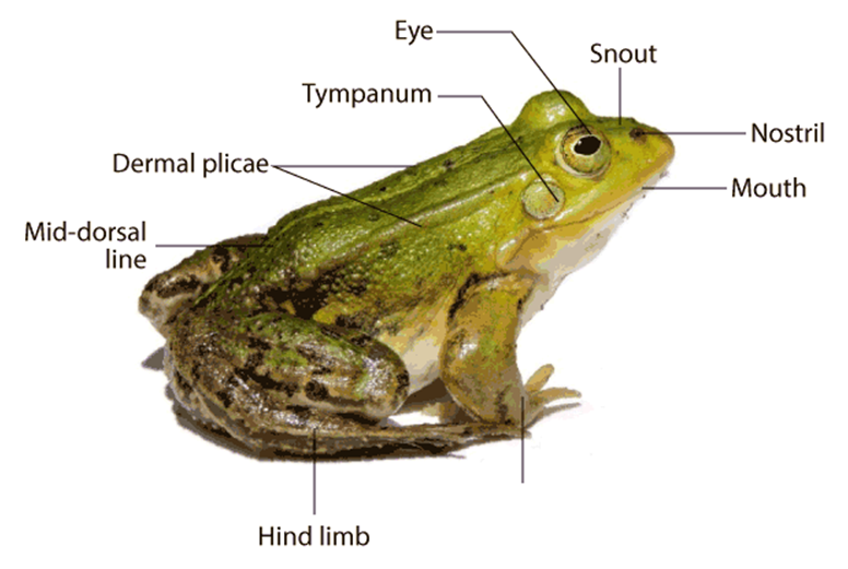
(ii) Anatomy: Frogs' bodily cavities house many organ systems with well-developed structures and functions, including the digestive, circulatory, respiratory, nervous, excretory, and reproductive systems. The alimentary canal and digestive glands make up the digestive system. Because frogs are carnivores, the alimentary canal is short, resulting in a shorter gut. The pharynx connects the mouth to the buccal cavity, which leads to the esophagus. The esophagus is a short tube that connects the mouth to the stomach, then to the intestine, rectum, and finally to the cloaca. Bile is produced by the liver and stored in the gall bladder. Pancreatic juice, which contains digestive enzymes, is produced by the pancreas, a digestive gland. The bilobed tongue is responsible for capturing food.
Food is broken down by the action of Hydrochloric acid and gastric secretions released from the stomach's walls. Chyme, or partially digested food, is transferred from the stomach to the duodenum, the first segment of the small intestine. Through a shared bile duct, the duodenum receives bile from the gall bladder and pancreatic secretions from the pancreas. Pancreatic fluids digest carbs and proteins while bile emulsifies fat. The intestine is where the final digesting takes place. The many finger-like folds in the inner wall of the intestine called villi and microvilli absorb digested food. The undigested solid waste goes through the cloaca and into the rectum. Frogs have two different strategies to breathe on land and in water.
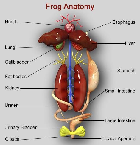
Skin serves as an aquatic respiratory organ in water (cutaneous respiration). Diffusion exchanges dissolved oxygen in the water through the skin. The buccal cavity, skin, and lungs serve as respiratory organs on land. Pulmonary respiration is the process of breathing through the lungs. The lungs are a pair of elongated, pink-colored sac-like structures located near the top of the trunk (thorax). Air enters the lungs via the nostrils, buccal cavity, and lungs. Gaseous exchange occurs through the skin during aestivation and hibernation. Frogs have a well-developed closed circulatory system. Frogs, like humans, have a lymphatic system. The heart, blood arteries, and blood make up the blood vascular system. Lymph, lymph channels, and lymph nodes make up the lymphatic system.
The heart is a muscle structure located in the upper body cavity. It has three chambers, two atria, and one ventricle, and is surrounded by a pericardium membrane. The right atrium is connected to the sinus venosus, a triangle structure. It receives blood through the vena cava, which are large veins. On the ventral side of the heart, the ventricle opens into a sac-like conus arteriosus. The arteries transport blood from the heart to all regions of the body (arterial system). The venous system collects blood from various regions of the body and transports it to the heart.
Frogs have a unique venous connection between the liver and gut, as well as the kidney and lower body.The first is known as the hepatic portal system, whereas the second is known as the renal portal system. Plasma and cells are found in the blood. RBCs (red blood cells) or erythrocytes, WBCs (white blood cells) or leucocytes, and platelets are the blood cells. RBCs are nucleated and contain the pigment haemoglobin, which is red in color. Lymph is not the same as blood. It is deficient in proteins and RBCs. During circulation, the blood transports nutrients, gases, and water to their proper locations. The pumping activity of the muscular heart is responsible for blood circulation. A well-developed excretory system is responsible for the removal of nitrogenous wastes. A pair of kidneys, ureters, cloaca, and urine bladder make up the excretory system.
On both sides of the vertebral column, they are compact, dark red, bean-like structures located a bit posteriorly in the body cavity. Uriniferous tubules or nephrons are structural and functional units that make up each kidney. Male frogs have two ureters that arise from their kidneys. The ureters serve as a urinogenital channel, leading to the cloaca. The ureters and oviduct open separately in the cloaca in females. The rectum, which likewise opens in the cloaca, has a thin-walled urine bladder ventral to it. The frog is a ureotelic animal because it excretes urea. Blood transports excretory wastes to the kidney, where they are sorted and expelled. The frog's control and coordination system is highly developed.
It involves both the nervous and endocrine systems. Hormones released by the endocrine glands are responsible for the chemical coordination of the body's many organs. The pituitary, thyroid, parathyroid, thymus, pineal body, pancreatic islets, adrenals, and gonads are the most significant endocrine glands in frogs. The nervous system is arranged into a central nervous system (brain and spinal cord), a peripheral nervous system (cranial and spinal nerves), and an autonomic nervous system (sympathetic and parasympathetic) (sympathetic and parasympathetic). The brain gives rise to ten pairs of cranial nerves. The brain is protected by a bony structure known as the brain box (cranium). The forebrain, midbrain, and hindbrain are the three parts of the brain. The olfactory lobes, paired cerebral hemispheres, and unpaired diencephalon makes up the forebrain.
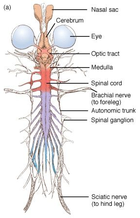
A pair of optic lobes distinguish the midbrain. The cerebellum and medulla oblongata make up the hindbrain. The medulla oblongata exits the foramen magnum and continues into the spinal cord, which is contained within the vertebral column. The sense organs of the frog are touch (sensory papillae), taste (taste buds), smell (nasal epithelium), vision (eyes), and hearing (ears) (tympanum with internal ears). Eyes and internal ears are well-organized structures, with the rest consisting of cellular aggregations around nerve endings. Frog eyes are a pair of spherical structures in the orbit of the skull. These are basic eyeballs (possessing only one unit). In frogs, the external ear is missing, leaving just the tympanum visible.
The ear is both a hearing and a balancing organ (equilibrium). Male and female reproductive systems in frogs are well-organized. A pair of yellowish ovoid testes are discovered adhering to the top section of the kidneys by a double fold of peritoneum called the mesorchium in male reproductive organs. The number of Vasa efferentia that emerge from the testes is 10-12. On their side, they enter the kidneys and open into Bidder's canal. Finally, it connects with the urinogenital duct, which runs from the kidneys to the cloaca. The cloaca is a small, midline chamber where faeces, urine, and sperms are passed to the outside. A pair of ovaries are among the female reproductive organs.
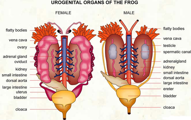
The ovaries are close to the kidneys but have no functional relationship with them. Separate oviducts emerge from the ovaries and open into the cloaca. At any given time, a mature female can lay between 2500 and 3000 ova. External fertilization takes place in water. Tadpoles are in the larval stage of development. Tadpoles go through metamorphosis to become adults. Frogs are advantageous to humans because they consume insects and protect crops. Frogs keep the ecology in balance since they are a vital link in the food chain and food web. Humansconsume the muscular legs of frogs in several regions.
