 ACME SMART PUBLICATION
introduction
- Books Name
- A TEXT OF BIOLOGY - CLASS XII
- Publication
- ACME SMART PUBLICATION
- Course
- CBSE Class 12
- Subject
- Biology
REPRODUCTION
Reproduction is defined as a biological process in which an organism gives rise to young ones (offspring) similar to itself.
The offspring grow, mature and in turn produce new offspring. Thus, there is a cycle of birth, growth and death.
Reproduction enables the continuity of the species, generation after generation. You will study later in Chapter (Principles of Inheritance and Variation) how genetic variation is created and inherited during reproduction.
There is a large diversity in the biological world and each organism has evolved its own mechanism to multiply and produce offspring.
Habitat and internal physiology of organism and several other factors are collectively responsible for how it reproduces.
There are two types of reproduction:
(1) Asexual reproduction
(i) Offspring is produced by a single parent with or without the involvement of gamete formation.
(ii) Gametic fusion is absent.
(2) Sexual reproduction
(i) Offspring is produced by two parents (opposite sex) with the involvement of gamete formation.
(ii) Gametic fusion is present (fertilization/syngamy).
asexual reproduction
- Books Name
- A TEXT OF BIOLOGY - CLASS XII
- Publication
- ACME SMART PUBLICATION
- Course
- CBSE Class 12
- Subject
- Biology
ASEXUAL REPRODUCTION
A single individual (parent) is capable of producing offsprings in asexual type of reproduction. As a result, the offsprings
produced are exact copies of each other and their parents. Such morphologically and genetically similar individuals are
called clones.
It is common among single-celled organisms, plants and animals with relatively simple organisation.
It is also called somatogenic reproduction because propagules are formed from somatic cells of the parent.
It occurs by fission, budding, sporulation, fragmentation, regeneration and vegetative reproduction.
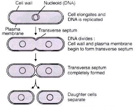
(a) Binary Fission
In this process, the parent organism divides into two halves, each half forming an independent daughter organism.
It means, the parent body as a whole forms the reproductive unit and the parent continues living as two daughter
individuals.
Therefore, the organisms that undergo binary fission are said to be immortal.
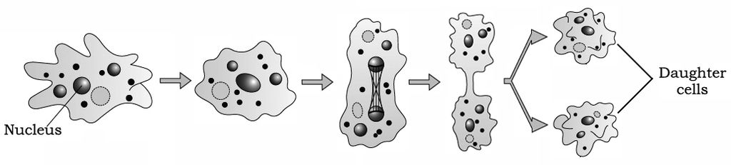
(ii) It occurs in Bacteria (Moneran), Amoeba and Paramecium (Protists).
Depending upon the plane of division, binary fission is of the following types:
(i) Simple Binary Fission (Irregular Binary Fission) : Division can occur through any plane, e.g.; Amoeba.
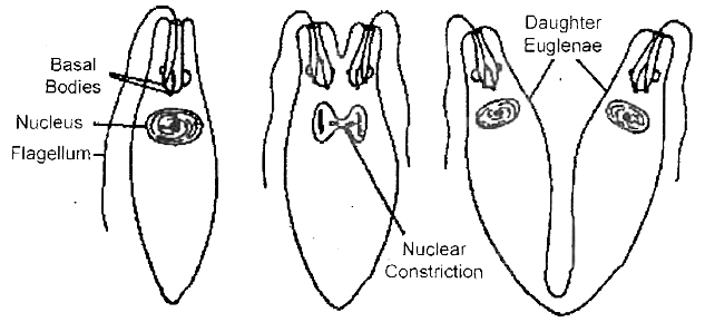
(ii) Longitudinal Binary Fission : The plane of fission passes along the longitudinal axis of the organism, e.g., Euglena.
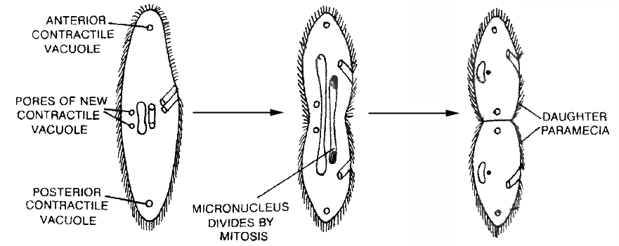
(iii) Transverse Binary Fission : The plane of this division runs along the transverse axis of the individual, e.g., Bacteria,
Paramecium, Diatoms.
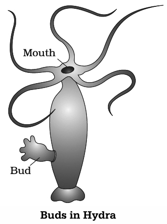
(b) Budding
In yeast, the division is unequal and a small bud (protuberance) is produced that remains attached initially to the
parent cell, later on the bud gets separated and mature into new yeast (cells).
Budding or sporulation is also shown by oidia of Rhizopus.
Buds are also reported in hydra.

(c) Spores: Members of the kingdom fungi and simple plants such as algae reproduce through special asexual
reproductive structure. The most commonly produced structures are conidia and zoospores.
Zoospores
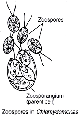
(i) These are motile and flagellated spores produced inside the zoosporangia under favourable conditions.
(ii) In Chlamydomonas(n), the protoplast of cell divides to form 8-16 zoospores. They are pyramid shaped and anteriorly
biflagellated, resembling the parent cell. The parent cell wall breaks and the zoospores are liberated in water. They enlarge
and behave as adult individuals.
(iii) Zoospores are also produced in the asexual life cycle of Achlya, Saprolegnia, Phytophthora and Ulothrix.
(iv) Zoospores of Cladophora glomerata are diploid.
Conidia
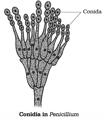
(i) In Penicillium, these spores are produced at the tips of special hyphal branches, called conidiophores.
(ii) There are two types of conidiophores :
a. Unbranched/monoverticillate
b. Branched/biverticillate
(iii) The branches of conidiophores are called rami and branches of rami are called metulae. Each metula bears 2-6 flask shaped structures called sterigmata (phialides). Each sterigma produces a chain of conidia.
(iv) Features of Conidia:
a. Pigmented
b. Uni or multinucleated
c. The conidia in the chain are arranged in basipetal manner.
(d) Vegetative Reproduction
(i) Vegetative reproduction does not involve meiosis and fusion of gametes, therefore it is considered ' as a type of
asexual reproduction .
(ii) New plants or individuals are produced from vegetative parts of plants and newly formed individuals are genetically
identical to the parent plant.
(iii) It is common method of reproduction in the flowering plants.
There are two types of vegetative reproduction.
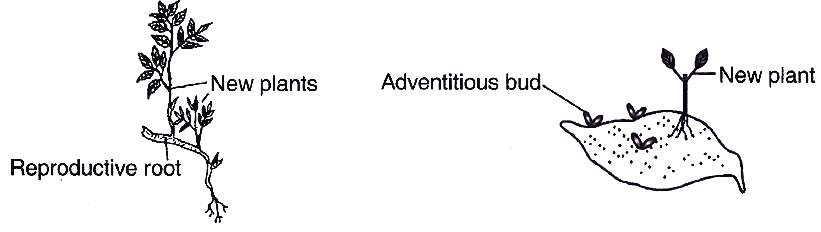
I. Natural Methods of Vegetative Reproduction:
These are methods of plant multiplication occurring naturally in which a somatic part of the plant detaches from the
body of the mother and develops into a new independent plant under suitable environmental conditions.
The detachable somatic part that functions in vegetative propagation is called vegetative propagule.
It carries one or more buds. Natural buds occur over the nodes of the stem. When placed in contact with damp soil,
the buds sprout, producing roots and new plants.
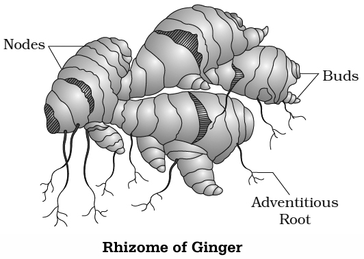
This potential is exploited by farmers, e.g., tubers of potato, rhizome of banana and ginger, bulbs, runners, offsets,
stolons, aerial stems etc.
Some propagules carry adventitious buds, e.g., normal and storage roots, leaves.
It occurs by following means:
STEM
Underground stems
Different types of underground stems like tuber, Germinating rhizome, bulb and corm can take part in Young shoot
vegetative propagation.
A portion of underground stem bearing bud Roots forms a new plant.
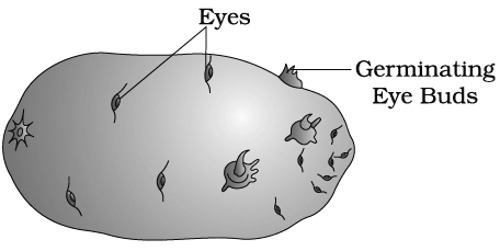
(i) Tuber : It is terminal portion of underground stem branch which is swollen on account of accumulation of food.
e.g., Potato, Artichoke
(ii) Rhizome: It grows obliquely or horizontally under soil surface. It is well branched and bears nodes, internodes,
buds and scale leaves. e.g., Banana, Turmeric, Aspidium, Adiantum, Ginger
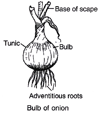
(iii) Bulb: Stem is unbranched, highly reduced and disc shaped. The bud is surrounded by many concentric scale leaves.
Leaf bases of inner ones are fleshy and edible and outer ones are dry known as tunic. e.g., Onion, Garlic, Narcissus
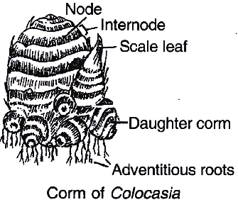
(iv) Corm: It grows vertically beneath the soil surface. It bears nodes, internodes, buds and scale leaves. e.g.,
Colocasia, Gladiolus, Freesia., Crocus, Amorphophallus
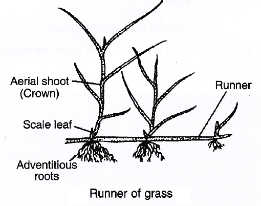
(3) Creeping stems:
(i) Runner: It is elongated, prostrate, sub-aerial branch with long internodes and roots at nodes. e.g., Grasses
(ii) Offset: Short horizontal branch which is one internode long and produces a cluster of leaves above and the cluster
of roots below is called offset. e.g., Eichhornia (Water hyacinth), Pistia.
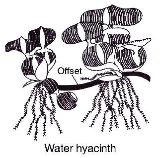
Water hyacinth or 'terror of Bengal' was introduced in Bengal because of its beautiful flowers and shape of leaves.
However, it turned out to be highly invasive aquatic weed that not only spread to all water bodies of Bengal but also
throughout India. It drains oxygen from the water, which leads to death of fishes and other animals. It is very
difficult to get rid off them since it can propagate vegetatively by offset at a phenomenal rate and spread all over the
water body in a short period of time.
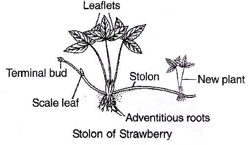
(iii) Stolon : It is subterranean long lateral branch arising from base of stem. It first grows obliquely upward and then
bends down to the ground surface.
e.g., Strawberry, Vallisneria Leaflets
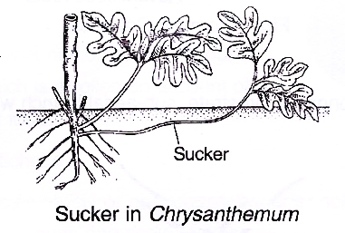
(iv) Sucker : It arises by axillary bud of underground part of stem. This lateral branch creeps below the soil surface,
grows obliquely upward and produces new shoot.
e.g., Chrysanthemum, Pineapple, Banana
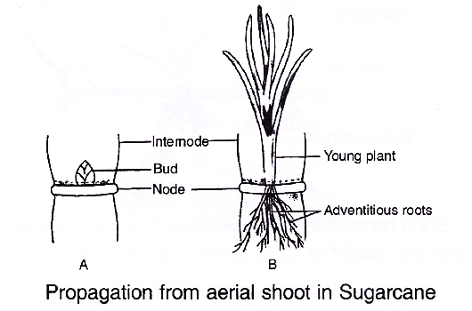
(4) Aerial shoots : Each segment of stem having at least one node can form a new plant.
e.g., Sugarcane, Opuntia
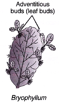
(5) Leaves: Leaves of several plants having adventitious buds help in vegetative reproduction. In Bryophyllum
adventitious buds arise from the notches present at margins of leaves.
e.g., Adiantum (walking fern), Begonia, Streptocarpus, Saintpaulia and Kalanchoe.
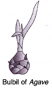
(6) Bulbils: These are fleshy buds which produce new plant. e.g., Agave, Oxalis, Ananas, Dioscorea, Lily, Chlorophytum
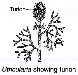
(7) Turions : Fleshy buds in aquatic plants helping in perennation, e.g., Potamogeton, Utricularia.
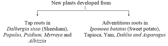
ROOTS
(i) Both tap roots and adventitious roots takes part in vegetative reproduction due to the presence of bud, known as radicle
bud.
(ii) Adventitious buds on root detaches and gives rise to new plant.

sexual reproduction
- Books Name
- A TEXT OF BIOLOGY - CLASS XII
- Publication
- ACME SMART PUBLICATION
- Course
- CBSE Class 12
- Subject
- Biology
SEXUAL REPRODUCTION
Sexual reproduction involves formation and fusion of gametes to form the zygote which develops to form the new organism.
Characteristics
1. Two fusion gametes can be produced by same individual or different individuals. So it can be both uniparental as well as biparental (mostly).
2. Offsprings produced are not identical to parents or amongst themselves.
3. It involves meiosis and syngamy.
4. It is a slow, elaborate or complex process. So multiplication is not so rapid.
All organisms have to reach a certain stage of growth and maturity in their life cycle before they reproduce sexually. For understanding this better lets study the different phases in the life cycle of organism.
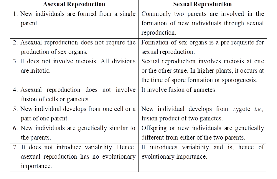
Table: Differences between Asexual and Sexual Reproduction
Phases in Life Cycle
Three phases are there in the organism's life cycle.
1. Juvenile phase
2. Reproductive phase
3. Senescent phase
1. Juvenile phase/pre-reproductive phase
During this phase organism will show growth so that it can attain certain maturity to perform the sexual reproduction.
This phase is known as vegetative phase in plants. It is of variable durations in different organisms.
2. Reproductive phase
Reproductive organs develop and mature during this phase. In the higher plants, end of juvenile phase or onset of reproductive phase is easily marked.
In the higher plants during this phase, there is formation of reproductive structures i.e., flowers.
The motto of this phase is to produce the offsprings which may be similar or dissimilar to parental generation. This phase is also of variable durations in different organisms.
Based upon flowering and fruiting pattern there are two types of flowering plants, i.e., monocarpic and polycarpic.
Monocarpic Plants :
They are plants which flower only once in their life. After flowering, they produce fruits and die. All annuals (e.g., Wheat, Rice, Marigold) and biennial plants (e.g., Radish, Carrot, Henbane), are monocarpic.
A few perennial plants are also monocarpic. Certain bamboo species (e.g., Bambusa tulda, Meloeanna bambusoides) live vegetatively for 50-100 years, flower and fruit abundantly and then die.
Strobilanthus kunthiana (vern. Neelakurinji) flowers once in 12 years.
The last time it flowered was September-October 2006. The flowering converted large hilly tracts of Kerala, Karnataka and Tamil Nadu into blue stretches that attracted a large number of tourists.
Polycarpic Plants :
They are perennial plants which after reaching maturity, flower repeatedly at intervals, e.g., Mango, Apple, Jackfruit, Grape vine, Orange.
Very few perennial plants bear flowers throughout the year, e.g., China rose (Shoe Flower). The period between two flowering phases is called Inter flowering period which is used for building up resources and is, therefore, a recovery phase. It is not the juvenile phase but is part of the mature phase.
3. Senescent Phase:
It is a post-reproductive phase. It involves structural and functional deterioration of body by accumulation of waste metabolites which ultimately leads to death.
In both plants and animals, hormones are responsible for the transitions between three phases. Interaction between hormones and certain environmental factors regulate the reproductive processes and the associated behavioural expressions of organisms.
Concept Builder
The females of placental mammals exhibit cyclical changes in the activities of ovaries and accessory ducts as well as hormones during the reproductive phase.
In non-primate mammals like cows, sheep, rats, deers, dogs, tiger, etc., such cyclical changes during reproduction are called oestrus cycle.
Where as in primates (monkeys, apes, and humans) it is called menstrual cycle.
Many mammals, especially those living in natural, wild conditions exhibit such cycles only during favourable seasons in their reproductive phase and are therefore called seasonal breeders.
Many other mammals are reproductively active throughout their reproductive phase and hence are called continuous breeders.
events in sexual reproduction
- Books Name
- A TEXT OF BIOLOGY - CLASS XII
- Publication
- ACME SMART PUBLICATION
- Course
- CBSE Class 12
- Subject
- Biology
EVENTS IN SEXUAL REPRODUCTION
After attainment of maturity, all sexually reproducing organisms exhibit events and processes that have remarkable fundamental similarity, even though the structures associated with sexual reproduction are indeed very different.
These sequential events may be grouped into three distinct stages, namely, the pre-fertilisation, fertilisation and the post-fertilisation events.
1. Pre-fertilization events
These are events in sexual reproduction which occur prior to the process of fertilization. The two main prefertilization events are gametogenesis and gamete transfer.
(a) Gametogenesis: It refers to the process of formation of gametes-male and female.
Categories of Gametes :
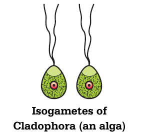
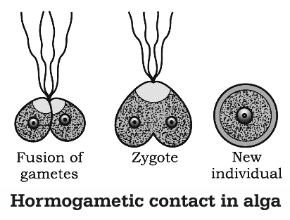
(i) Isogametes : When the fusing gametes are morphologically similar they are known as isogametes or homogametes.
e.g., (i) Algae: Chlamydomonas debaryana, Ulothrix
(ii) Fungi : Synehytrium, Rhizopus
(ii) Heterogametes: When the fusing gametes are morphologically distinct types, they are known as heterogametes. It is the feature of majority of sexually reproducing organisms.
e.g., (i) Algae: Volvox, Chara, Fucus
(ii) All Bryophytes, Pteridophytes, Gymnosperms and Angiosperms.
In such organisms, male gamete is called antherozoid or sperm and the female gamete is called egg or ovum.
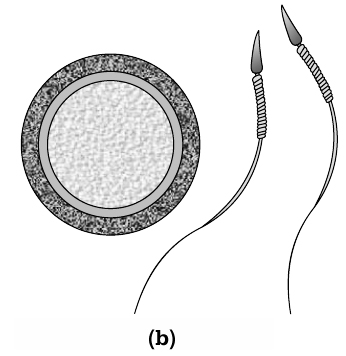
Sexuality in organisms:
Sexual reproduction in organisms generally involves the fusion of gametes from two different individuals. But this is not always true.
Plants may have both male and female reproductive structures in the same plant (bisexual) or on different plants (unisexual).
In several fungi and plants, terms such as homothallic and monoecious are used to denote the bisexual condition and heterothallic and dioecious are the terms used to describe unisexual condition.
A. Sexuality in animals
The individuals of all species either male or female (unisexual)?
Or are there species which possess both the reproductive organs (bisexual)?
You probably can make a list of several unisexual animal species.
Earthworms, sponge, tapeworm and leech, typical examples of bisexual animals that possess both male and female reproductive organs, are hermaphrodites.
Cockroach is an example of a unisexual species.
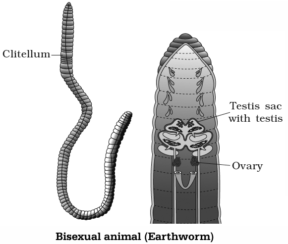
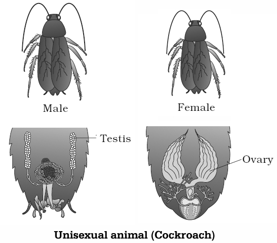
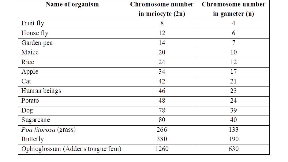
Table: Chromosome numbers in meiocytes (diploid, 2n) and
gametes (haploid, n) of some organisms.
B. Sexuality in Plants
In most of the lower sexually reproducing organisms, two fusing gametes are morphologically similar.
If these gametes belong to the same parent then such organisms are called homothallic, e.g., fungi (Mucor mucedo). When these gametes belong to different parents then these organisms are called heterothallic.
Higher Organisms:
In higher plants there is well developed sex organs and there is clear distinction between male and female sex organs.
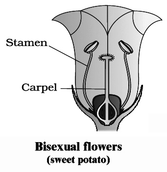
Angiosperms possess flowers as reproductive structures. The male sex organ is called stamen and female sex organ is carpel or pistil.
If male and female sex organs occur in the same flower then these plants are called bisexual, e.g., China rose. If flowers possess only stamen or carpel then these plants are called unisexual.
When male flower (staminate) and female flower (pistillate) are present on same plant body such plants are monoecious, e.g., Acalypha, cucurbits, coconut and maize.
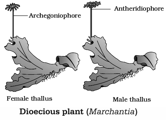
However, if they are present on separate plant body then these plants are known as dioecious, e.g., date palm, papaya and mulberry.
In some of the lower plants also the monoecious and dioecious condition occur. For knowing this, we will study the sexuality in Chara and Marchantia.
Sexual Reproduction in Chara and Marchantia :
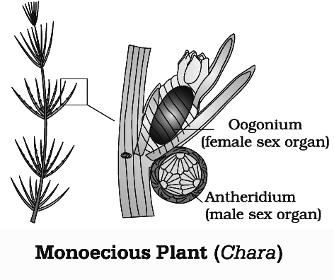
The Chara is a green alga. It is oogamous. The sex organs are highly specialised. Some workers prefer to call the male sex organ as antheridium and female as oogonium, while others did not favour this terminology.
They call the male sex organ as globule and the female as nucule and this terminology is largely followed in Chara. These sex organs are exceptionally multicelled and covered by jacket.
The jacket of nucule is formed by tube cells and the jacket of globule is formed by shield cells. The nucule has a cap of 5 coronary cells.
The sex organs are borne on the adaxial surface of the short lateral branch almost on each node. The nucule occupies an upper position than the globule.
While most of the species of Chara are monoecious, C. wallichii is dioecious. The globule matures prior to nucule (protandrous condition).
Each antheridium produces many band shaped, spirally coiled, biflagellate antherozoids. The oogonium contains a single egg. The egg is laden with starch and oil globules.
In Marchantia, the archegonia are borne on special branches called archegoniophores or the female receptacles. The archegonia may be stalked or sessile.
Cell division during gamete formation
Gametes are haploid though the parent plant body producing these gametes may be either haploid or diploid.
A haploid parent produces the gametes by mitotic division however organisms having diploid body the gametes are formed through reductional division, i.e., meiosis.
In these organisms specialised cells called meiocytes or gamete mother cells undergo meiosis. At the end of meiosis only one set of chromosomes gets incorporated into each gamete.
(b) Gamete transfer :
After the formation of male and female gametes, compatible gametes must be physically brought together to facilitate fusion (fertilisation or syngamy).
In few fungi and algae, both types of gametes are motile. In heterogametic condition, the female gamete is non motile. So there is a need of a medium through which the male gametes move.
Water is the medium for gamete transfer in algae, bryophytes and pteridophytes. A large number of the male gametes however, fail to reach the female gametes.
To compensate this loss of gametes, the number of male gametes produced in several thousand times the number of female gametes produced.
In seed plants, pollen grains are the carrier of male gametes and ovule has the egg.
2. FERTILISATION
The most vital event of sexual reproduction is the fusion of gametes. This process is called syngamy or fertilisation which results in the formation of a diploid zygote.
Syngamy can occur in external medium as well as inside the body of organism.
On this basis syngamy can be distinguished into two types :
(a) External fertilisation :
Syngamy occurs outside the body of organism in external medium (water). It is shown by majority of aquatic organisms like most of algae, fishes as well as amphibians.
Organisms exhibiting external fertilisation show great synchrony between the sexes and release a large number of gametes into the surrounding medium in order to enhance the chances of syngamy.
The disadvantage associated with it is that the offsprings are extremely vulnerable to predators.
(b) Internal fertilisation:
Syngamy occurs Inside the body of organisms. It is present in majority of plants like bryophytes, pteridophytes, gymnosperms and angiosperms.
It occurs in few algae like Spirogyra. In all these organisms egg is formed inside the female body where syngamy occurs.
The male gamete is motile and has to reach the egg in order to fuse it. In order to enhance the chances of syngamy large number of sperms are produced in these organisms and to compensate for this there is significant reduction in number of eggs produced.
Concept Builder
Sexual reproduction is divided into two types.
Zooidiogamy :
It is a type of sexual reproduction in which transfer of male gamete occurs through the medium i.e., water. It occurs in several simple plants like algae, bryophytes and pteridophytes.
Siphonogamy :
It is a type of sexual reproduction in which male gamete carrier is pollen grain and transfer of male gamete occurs through pollen tube. It is the feature of seeded plants like gymnosperms and angiosperms. Pollen grains are produced in anthers, therefore have to be transferred to the stigma before it can lead to fertilisation.
External and internal fertilisation should not be confused with exogamy and endogamy.
Exogamy: Two fusing gametes belong to different individuals. Also known as cross fertilisation.
Endogamy: Two fusing gametes belong to same individual. Also known as self-fertilisation.
3. POST-FERTILISATION EVENTS
Events in sexual reproduction after the formation of zygote are called post-fertilisation events.
Zygote :
It is the first cell of the new generation in all sexually reproducing organisms. Zygote is always diploid.
It is formed in the external aquatic medium in those organisms which perform external fertilization. Zygote is produced inside the body in cases where fertilization is internal.
Zygote is a vital link between two successive generations. It ensures the continuity of race from generation to generation.
The body of all multicellular organisms develops from the single-celled zygote. All the cells of the body, therefore, contain the same genetic traits as present in the zygote.
Embryogenesis
A. In Plants :
Embryogenesis is the process of development of embryo from zygote. Embryo is a multicellular stage in the life cycle of a plant or animal prior to formation of an independent individual.
In embryogenesis, the zygote undergoes repeated cell divisions through mitosis. The divisions help in growth of the embryo.
Cells undergo differentiation attaining specific shape, size and function. Cell differentiation occurs at specific locations resulting in production of different tissues, organs and organ systems.
Development of different external and internal structures is called morphogenesis. In flowering plants, zygote develops into embryo.
The food for development of embryo comes from a special tissue known as endosperm. Ultimately, the fertilized ovule matures into a seed.
A number of seeds develop in an ovary depending upon the number of ovules. Meanwhile, wall of the ovary also proliferates. It produces pericarp. The pericarp can be dry or fleshy.
The ripened ovary with pericarp and seeds is called fruit. As the fruit begins to develop, sepals, petals, stamens, style and stigma normally sheds. After dispersal, the seeds, upon reaching suitable substratum germinate and form new plants.
B. In Animals :
Animals are categorised into oviparous and viviparous based on whether the development of the zygote take place outside the body of the female parent or inside.
Whether they lay fertilised/unfertilised eggs or give birth to young ones.
In oviparous animals like reptiles and birds, the fertilised eggs covered by hard calcareous shell are laid in a safe place in the environment; after a period of incubation young ones hatch out.
On the other hand, in viviparous animals (majority of mammals including human beings), the zygote develops into a young one inside the body of the female organism.
After attaining a certain stage of growth, the young ones are delivered out of the body of the female organism.
Because of proper embryonic care and protection, the chances of survival of young ones is greater in viviparous organisms.
II. Artificial Methods of Vegetative Reproduction:
Artificial methods are man-made special techniques in which, part of somatic body of a plant is made to develop into new independent plant.
Artificial methods are used to propagate desired varieties according to human requirements, Rainy and spring seasons are the best periods for vegetative propagation.
The various horticultural methods of vegetative propagation are as follows:
1. Cuttings:
Cuttings are cut pieces of stem, leaves and root which are planted in nurseries in natural polarised fashion, Pre-requisite to successful cutting is induction of rooting, For this, root promoting chemicals like IBA, NAA are used,
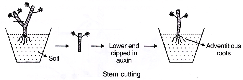
(i) Stem Cuttings :
It is a common artificial method of plant propagation, 20-30 cm long pieces of one year old stems are cut and their lower ends are dipped in dilute auxin for several minutes before planting in the soil. The lower ends develop adventitious roots, Buds present over the exposed parts sprout and form the shoot system,
e,g., Rosa, Duranta, Citrus, Clerodendron, Thea, Bougainvillea, Croton and China rose
(ii) Leaf Cuttings: Snake plant (Sansevieria) can be propagated by leaf cuttings, Leaves are cut transversely into two or three parts and planted in vertical position in the soil. For successful leaf cutting, besides induction of rooting, formation of adventitious buds is also important.
(iii) Root Cuttings : They are long pieces of roots which are used to artificially propagate new plants, Ability to form adventitious roots and adventitious buds are pre-requisites. Root cuttings are used in propagation of Lemon, Tamarind, Blackberry and Raspberry.
2. Layering:
(i) It is a type of rooting-cutting method in which adventitious roots are induced to develop on a soft stem while it is still attached to the plant.
(ii) It is carried out on one year old basal shoot branches commonly during early spring or early rainy season.
(iii) A soft basal branch is defoliated in the middle where a small injury or cut is also given, like tongueing (oblique cut), notching (V-shaped cut), ringing (removal of a ring of bark). The injured defoliated part is pegged in the soil to develop adventitious roots. The pegged down branch of the plant is called layer. Later on as the roots develop, the layer is separated and planted.
(iv) Layering is of following types:
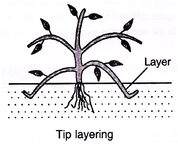
(a) Tip Layering : A shoot is bent down in the soil in such a way that its basal end is slanting while the tip is upright. Soil is pressed. It induces root formation and later growth of shoot tip.
e.g., Blackberry, Raspberry.
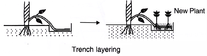
(b) Trench Layering : The basal branch is pegged in a horizontal position in a trench made in soil. It develops a number of vertical shoots. e.g., Walnut, Mulberry.
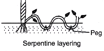
(c) Serpentine Layering : The basal branch is pegged at several places in soil at regular intervals, so as to form many plants. e.g., Clematis
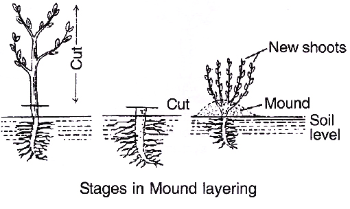
(d) Mound (Stool) Layering
The shoot is pruned and its lower part is covered by soil but the tip is kept outside the soil. When a number of new shoots develop, soil and saw dust are poured over the base to form a mound. Each shoot develops roots. Rooted shoots are separated and planted.
e.g., Currant, Gooseberry, Apple, Pear and Jasmine
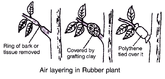
(e) Gootee (Air Layering)
(i) It is an ancient horticultural technique for propagation of tropical and subtropical trees and shrubs where soft branches do not occur near the soil.
(ii) During early monsoon rains, 3-5 cm long ring of bark is removed from the basal region of a healthy and woody branch. It is covered by a thick plaster of grafting clay. Grafting clay is made of 1 part cow dung, 1 part finely cut hay or moss and two parts clay. To it is added water and a small quantity of root promoting hormones like IAA, IBA or NAA. It is then wrapped in polythene. After 2-3 months, roots appear. The shoot is now cut below the covered part and used for planting.
e.g., Litchi, Pomegranate.
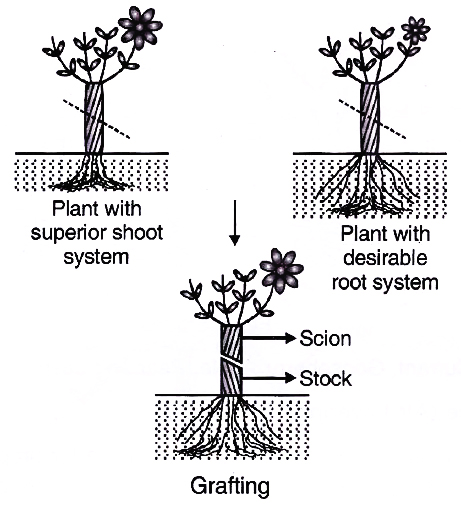
3. Grafting:
(i) Grafting is a technique of connecting two parts, usually a root system and a shoot system of two different plants in such a way that they unite and later develop as a composite plant.
(ii) It is used only in cambium containing eustelic plants.
(iii) A small shoot of plant with superior traits is employed. It is called graft or scion. It should have one to several buds. The root system of the other plant is allowed to remain intact. It is called stock (under stock). The shoot of the stock is often cut 10-30 cm above the base of the root. Leaves and buds present over the stump of stock are removed.
(iv) In grafting, scion is fixed over the stock in a manner that cambia of the two come in contact. The union is covered with grafting wax. It is then tied with the help of a bandage, tape, rubber or nail. The buds of the stock are not allowed to sprout.
They are removed as soon as they are noticed.
e.g., Mango, Apple, Pear, Citrus, Guava, Plum, Peach, Pine etc.
The various types of grafting are as follows:
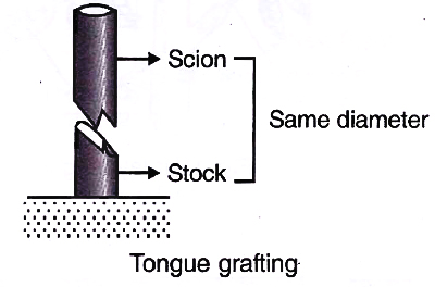
(a) Tongue (Slice or Whip) Grafting
Oblique sloping cut or notch is given to both stock and scion. The two perfectly fit upon one another. They are tied together.
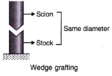
(b) Wedge Grafting
V-shaped notch is given to stock while wedge like cut is given to scion.
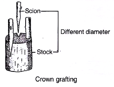
(c) Crown Grafting
Many scions are selected and shaped at the base to form wedge. Many slits are formed on the sides of stock. Scions are inserted in the slits and are bandaged.
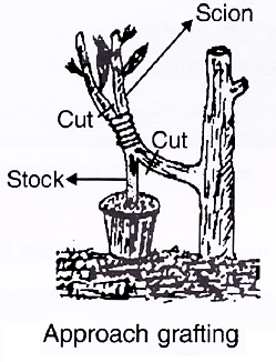
(d) Approach Grafting
Two independently growing plants are brought together. The shoots of the two are given cuts at the same level for a distance of 2.5 -5.0 cm. The cuts are in the form of smooth slices of bark, tongue shaped cuts or deeper vertical cuts. In this grafting, the scion is cut below the graft while stock is cut above the graft after the establishment of union.
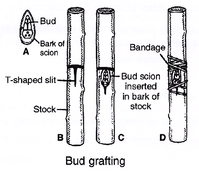
(e) Bud Grafting
Scion is a bud with a small piece of bark and cambium. Stock is given a T-shaped cut. Bark is lifted to expose cambium. Bud is inserted and the bark is allowed to come back to its original position. Only the bud is exposed. The joint is treated with grafting wax and bandaged. Bud sprouts after 3-5 weeks. Bud grafting is commonly practised in apple, peach and rose.
4. Micropropagation:
Micropropagation is the raising of new plants from a small plant tissue with the help of tissue culture technique.
Tissue culture is the technique of maintaining and growing cells, tissues, etc. and their differentiation on artificial medium under aseptic conditions inside suitable containers.
Concept Builder
N.Grew first pointed out that flowers are reproductive organs of plants.
Largest male gamete is seen in Cycas.
Citric acid helps in attraction of male gametes towards the archegonium in Lycopodium, a pteridophyte.
Parthenospores: A spore is formed directly from a gamete. It is also called azygospore.
Parthenogamy : It is the union of two incompatible gametes like two female gametes.
Parthenoapogamy: It is the fusion of vegetative nuclei.
Parthenocarpy (Noll, 1902) : It is formation of fruits without fertilization. It is useless in case of plants where seeds provide the economic produce, e.g., Almond , Walnut, Coconut, Pomegranate.
structure of flower
- Books Name
- A TEXT OF BIOLOGY - CLASS XII
- Publication
- ACME SMART PUBLICATION
- Course
- CBSE Class 12
- Subject
- Biology
FLOWER –
A Fascinating Organ of Angiosperms
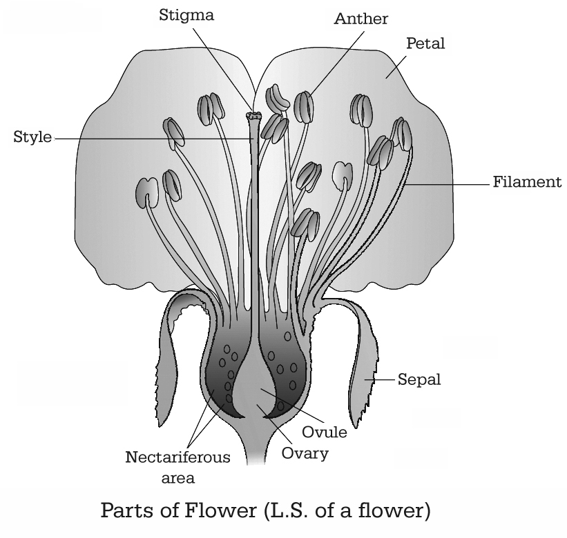
Flowers are objects of aesthetic, ornamental, social, religious and cultural value. To a biologist, a flower is a modified condensed shoot to perform sexual reproduction in angiosperms.
A typical angiospermic flower consists of four whorls of floral appendages attached on the receptacle - Calyx, Corolla, Androecium and Gynoecium.
Pre fertilisation: Structure and Events
- Books Name
- A TEXT OF BIOLOGY - CLASS XII
- Publication
- ACME SMART PUBLICATION
- Course
- CBSE Class 12
- Subject
- Biology
PRE-FERTILIZATION : Structures and Events
Much before the actual flower is seen on a plant, the decision that the plant is going to flower has taken place. A number of hormonal and structural transformations occur prior to initiation of flowering.
Shoot apical meristem is transformed into reproductive meristem.
Reproductive meristem grows to form inflorescence axis over which floral primordia develop.
The primordia grow into floral buds and then flowers. In the flower, the androecium and gynoecium differentiate and develop.
Stamen, Microsporangium and Pollen grain
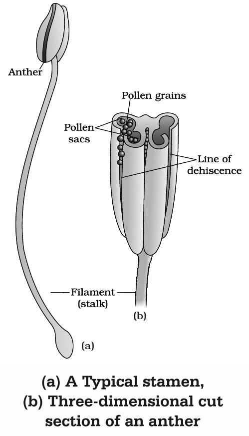
(a) Stamen or Microsporophyll (Male sex organ)
It consists of two parts :
(i) Anther
It is broader knob-like fertile part.
A typical angiospermic anther has two lobes and such anther is called dithecous.
Dithecous anther is a four sided (tetragonal) structure consisting of four microsporangia located at the corners, two in each lobe.
(ii) Filament
It is sterile, long and slender stalk.
The proximal end of the filament is attached to the thalamus, petal or tepal.
Concept Builder
(i) Floriculture is the science of cultivation, breeding, marketing and arrangement of flowers.
(ii) In members of Malvaceae, anther consists of one lobe and two microsporangia. Such anthers are monothecous and bisporangiate.
(iii) In Arceuthobium, the smallest dicot parasite, anther consists of one microsporangium, i.e., monothecous and monosporangiate
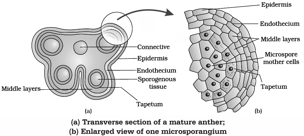
(b) Structure (T.S.) of Anther
A young anther consists of homogenous mass of meristematic cells called primary sporogenous cells surrounded by anther wall. Primary sporogenous cells form microspore mother cells (2n) inside the microsporangium.
Anther Wall Layers : Anther wall consists of following layers :
(1) Epidermis : Outermost single layered and protective in function. The epidermis of Arceuthobium develops some fibrous thickenings and is called exothecium.
(2) Endothecium : Cells of this layer have -cellulosic fibrous bands arising from inner tangential wall which help in dehiscence of anther due to their hygroscopic nature. Fibrous bands are absent in hydrophytes, e.g., Hydrocharitaceae.
(3) Middle layer : Cells of this layer are ephemeral and are 1-3 layered. It degenerates at maturity.
(4) Tapetum : This is the innermost layer of anther wall which surrounds the sporogenous tissue. Tapetal cells nourishes the developing pollen grains. Cells of the tapetum possess dense cytoplasm and generally have more than one nucleus. They are polyploid. The tapetal cells show increase in their DNA content.
Concept Builder
Increase in DNA content of tapetum may be 'achieved by the following ways:
(i) Endomitosis : It involves DNA replication and splitting of chromosomes through endoprophase, endometaphase, endoanaphase and endotelophase.
(ii) Formation of restitution nuclei : It involves normal mitosis upto anaphase but the chromosomes at two poles get surrounded by a common nuclear membrane so as to form a restitution nucleus.
(iii) Polyteny : If DNA replication is not accompanied by splitting of chromosomes, polytenic chromosomes are formed.
The tapetum is of two types :
(a) Secretory or glandular tapetum : These cells secrete sporopollenin, pollenkitt and compatibility proteins. These cells provide Ubisch bodies which help in the ornamentation of exine, as they have a chemical called sporopollenin which is deposited on them.
(b) Amoeboid or plasmodial or invasive tapetum : Cells undergo breakdown and their entire protoplasts move in the centre to nourish microspores.
(c) Microsporogenesis
The process of formation of microspores from a pollen mother cell (PMC) or microspore mother cell (MMC) through meiosis is called microsporogenesis. As each cell of the sporogenous tissue is capable of giving rise to a microspore tetrad, therefore each one is a potential pollen mother cell.
Cytokinesis, after the meiotic divisions in PMCs, is of two types:
(i) Successive : In this type, cytokinesis occurs after each meiotic division, thus isobilateral tetrad of microspores is formed, e.g., monocots. Successive type of cytokinesis is advanced type.
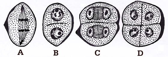
during microsporogenesis, (A-B) Dividing mother cell, (C) Dividing dyad, (D) Tetrad
(ii) Simultaneous : It occurs after complete meiotic (I and II) division, thus tetrahedral tetrad of microspores is formed, e.g., dicots.
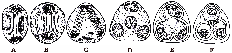
The microspores, as they are formed, are arranged in a cluster of four cells, called microspore tetrad. Usually the arrangement of microspores in a tetrad is tetrahedral or isobilateral.
However, T-shaped, linear and decussate tetrads are also found. In Aristolochia elagans, all the five types of tetrads are present.
A. Tetrahedral, B. Isobilateral, C. T-shaped, D. Linear, E. Decussate
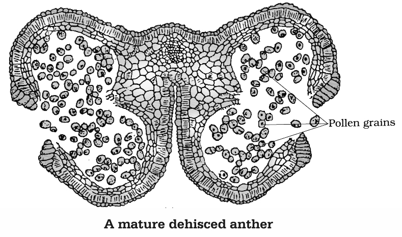
As the anthers mature and dehydrate, the microspores dissociate from each other and develop into pollen grains. These are released with the dehiscence of anther.
Concept Builder
(i) R. Camerarius described sexual reproduction for the first time in plants.
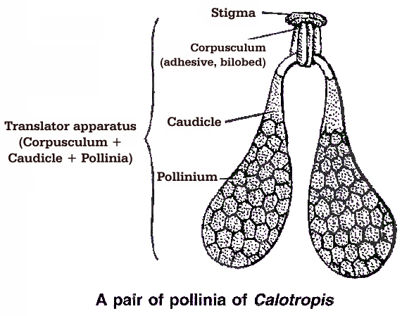
(ii) 'In family Asclepiadaceae (Calotropis) and Orchidaceae, all the microspores in a sporangium adhere together in a single mass known as pollinium.
(iii) In Calotropis, the pollinia of adjacent anthers of different stamens are attached by thread like caudicles (retinaculi) to a sticky disc called corpusculum. The whole structure is called translator.
(iv) After their formation, the microspores are separated from tetrad, but in Elodea, Drosera, Typha, the microspores do not separate from each other, thus developing into compound pollen grains.
(v) In family Cyperaceae, out of 4 microspores formed, 3 degenerate, so ultimately one MMC (2n) produces only one microspore or pollen grain.
(vi) Sometimes more than four microspores are produced from one microspore mother cell. It is called polyspory, e.g., Cuscuta reflexa.
(d) Pollen Grain
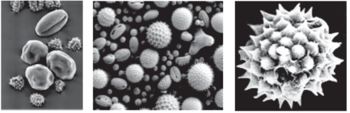
(i) These are generally spherical measuring about 25-50 µm in diameter. The cell wall of pollen grain is called sporoderm which consists of two layers. The hard outer layer called the exine and inner thin layer is called the intine.
(ii) Intine : It is made of cellulose and pectin.
(iii) Exine : The exine is made up of sporopollenin is a fatty substance and one of the most resistant organic material known. It is not affected by high temperature, strong acids or alkali. No enzyme is known to degrade it. Because of the presence of sporopollenin, pollen grains of the past plants are well preserved as fossils. Exine is made of two layers :
(a) Ektexine : It is highly sculptured and is differentiated into outer tectum, middle baculum and innermost foot layer. Tectum provides a characteristic sculpturing or designs over the surface of pollen grain. The design helps the experts to identify the pollen grain as to its class, family, genus or species.
(b) Endexine: It is not sculptured.
(iv) Pollen grain exine has prominent apertures called germ pores where sporopollenin is absent. Pollen grains can be monocolpate (having one germ pore called germinal furrow, e.g., monocots), bicolpate (2 germ pores) and tricolpate (3 germ pores, e.g., dicots).
(v) The pollen kitt is a sticky layer found on the outer side of exine of mature pollen grains of many insect pollinated species, it is made of carotenoids and lipids. Pollenkitt material is contributed by the tapetal cells. Pollen kitt acts as an insect attractant and can help against UV.
(vi) Pollen grains of many species (especially anemophilous plants) cause severe allergies and bronchial affictions in some people. Weed Parthenium hysterophorus (carrot grass) came to India as a contaminant with improved wheat. The weed has become a major cause of pollen allergy. Hay fever is an allergic reaction due to the presence of pollen in the air. Plants commonly causing hay fever are Amaranthus, Chenopodium and Parthenium.
(vii) Pollen grains are rich in nutrients. They are taken as tablets and syrups to improve health. Pollen consumption has been claimed to enhance the performance of athletes and race horses.
(viii) The period for which pollen grains retain the ability to germinate is called Pollen Viability. It is highly variable and to some extent depends upon the environmental factors like temperature, humidity.
In cereals, like rice, wheat etc, pollen viability is minimum upto 30 minutes, while in Rosaceae, Leguminosae and Solanaceae it is upto several months.
(ix) Pollen grains can be cryopreserved in liquid nitrogen (-196°C) and used as pollen banks.
Concept Builder
(i) The study of pollen grain is called Palynology. (Term given by Hyde and Williams).
(ii) In Hyacinthus, Nemec observed 8-nucleated embryo sac type organisation in pollen grain. This is called Nemec phenomenon.
(iii) Smallest pollen grain: Myosotis (2.5 -3.5 µ).
(iv) Largest pollen grain : Mirabilis (250 µ).
(v) Longest or Filamentous pollen grain : Zostera (2500 µ).
(e) Development of Male Gametophyte
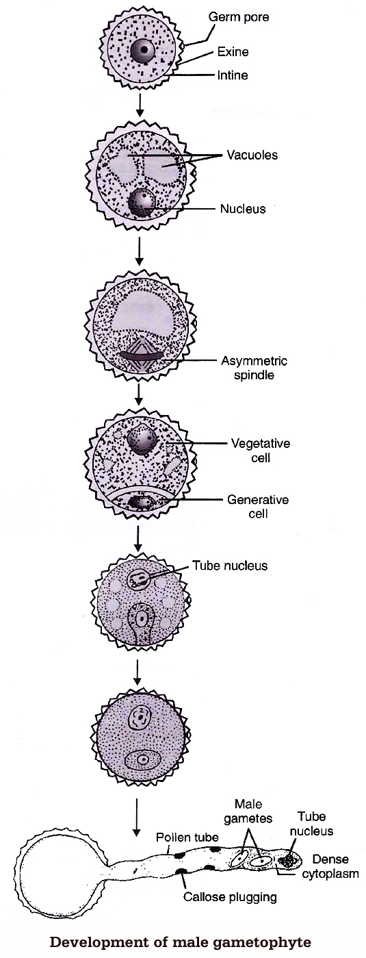
The microspore is the first cell of male gametophyte. The germination of microspore starts in situ (in the mother place). Microspore may be best defined as partially developed male gametophyte.
Microspores divide mitotically into large tube cell and small generative cell Pollination takes place at two celled stage in 60% of angiosperms (in some cases at three-celled stage). The further development of these male gametophyte takes place on the stigma.
Pollen grain expands by absorbing the liquid from the moist surface of stigma. Stigma provides, boron, sugar, amino acids etc. The intine comes out in the form of pollen tube, from germ pores.
The pollen grains are either monosiphonous (with one pollen tube) or polysiphonous (with more than one pollen tubes), e.g., members of Cucurbitaceae and Malvaceae.
The generative nucleus divides mitotically to form two male gametes called sperm. The male gametes are non-motile and amoeboid. They are slightly unequal in size, such a pollen will be called three celled pollen or mature male gametophyte.
The function of pollen tube is to carry aflagellated sperm. Tube nucleus enters first in the pollen tube and is a vestigial structure and soon disintegrates. Growth of the pollen tube is chemotropic apicle and entire cytoplasm of pollen grain is confined to tip of the pollen tube.
Concept Builder
(i) The pollen tube was first observed by G.B. Amici (1824) in Portulaca.
(ii) Longest pollen tube occurs in Zea mays.
(iii) B-Ca-inositol sugar complex act as chemotropic agent for pollen tube growth.
The Pistil, Ovule and Embryo sac
(a) Pistil (Female Sex Organ)
The gynoecium may consist of a single pistil (monocarpellary) or may have more than one pistil (multicarpellary).
If there are more than one, the Pistils may be fused together (Syncarpous) or may be free (apocarpous).
There are three parts of each pistil -the stigma, style and ovary. The stigma is upper broader region which is specialised for receiving pollen grains.
The style is long stalk-like structure and ovary is basal swollen ovule containing region.
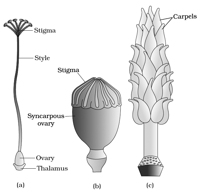
(c) A multicarpellary, apocarpous gynoecium of Michelia
(b) Ovule (Integumented Megasporangium)
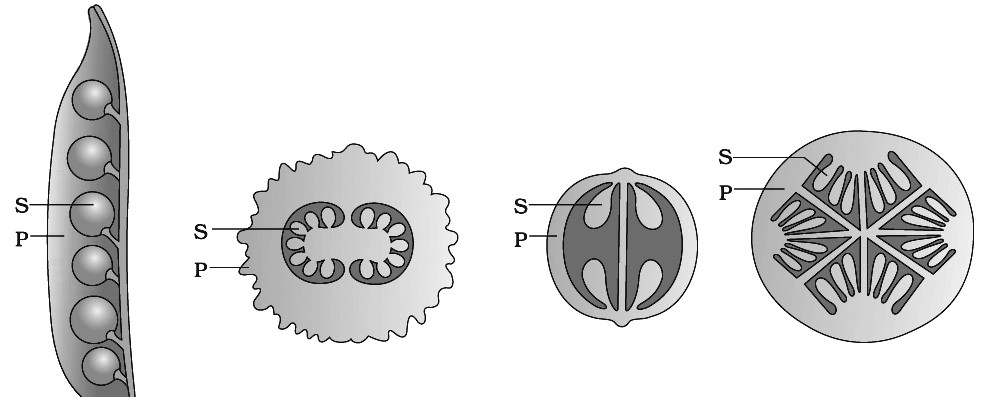
(i) Ovary has an ovarian cavity with one or more chambers (lobules). The placenta is located inside the ovarian cavity. One or many ovules are present inside the ovary.
Plants with one ovule in an ovary: (i) Wheat, (ii) Paddy, (iii) Mango
Plants with many ovules in an ovary: (i) Papaya, (ii) Water melon, (iii) Orchids
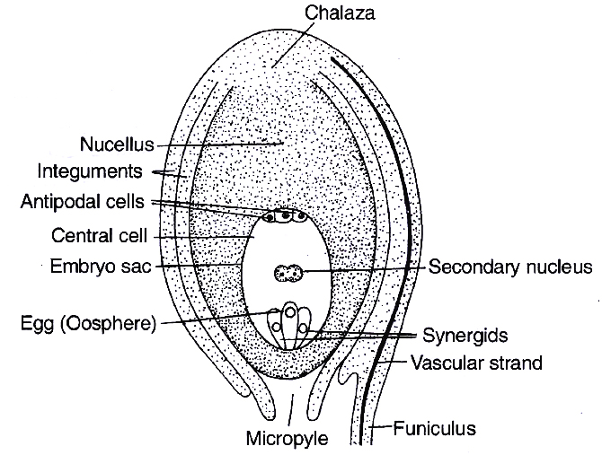
(ii) The ovule is a small structure attached to the placenta by means of a stalk called funicle. The point of attachment of the funicle with the main body of the ovule is called hilum. Thus, hilum represents the junction between ovule and funicle. Sometimes funicle gets fused with the body of the ovule along one side and forms a ridge known as raphe. The basal region of the ovule is known as Chalaza.
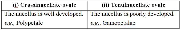
(iii) Main body of an ovule is called nucellus (megasporangium) which consists of a mass of parenchymatous tissue. Cells of nucellus have abundant reserve food materials. Depending upon the development of nucellus, ovules are of two types:
containing developed embryo sac (female gametophyte)
(iv) The nucellus is invested all around by one or two layered protective covering called integuments, except apex where a small passage is formed known as micropyle. On the basis of number of integuments, ovules are of following types:
(a) Unitegmic: Ovules with one integument, e.g., members of gamopetalae and gymnosperms.
(b) Bitegmic : Ovules with two integuments, e.g., members of polypetalae and monocots.
(c) Ategmic : Ovules are without integument, e.g., Santalum, Loranthus (Parasites) and Liriosoma.
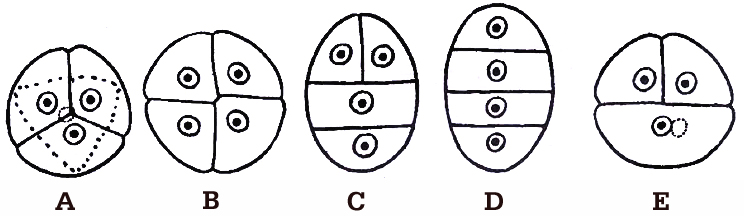
(v) On the basis of the relative position of micropyle, body of the ovule and funicle, i.e., the degree of curvature, there are six types of ovules:
(a) Orthotropous : The micropyle, chalaza and funicle are in a straight line. This is the most primitive type of ovule, e.g., Piper, Polygonum, Cycas.
(b) Anatropous : The ovule turns 180º angle. Thus it is inverted ovule. Micropyle lies close to hilum or at side of hilum, e.g., found in 82% of angiosperm families.
(c) Hemianatropous: Ovule turns at 90º angle upon the funicle or body of ovule is at right angle to the funicle, e.g., Ranunculus.
(d) Campylotropous: Ovule is curved more or less at right angle to funicle. Micropylar end is bend down slightly, e.g., in members of Leguminosae, Cruciferae.
(e) Amphitropous: Ovule as well as embryo sac is curved like horse shoe, e.g., Lemna, Poppy, Alisma.
(f) Circinotropous: The ovule turns at more than 360º angle, so funicle becomes coiled around the ovule, e.g., Opuntia (Cactaceae), Plumbaginaceae.
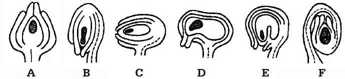
A. Orthotropous, B. Anatropous, C. Hemianatropous,
D. Campylotropous, E. Amphitropous, F. Circinotropous
Concept Builder
(i) Third integument in the form of aril develops from base of ovule or funicle in many plants, e.g., Litchi, Asphodelus and Inga dulce. In litchi and Inga dulce, aril is fleshy and edible.
(ii) In some ovules, e.g., Zostera, a group of thickened cells is found in the chalazal region just below the embryo sac, called hypostase. When a similar group of specialized cells is found in the nucellar region just above the embryo sac, it is called epistase.
(iii) In castor (Ricinus), proliferation of the outer integumentary cells at micropylar region is called caruncle or strophiole. It performs two functions :
a. It acts as water absorbing pad.
b. It is made up of sugary substance that attract and helps in the seed dispersal by ants (myrmecochory).
(iv) The placental or funicular outgrowth present at, the micropylar end is called obturator. It directs the passage of pollen tube towards the ovule.
(c) Megasporogenesis
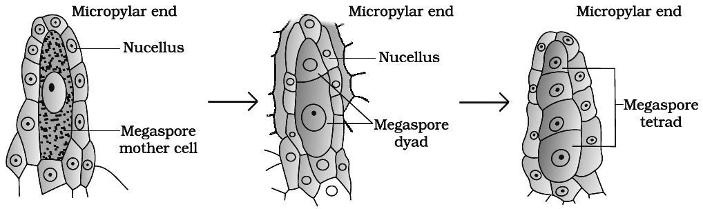
(i) Any of the cells of nucellus hypodermis towards the micropylar end gets differentiated from the other cells. This cell is called archesporial cell. In crassinucellate ovules, it undergoes periclinal division to form an outer primary parietal cell and inner primary sporogenous cell. The later behaves as megaspore mother cell (MMC). The archesporial cell directly behaves as megaspore mother cell in tenuinucellate ovules.
(ii) The megaspore mother cell is large sized containing dense cytoplasm and a prominent nucleus. Some carbohydrate storing bodies appear between cell wall and plasma membrane of megaspore mother cell in Lilium, called paramular bodies.
(iii) The MMC (2n) undergoes meiosis and forms a linear tetrad of 4 haploid megaspores. The process of formation of megaspores from the MMC is called megasporogenesis.
(d) Female Gametophyte or Embryo sac
(i) P. Maheshwari classified the embryo sac on the basis of number of megaspore nuclei participating in embryo sac formation into following types :
Monosporic embryo sac : Only one megaspore nucleus forms embryo sac, e.g., Polygonum, Oenothera.
Bisporic embryo sac: Two megaspore nuclei take part in development of embryo sac, e.g., Allium, Endymion.
Tetrasporic embryo sac: All the four megaspore nuclei take part in development of embryo sac, e.g., Adoxa, Plumbago, Drusa, Fritillaria, Paenaea, Plumbagella, Peperomia.
(ii) Development of Monosporic Embryo sac (Polygonum type)
(A) In majority of angiosperms, one of the megaspore is functional while the other three degenrate. Only the functional megaspore (n) develops into the female gametophyte. This process of embryo sac formation from a single megaspore is termed monosporic development.
(B) Polygonum type of embryo sac is found in 80% flowering plants. This development has been studied in (Polygonum by Strasburger). The nucleus of chalazal functional megaspore (4th from micropyle) divides by three mitotic divisions to form 8 nuclei, four towards each pole. One nucleus from each pole moves to the middle and they form polar nuclei. These mitotic divisions are strictly free nuclear, i.e., nuclear divisions are not followed immediately by cell wall formation.
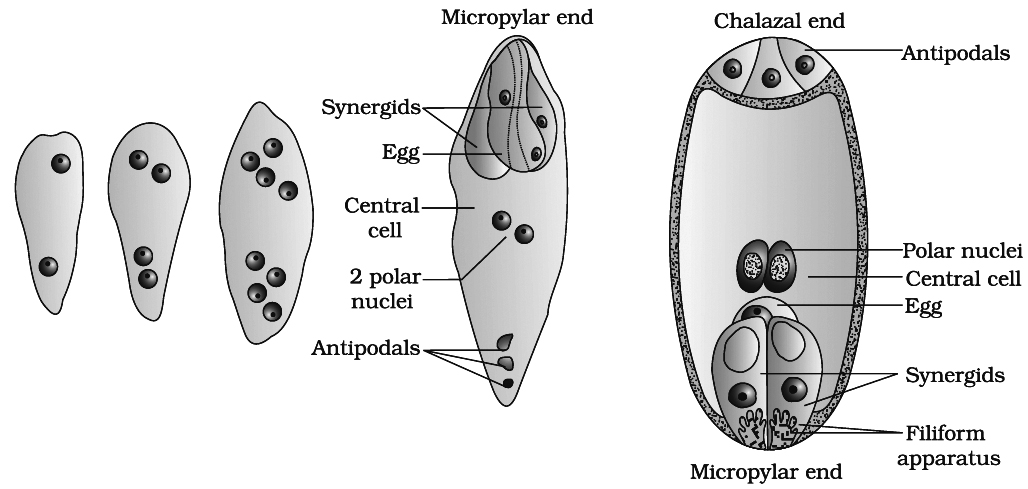
At this stage, following changes occur:
(a) Three of the nuclei (n) get organised as cells at micropylar end forming egg apparatus. One is the egg cell (n) and two are synergids (n).
(b) Three nuclei get organised as antipodal cells (n) at chalazal end
(c) Two nuclei in the centre are called polar nuclei (n)
This constitutes a 7-celled and 8-nucleated embryo sac.
Organization of Embryo Sac
(a) Synergids or helper cells or co-operative cells: These cells generally possess a micropylar nucleus and a chalazal vacuole. The electron microscopic studies have revealed that the synergids lack a cell wall on their chalazal side at maturity. They are characterised by the presence of a 'filiform apparatus at the' micropylar tip. It is in the form of finger like projections, each projection comprising a core of microfibrils enclosed in a sheath. Usually one synergid starts to degenerate just with pollination. The synergids perhaps secrete some chemotropic substance and thus, direct the pollen tube growth inside embryo sac.
(b) Egg : The egg shows cytoplasmic polarity opposite to synergid and its wall is thicker at the micropylar end. Usually the egg has a micropylar vacuole and a chalazal nucleus. Plasmodesmata connection is present in between egg and synergids.
(c) Antipodals or vegetative cells : These are vegetative cells of embryo sac. In most of the plants there are three antipodal cells.
(d) Central Cell: It is the largest cell of the embryo sac. It initially contains two polar nuclei which fuse just before, fertilization to form a secondary nucleus or definitive nucleus (2n).
Concept Builder
(i) Synergids are absent in Plumbago and Plumbagella. Number of synergid is one in Peperomia.
(ii) In Zea mays 20 antipodal cells are present, in Sasa paniculata they are 300 in number. This is perhaps the highest number recorded for any plant. These are absent in Oenothera type.
pollination
- Books Name
- A TEXT OF BIOLOGY - CLASS XII
- Publication
- ACME SMART PUBLICATION
- Course
- CBSE Class 12
- Subject
- Biology
POLLINATION
In flowering plants, male and female gametes are produced in the pollen grain and embryo sac respectively. These gametes are non-motile, therefore, they have to be brought together for fertilisation. Pollination is the mechanism to achieve this objective. The transfer of pollen grains to the stigma is called pollination. Depending upon the source of pollen, pollination is of three types :
(1) Autogamy :
If the pollen grains are transferred from anther to the stigma of the same flower, the process is called self pollination or autogamy.
Contrivances for autogamy
(i) Bisexuality - Flower should be bisexual, e.g., Catharanthus
(ii) Homogamy - Male and female reproductive parts in a bisexual flower mature at the same time, e.g., Mirabilis.
(iii) Cleistogamy - Sometimes bisexual flowers remain closed and never open, such flowers are known as cleistogamous. In such flowers, the anthers and stigma lie close to each other. When the anthers dehisce, the pollen grains come in contact with stigma and pollination takes place. Thus, cleistogamous flowers are invariably autogamous as there is no chance of cross-pollen landing on the stigma. So one of the advantage of cleistogamy is, it ensures seed-set even in the absence of pollinators, e.g., Arachis hypogea (Groundnut). Some plants like Viola (common pansy), Oxalis and Commelina produce chasmogamous flowers (open flowers) as well as cleistogamous flowers.
(iv) Bud pollination -When self pollination occurs in the bud stage before the opening of flowers, e.g., Pisum, Wheat, Rice.
(2) Geitonogamy:
Pollination taking place between the two flowers of the same plant (genetically self pollination but ecologically or functionally cross pollination), e.g., Maize.
(3) Xenogamy :
When the pollen grains are transferred from the anther to the stigma of the flower of a different plant, it is called as xenogamy. (It is cross pollination, both genetically and ecologically).
Outbreeding Devices (Contrivances for cross pollination)
Majority of flowering plants produce hermaphrodite flowers and pollen grains are likely to come in contact with the stigma of the same flower. Continued self-pollination results in inbreeding depression, therefore angiosperms have developed following devices to, discourage self pollination and to encourage cross-pollination.
(i) Dichogamy: In bisexual flowers, the two sexes mature at different timings. When anthers mature first it is called protandry, e.g., Sunflower, Cotton, Salvia. When gynoecium matures first it is called as protogyny, e.g., Ficus, Aristolochia, Magnolia.
(ii) Incompatibility: It is the inability of certain gametes, even from genetically similar plant species, to fuse with each other. This is also called intra-specific incompatibility, self-sterility or self-incompatibility.
|
Concept Builder
|

Incompatibility may involve morphological or physiological mechanisms, therefore there are two types of this device :
(a) Morphological self-incompatibility : It occurs in flowers having heterostyly. Flowers are dimorphic or trimorphic with regard to the length of style. Thus facilitate cross pollination.
e.g., Primula (Primrose), Jasminum, Lythrum

(b) Physiological self-incompatibility :
It is controlled by multiple alleles of S-gene. There are two types of physiological self-incompatibility :
(i) If incompatibility is due to the genotype of the sporophytic/stigmatic tissues, is termed sporophytic incompatibility (SSI), e.g., Brassicaceae, Asteraceae. (ii) The incompatibility due to the genotype of the pollen is termed gametophytic incompatibility (GSI), e.g., Solanaceae, Liliaceae, Poaceae. This may be due to prevention of pollen germination, deorientation of pollen tube, or even failure of nuclear fusion. A plant carries two such alleles e.g., S1S2, S2S3, S1S3, S2S4, S3S5. A pollen carries only one allele. If it happens to be one of the two alleles of pistil, the rejection may occur on stigma surface (SSI) or in the style (GSI).
(iii) Unisexuality or dicliny : It is the formation of unisexual flowers. Thus, cross pollination becomes obligatory. It can be seen in monoecious plants where both male and female flowers are produced on same plant, e.g., maize, castor as well as dioecious plants where male and female flowers are produced on different plants, e.g., Vallisneria, Papaya. Monoecious condition prevents autogamy but not geitonogamy while dioecious condition prevents both autogamy and geitonogamy.
(iv) Herkogamy : It is the presence of natural and physical barrier between androecium and gynoecium, e.g., in Calotropis, gynoecium is fused with pollinium (anthers) and form gynostegium.
(v) Prepotency : Pollen grain of one flower germinates more rapidly over stigma of another flower as compared to its own flower, e.g., Apple, Grape.
Agencies for cross pollination :
Pollinating agency may be biotic (animals) or abiotic (wind and water). Majority of plants use biotic agents for pollination.
A. Abiotic agents
(a) Anemophily: (Pollination by wind).
 It is common amongst abiotic pollinations. It is a non-directional and wasteful process. The female flowers have large, feathery or brush like stigmas to catch the pollen grains. Anemophilous flowers are small and inconspicuous with long and versatile stamens. Pollen grain are dry, powdery, light and non-sticky. The flowers often have a single ovule in each ovary and numerous flowers packed into an inflorescence. Anemophily is quite common in grasses.
e.g., Maize, wheat, sugarcane, bamboo, Pinus (winged pollen), Papaya.
(b) Hydrophily : (Pollination by water).
It is quite rare in angiosperms and is limited to about 30 genera, mostly monocotyledons. All aquatic plants are not hydrophilous. In a majority of aquatic plants like water hyacinth and water lily, the flowers emerge above the level of water and are pollinated by insects or wind.
|
Hydrophyte |
Type of pollination |
|
1. Alisma |
Entomophily |
|
2. Lotus |
Entomophily |
|
3. Potamogeton |
Anemophily |
|
4. Myriophyllum |
Anemophily |
Hydrophily is of two types:
(1) Epihydrophily : Pollination at the surface of water
Example : Vallisneria (Tape grass, Ribbon weed)
Vallisneria is a dioecious rooted submerged aquatic plant in which male flowers are small and light weight. Female flowers have very long coiled pedicels which uncoil when they become mature. Male flowers float at the surface of water. As soon as the male flowers touch the female flowers, anther lobes burst, stigma receives the pollen grains and pedicels coil again.

(2) Hypohydrophily : Pollination inside the water.
Examples: Zostera (sea grass), Ceratophyllum
Zostera is a marine water plant. Female flowers remain submerged in water and the pollen grains are long, ribbon like and they are carried passively inside the water, some of them reach the stigma and achieve pollination.
(i) Pollen grains are protected from wetting by a mucilaginous covering in most of the hydrophilous species.
(ii) Anemophilous as well as hydrophilous flowers are not very colourful and do not produce nectar.
(iii) Pollen grains coming in contact with the stigma is a chance factor in both wind and water pollinating plants. To compensate for these uncertainities and associated loss of pollen grains, the flowers produce enormous amount of pollen when compared to the number of ovules available for pollination.
B. Biotic Agents
(a) Entomophily: (Pollination by insects)
(1) 80% pollination occurs by insects (chief pollinators) and honey bee is main pollinator among insects. All the flowers pollinated by bees are brightly coloured, have a sweet smell and produce nectar. Entomophilous flowers produce a small amount of pollen which have a spinous and may have a sticky exine due to presence of pollenkitt. The stigmas of such flowers are long, rough and sticky. If the entomophilous flowers are small, a number of flowers are clustered into an inflorescence to make them conspicuous. Moth pollinated plants are white flowered and fragrant. The flowers pollinated by flies and beetles secrete foul odours to attract these animals.
(2) When petals are not conspicuous, other parts may become coloured or showy to attract the insects, e.g., bracts in Bougainvillea, leaves in Euphorbia pulcherrima (Poinsettia), one sepal in Mussaenda acts like advertisement flag, anthers are coloured in Mimosa.
(3) To sustain insect visits, the flowers have to provide rewards to the insects. Nectar and pollen grains are the usual floral rewards. In plants like Papaver, Rosa, Clematis, etc., edible pollen grains are produced. Some of the pollen grains stick to the back of insects while feeding on the edible pollen grains. Some species provide safe place to insect for laying eggs, e.g., Amorphophallus (the flower is 6 feet in height), Yucca.
(4) Some special cases of entomophily are as follows:
(i) Yucca : The flowers at Yucca and pollinating moth, i.e., Pronuba yuccasella / Tegaticula show a very close relationship. The insect punctures the ovary for egg laying. The insect creeps on the style and deposits pollen ball in between the stigmatic lobes, hence bringing about pollination. The larvae of the moth come out of the eggs as the seeds start developing.
(ii) Salvia or Sage plant : It shows lever mechanism or turn pipe mechanism. Anthers are distractile; lower lobe is sterile and upper lobe is fertile. The flower is bilabiate and protandrous. The insect sits on the lower lobe of the corolla; the upper fertile lobe of anther touches the body of insect. It ruptures and pollen grains are shed on the back of the insect. When this insect visits the other flowers, pollination is affected.

(iii) Ficus sp. : Trap Door mechanism in Ficus carica. The receptacle forms a cup having a cavity. The cavity has a pore called ostlole near which male flowers are present. Female flowers are present at the bottom and gall (sterile) flowers are present between these types. The pollinating insect is Blastophaga or gall insect. The insect lays eggs in the bottom, the larvae feed on the ovules of gall flowers. When the young insects crawl out of the inflorescence, their bodies are laden with pollen grains. They enter new hypanthodium and affects the pollination.

(iv) Calotropis : It shows translator or clip mechanism. Pollen grains are present in pollinia. The pollinia are attached to a rough and sticky disc called corpusculum. When the insect visits the flower, pollinia get entangled in the legs of the insect. When this insect visits other flowers, pollinia are transferred.

(v) Centaurea : It belongs to Compositae family and exhibits piston mechanism. It has syngenesious and epipetalous androecium. The anther lobes dehisce introsely. When filaments contract on being touched by the insect, the stigma pushes the pollen grains out of the anther tube which stick to the body of the insect. When the insect visits some other flower, pollination is affected.
(vi) Aristolochia : The upwardly directed protogynous flowers have downwardly directed stiff hairs in the middle of the corolla tube. These form the trap. This is called pit fall mechanism or fly trap mechanism.

(vii) Orchid : Pollination occurs by wasp (Pseudocopulation mechanism), e.g. , Ophrys (orchid) and Colpa (wasp). The flower of Ophrys resemble in shape, colour and odour to female of wasp Colpa aurea, thus showing mimicry. It is a case of co-evolution also.
(5) Many insects may consume pollens or the nectar without bringing about pollination. Such floral visitors are referred to as pollen/nectar robbers.
Pollen robbers create a hole at the base of corolla tube and draw nectar from a flower whose design is not suitable for them. Other insects often take advantage of the same.
(b) Ornithophily: (Pollination by birds).
Flowers are brightly coloured but odourless and produce plenty of nectar and large quantity of pollens, e.g., Bombax (red silk cotton), Callistemon (bottle brush), Sterlitzia, Erythrina (coral tree) etc. Honey bird, humming bird and sun bird are common pollinators.
(c) Cheiropterophily: (Pollination by bats).
Bats pollinate the flowers of tropical regions, e.g., Anthocephallus (Kadamb), Kigelia (Sausage tree), Adansonia (Baobab tree).
(d) Malacophily: (Pollination by snails), e.g., Arum; Lilies, Arisaema, Lemna.
(e) Ophiophily: (Pollination by snakes), e.g., Santalum, Michelia.
(f) Some larger animals have been reported as pollinators in some species, e.g., Lemur (Primate) in Ravenela plant, Lizard in Flax
|
Self Assessment
|

Q.21 Polygonum type of embryo sac is
(1) 8-celled (2) 15-nucleated (3) Haploid (4) Exosporic type
Q.22 Mark the odd option (w.r.t. contrivances of autogamy)
(1) Homogamy (2) Cleistogamy (3) Dicliny (4) Bud pollination
Q.23 Which of the following pollination is common amongst abiotic agents?
(1) Hydrophily (2) Entomophily (3) Ornithophily (4) Anemophily
Q.24 Epihydrophily is found in
(1) Tape grass (2) Sea grass (3) Lotus (4) Alisma
Q.25 Mark the incorrect match (w.r.t. pollination)
(1) Salvia Turn pipe mechanism (2) Ficus Fly trap mechanism
(3) Aristolochia Pit fall mechanism (4) Calotropis Clip mechanism
Q.26 Which of the following plant provide safe place to insect for laying eggs?
(1) Sage plant (2) Ophrys (3) Centaurea (4) Amorphophallus
Q.27 Pollination occurs by pseudocopulation mechanism in
(1) Ophrys (2) Fig (3) Mango (4) Zea mays
Q.28 Lemur, a large animal, acts as pollinator in
(1) Flax (2) Ravenela (3) Capsella (4) Hydrilla
Q.29 Which of the following type of pollination is present in Santalum?
(1) Ornithophily (2) Ophiophily (3) Malacophily (4) Entomophily
Q.30 In ornithophily, flowers are
(1) Dull coloured (2) Nectarless (3) Odourless (4) Very small
Ans. Q.21 (3), Q.22 (3), Q.23 (4), Q.24 (1), Q.25 (2), Q.26 (4), Q.27 (1), Q.28 (2), Q.29 (2),
Q.30 (3)
POLLEN-PISTIL INTERACTION
Pollination does not guarantee the transfer of the right type of pollen. Often, pollen of the wrong type, either from other species or from the same plant (if it is self-incompatible), also land on the stigma.
The pistil has the ability to recognise the pollen, whether it is of the right type (compatible) or the wrong type (incompatible). The ability of the pistil to recognise the pollen followed by its acceptance or rejection is the result of a continuous dialogue between pollen and the pistil.
This dialogue is mediated by chemical components of the pollen interacting with those of the pistil. If the reaction is favourable, the pollen grain germinates on the stigma to produce pollen-tube through one of the germ pores.
Plants in which the pollen grain are shed at 2-celled stage, the generative cell divides and forms the 2 male gametes during the growth of pollen tube in the stigma. If pollen grains are shed at 3-celled stage, pollen tubes carry the 2-male gametes from the beginning.
(1) Entry of the pollen tube into an ovule
(A) Pollen tube, after reaching the ovary, enters the ovule through the micropyle. It is called as porogamy as seen in most of the flowering plants.
(B) In some plants, e.g., Casuarina, the pollen tube enters an ovule through chalaza and it is called as chalazogamy.

(a) Pollen grains germinating on the stigma; (b) Pollen tubes growing through the
style; (c) L.S. of pistil showing path of pollen tube growth
(C) Some times the pollen tube enters into an ovule through integuments or funicle. It is called as mesogamy. e.g,. Cucurbita.
(2) Entry of the pollen tube into the embryo sac :
Irrespective of the place of entry of pollen tube into the ovule, the tube invariably enters the embryo sac at micropylar end, i.e., degenerating synergid cell. Entry of pollen tube in the embryo sac is under chemotropic guidance (synergids have filiform apparatus and secrete some chemicals).

(A) enlarged view of an egg apparatus showing entry of pollen tube into a synergid;
(B) Discharge of malegametes into a synergid and the movements of the sperms,
one into the egg and the other into the central cell
(3) All these events from pollen deposition on the stigma until pollen tubes enter the ovule are together referred to as pollen-pistil interaction. This interaction is a dynamic process.
(4) Pollen germination can be studied by dusting pollen (e.g., pea, chick pea, Grotalaria, balsam, Vinca) on a glass slide containing a drop of 10% sugar solution with boric acid, Ca, Mg and K salts. After 15-30 minutes, pollen tubes will' be observed to come out of the pollen grains. So, this germination of pollen grain in laboratory is called hanging drop method.
(5) Artificial hybridisation : It is one of the major approaches of crop improvement in which crosses are made between different varieties, species and genera in order to combine their desirable characters in a single superior variety. Emasculation and bagging are two precautionary measures in this hybridisation. Emasculation is the removal of anthers from the floral buds of female parent bearing bisexual flowers. Bagging is the covering of both emasculated as well as non-emasculated flowers with butter paper or polythene to prevent contamination of its stigma with unwanted pollen. When the stigma of bagged flower attains receptivity, mature pollen grains collected from anthers of the male parent are dusted on the stigma and the flowers are rebagged, and the fruits allowed to develop.
double fertilization
- Books Name
- A TEXT OF BIOLOGY - CLASS XII
- Publication
- ACME SMART PUBLICATION
- Course
- CBSE Class 12
- Subject
- Biology
DOUBLE FERTILIZATION
Pollen tube enters the degenerating synergid and bursts, thereby releasing two male gametes. One
male gamete fuses with the egg to form diploid zygote. This fusion is called syngamy or
generative fertilization.

The other male gamete fuses with secondary nucleus (2n, i.e., fused polar nuclei of both polar
nuclei) to form primary endosperm nucleus (3n). This fusion is also called triple fusion because
three nuclei takes part in this fusion. It is also known as vegetative fertilization or
pseudofertilization or trophomixis.

Since two types of fusions, i.e., syngamy and triple fusion take place in an embryo sac the
phenomenon is termed double fertilisation. This event occurs in flowering plants only. Five nuclei
are involved in double fertilization.
Concept Builder
(i) The syngamy was discovered by Strasburger.
(ii) Triple fusion and double fertilization was discovered by S.G. Nawaschin and Guignard in Lilium and Fritillaria.
(iii) Entry of more than one pollen tubes in an ovule leading to the occurrence of supernumerary male nuclei (polyspermy).
(iv) When two pollen tubes enter an ovule and release their contents, it is possible that the egg may be fertilized by male gamete from one tube and triple fusion may involve participation of male gamete from another tube. This phenomenon is called as heterofertilization, e.g., Zea mays.
(v) When the entry of male gamete is not accompanied by fusion,the phenomenon is called as semigamy.
(vi) X-bodies are darkly stained DNA containing bodies found in synergid receiving pollen tube.
(vii) The percentage of pollen germination and tube growth is better in larger populations. This is referred to as "population effect" or "crowding effect".
post fertilization
- Books Name
- A TEXT OF BIOLOGY - CLASS XII
- Publication
- ACME SMART PUBLICATION
- Course
- CBSE Class 12
- Subject
- Biology
POST-FERTILIZATION: Structures and Events
Development of endosperm and embryo, maturation of ovules into seeds and ovary into fruit are collectively termed as post-fertilization events.
A. Endosperm
This is a product of triple fusion and develops from central cell of embryo sac. It is generally a triploid tissue. The cells of this tissue are filled with reserve food materials and are used for the nutrition of the developing embryo.
It is absent in families such as Orchidaceae, Podostemaceae and Trapaceae.
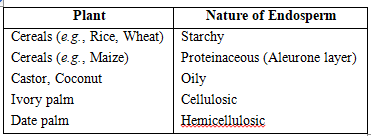
The stony endosperm is present in betel nut (Areca nut) and date palm (Phoenix dactylifera).
Diploid endosperm is found in Oenothera.
Types of endosperm :
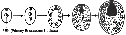
(i) Nuclear Endosperm : It is the most common type of endosperm in angiospermic plants. During the development of this endosperm, at first PEN undergoes successive nuclear divisions without wall formation (free nuclear division). In this manner, large number of free nuclei are produced. Finally, wall formation begins and it makes the endosperm a multicellular tissue, e.g., Cotton, Maize, Capsella, Coconut (milk).
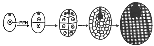
(ii) Cellular Endosperm: Primary endosperm nucleus divides many times and each division is followed by wall formation, e.g., Petunia, Utricularia, Coconut (copra).
(iii) Helobial Endosperm: It is an intermediate between nuclear and cellular type, e.g., members of order helobiales (Monocot).

Concept Builder
(i) The seeds with double endosperm is found in coconut (Cocos nucifera), (a) liquid endosperm, (b) cellular endosperm.
(ii) Xenia : The effect of foreign pollen on endosperm character is called xenia. This term was given by Focke. This was first observed in maize endosperm colour.
(iii) Metaxenia : Discovered by Swingle. The effect of foreign pollen on somatic tissue lying outside the endosperm is known as metaxenia, e.g., in date palm size of fruits and maturity time depends upon foreign pollen. .
(iv) Ruminate Endosperm : Endosperm with irregular surface, also known as chewed endosperm, e.g., Passiflora, Annona, Myristica.
(v) Mosaic Endosperm : Endosperm with sugary and starchy parts forming different colour patches of yellow and white, e.g., Maize.
B. Embryo
The development of an embryo from a zygote is called embryogeny. Embryo develops at the micropylar end of the embryo sac where the zygote is situated. Most zygotes divide only after certain amount of endosperm is formed.
This is an adaptation to provide assured nutrition to the developing embryo. It means endosperm development precedes embryo development.
Early stages of embryogeny are similar in both monocotyledons and dicotyledons.
(a) Development of Embryo in Dicots
The normal type of dicot embryo development has been studied in Shepherd's purse (Capsella bursa-pastoris) which belongs to family Cruciferae.
This is called as Crucifer or Onagrad type of embryo development. The development of embryo is endoscopic. Zygote (oospore) divides into two unequal cells, larger suspensor cell towards micropyle and a smaller embryonal cell (terminal cell) towards antipodal region.
The suspensor cell undergoes transverse divisions forming 6-10 celled long suspensor. The first cell of the suspensor (towards micropyle) is large and called haustorium or vesicular cell.
The last cell of suspensor (towards embryo cell) is known as hypophysis. It forms radicle tip.
Embryonal cell divides twice vertically and once transversely to produce a two-tiered eight-celled embryo. The epibasal tier forms two cotyledons and a plumule while the hypobasal tier produces only hypocotyl and most of the radicle.
For this the octant embryo undergoes periclinal divisions producing protoderm, procambium and ground meristem. It is initially globular but with the growth of cotyledons it becomes heart-shaped and then assumes the typical shape, e.g., Capsella bursa-pastoris.
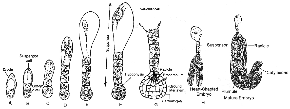
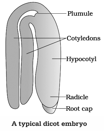
A typical dicot embryo consists of an embryonal axis and two cotyledons. The portion of embryonal axis above the level of cotyledons is the epicotyl, which terminates with the plumule (stem tip).
The cylindrical portion below the level of cotyledons is hypocotyl that terminates, at its lower end in the radicle. In orchids, Orobanche and Utricularia, the embryo does not show distinction of plumule, cotyledons and radicle.
(b) Development of Embryo in Monocot
The normal type of monocot embryo development has been studied in Luzula forsteri and is called Segittaria type. Suspensor is single celled in monocots.
The zygote of oospore divides transversely producing a vesicular suspensor cell towards micropylar end and embryo cell towards the chalazal end.
The embryo cell divides transversely again into a terminal and a middle cell. The terminal cell divides vertically and transversely into globular embryo.
It forms a massive cotyledon and a plumule.
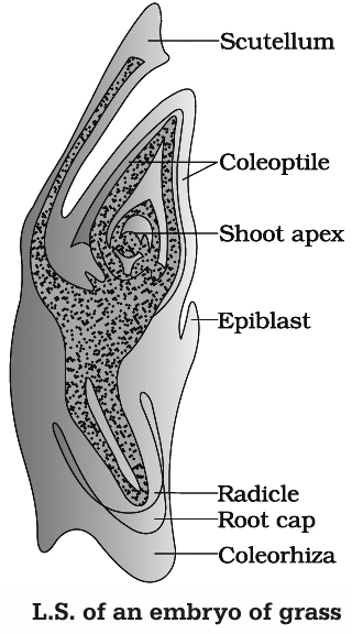
Growth of cotyledon pushes the plumule to one side. Remains of second cotyledon occur in some grasses. It is called epiblast. The single cotyledon of monocots is called scutellum. It is shield shaped and lateral in position but appears terminal.
The middle cell gives rise to hypocotyl and radicle. Radicle is enclosed in an undifferentiated sheath called coleorhiza. Epicotyl has a shoot apex and a few leaf primordia enclosed in a hollow foliar structure, the coleoptile.
C. Seed
It is often described as fertilised ovule. A seed typically consists of seed coat(s), cotyledon(s) and an embryonal axis. Mature seeds are of two types:
(a) Non endospermic or exalbuminous seeds:
In seeds like gram, pea, groundnut etc. the endosperm is completely consumed by developing embryo, thus the seeds are called non-endospermic or exalbuminous.
(b) Endospermic or albuminous seed :
In monocots and castor bean (dicots) embryo does not consume all endosperm. So, it persists in the mature seed. Such seeds are called endospermic or albuminous seeds. In these seeds, food is stored in endoseprm, e.g., wheat, maize, barley, sunflower, coconut.
Mostly nucellus is consumed after fertilisation due to absorption of food by the endosperm and embryo, but in some seeds remnants of nucellus are of persistant nature.
This residual, persistent nucellus is called perisperm. Such seeds are called perispermic seeds, e.g., Black pepper (Piper nigrum), beet.
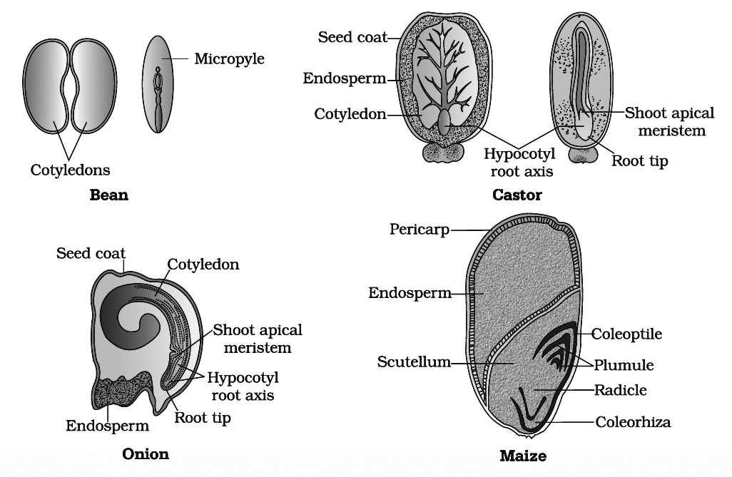
Integuments of ovules harden as tough protective seed coats. The outer seed coat called testa develops from outer integument and inner seed coat called tegmen is derived from inner integument.
The micropyle remains as a small pore in the seed coat. Micropyle facilitates entry of oxygen and water into the seed during germination.
As the seed matures, its water content is reduced and seeds become relatively dry (10-15% moisture by mass).
The embryo may enter a state of inactivity called dormancy or if favourable conditions like adequate moisture, oxygen and suitable temperature are available, they germinate.
The transformation of ovules into seeds and ovary into fruit proceeds simultaneously. The ovary wall develops into the fruit wall, called pericarp.
The fruit may be fleshy (guava, orange, mango) or dry (groundnut, mustard). In some cases thalamus and other floral parts show proliferation alongwith development of ovary wall.
Such fruits are called false fruits, e.g., Apple, Strawberry, Cashew nut. Most fruits develop from the ovary and are called true fruits.
In some plant species, fruits develop without fertilisation. Such fruits are seedless and are called parthenocarpic fruits, e.g., Banana. Parthenocarpy can be induced through the application of growth hormones like Auxins.
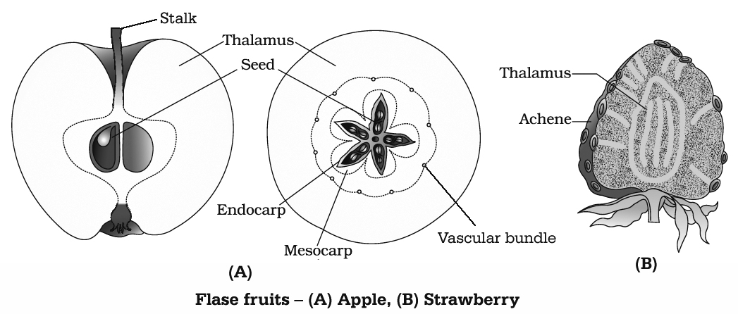
The first stimulus for fruit development comes from pollination while second stimulus is received from developing seeds and the third stimulus is provided by the availability of nutrients.
Advantage of seeds to Angiosperms
Seeds have better adaptive strategies for dispersal to new habitats and help the species to colonize in other areas. As they have sufficient food reserves, young seedling are nourished until they are capable of photosynthesizing on their own.
The hard seed-coat provide protection to young embryo. Being product of sexual reproduction, they generate new genetic combinations leading to variations.
Seed forms basis of our agriculture. Dehydration and dormancy of mature seeds are crucial for storage of seeds which can be used as a food throughout the year and also to raise crops in next season. Seed viability varies from species to species. '
Oldest viable seeds of lupine, Lupinus arcticus were excavated from Arctic Tundra. The seed germinated and flowered after an estimated record of 10,000 years of dormancy.
A recent record of 2000 years old viable seed is of date palm (Phoenix dactylifera), discovered during the archeological excavation at King Herod's Palace near Dead Sea.
The ability of a seed to retain the power of germination is called viability. It can be tested by using 0.1 % solution of triphenyl tetrazolium chloride (TTC).
Orchid fruits contain thousands of tiny seeds. Similar in the case in fruits of Ficus and parasitic species such as Orobanche and Striga.
apomixis and polyembryony
- Books Name
- A TEXT OF BIOLOGY - CLASS XII
- Publication
- ACME SMART PUBLICATION
- Course
- CBSE Class 12
- Subject
- Biology
APOMIXIS AND POLYEMBRYONY
Apomixis is the formation of new individuals directly through asexual reproduction without involving the formation and fusion of gametes.
It is of two types :
(i) Agamospermy and
(ii) Vegetative Propagation
Agamospermy is a mode of apomixis in which seeds are formed but are asexual in nature as the embryo develops directly without gametic fusion.
There are several types of agamospermy :
(i) Adventitive Embryony (Sporophytic budding) : Embryo arises from diploid sporophytic cells such as nucellus or integuments (other than egg), e.g., Citrus, Opuntia, Mango.
(ii) Recurrent Agamospermy : In this method, a diploid embryo sac is formed from megaspore mother cell which has a diploid egg or oosphere. The diploid egg grows parthenogenetically into diploid embryo, e.g., Apple, Pear, Allium. Diploid embryo sac can develop directly from either the diploid megaspore mother cell (diplospory) or diploid nuclear cell (apospory).
(iii) Non-recurrent Agamospermy : Embryo develops parthenogenetically from haploid egg, e.g., Banana.
(iv) Apogamy: It is formation of sporophyte or embryo directly from cells of gametophyte. In higher plants, only diploid apogamy is successful. In lower plants, haploid apogamy is equally successful.
Polyembryony is the phenomenon of having more than one embryo in a seed, discovered in Citrus by Leeuwenhock. There may be more than one egg cell in an embryo sac or more than one embryo sac in an ovule.
All the egg cells may get fertilised. Synergids and antipodal cells may also form embryos (mixed polyembryony). Occurrence of polyembryony due to fertilisation of more than one egg is called simple polyembryony.
Formation of extra embryos through sporophytic budding is called adventitive polyembryony.
Polyembryony is quite common in onion, groundnut, mango, lemon and orange. In some of these cases stimulus of pollination may be required. Citrus seed has 2-40 embryos, one normal and the rest adventitive mostly nucellar.
Importance of Apomixis:
Hybrid varieties of several of our food and vegetable crops are being extensively cultivated because they provide higher and better yield. However, there is one major drawback.
Hybrid seeds have to be produced every year because seeds collected from hybrid plants, if sown subsequently, do not maintain hybrid characters due to segregation of traits.
Production of hybrid seeds every year is costly thus increasing the cost of crop production. This can be avoided if apomixis is introduced in hybrid seeds.
Concept Builder
(i) The study of seed is called spermology.
(ii) Chalazosperm is perisperm like tissue in chalazal region. It is a substitute for endosperm e.g. Cynastrum.
(iii) Seed viability in Oxalis -few days
(iv) Apomixis is common in some species of Asteraceae and grasses.
male reproductive system
- Books Name
- A TEXT OF BIOLOGY - CLASS XII
- Publication
- ACME SMART PUBLICATION
- Course
- CBSE Class 12
- Subject
- Biology
HUMAN REPRODUCTION
Human beings are unisexual.
The growth, maintenance and function of the gonads are regulated by gonadotropins secreted from anterior lobe of pituitary gland.
The organs which neither produce gametes nor secrete sex hormones but perform important functions in reproduction are called secondary sex organs.
The latter include the prostate, seminal vesicles, vas deferentia and penis in males, and the fallopian tubes, uterus, vagina and mammary glands in females.
The characters which distinguish the male from the female externally are called accessory or external sex character. They are also called secondary sex characters.
Secondary Sexual Features in Man and Woman
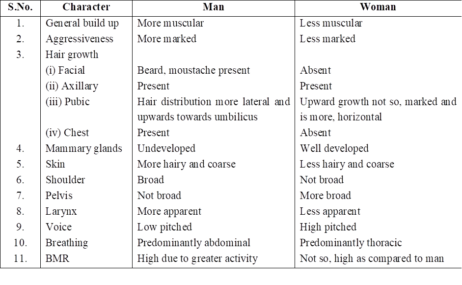
MALE REPRODUCTIVE SYSTEM
The male reproductive system of mammals consists of a pair of testes, several accessory glands, a duct system and a mating organ called the penis. Testis is the primary male sex organ. It produces spermatozoa and secretes the male sex hormone testosterone.
The human testis measures about 5 cm, 3 cm and 2.5 cm, respectively, in length, thickness and width. It is covered by thick, fibrous, connective tissue called tunica albuginea.
In man, both testes normally remain suspended in a pouch called scrotum outside the abdominal cavity. This keeps the testes at a low temperature than the body temperature (about 2°C below)-this is essential for the maintenance and normal functioning of the spermatogenic tissue of the testes.
Testes descend in the scrotal sac when foetus is about 7 months old and this occurs under the influence of FSH and testosterone.
If testes fail to descend, than the condition is called cryptorchidism that leads to sterility. Scrotum remains connected with the abdomen or pelvic cavity by the inguinal canal. Blood vessels, nerves and conducting tubes pass through inguinal canal.
Cremaster muscles and connective tissue form spermatic cord and surround all structures passing through inguinal canal. Cremaster muscles and dartos muscles of the scrotal sac help in the positioning of testes.
Whenever the outside temperature is low, these contract to move the testes close to the abdominal cavity/pelvic cavity. When outside temperature is high, these relax moving the testes away from the body.
In some seasonally breeding mammals, testes descend into the scrotum during breeding season but ascend back into the abdomen in the non-breeding season. e.g. rats and bats.
ANATOMY OF TESTIS
Each testis contains numerous tiny, highly convoluted tubules, called seminiferous tubules. They constitute the spermatogenic tissue of the testis.
Cells lining these tubules give rise to spermatozoa which are released into the lumen of the tubule.
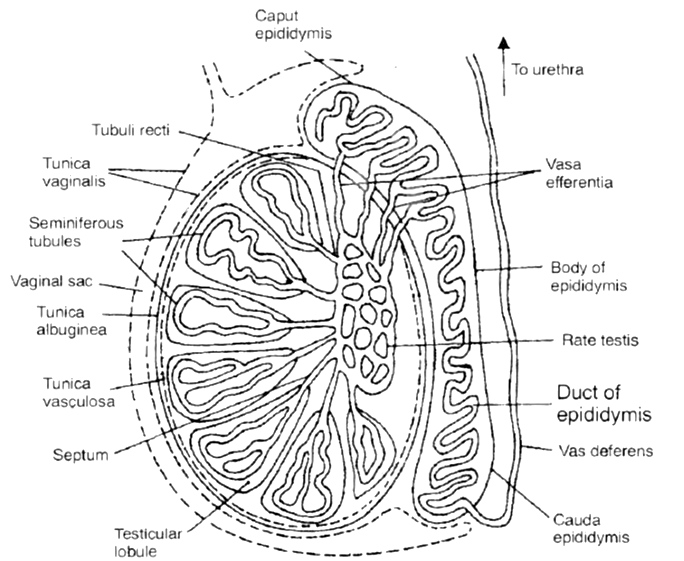
In between spermatogenic cells, Sertoli or sustentacular or nurse cells are present which provide nourishment to the developing spermatozoa and regulate spermatogenesis by releasing inhibin to check FSH overactivity.
The other functions of sertoli cells include
(i) Provide nourishment to the developing spermatozoa,
(ii) Absorb the parts being shed by developing spermatozoa
(iii) Release anti mullerian factor (AMF) to prevent development of mullerian duct/oviduct
(iv) Release of androgen binding protein (ABP).
Groups of polyhedral cells called Interstitial cells or Leydig cells, are located in the connective tissue around the seminiferous tubules.
They constitute the endocrine tissue of the testis. Leydig cells secrete testosterone into the blood.
Seminiferous tubules unite to form several straight tubules called tubuli recti which open into irregular cavities in the posterior part of the testis.
It is highly anastomosing labyrinth of cuboidal epithelium lined channels called rete testis.
Several tubes called vasa efferentia arise from it and conduct spermatozoa out from the testis (Seminiferous tubule to vasa efferentia form intratesticular genital duct system).
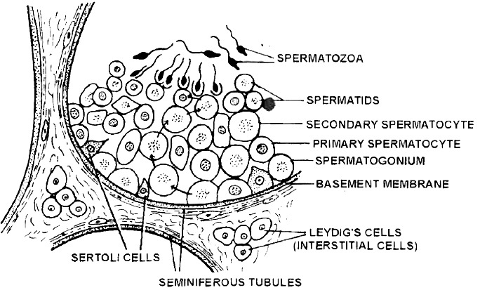
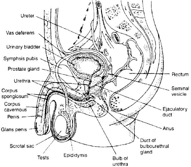
The extratesticular duct system consists of tubes which conduct sperms from the testes to the outside.
It starts with vasa efferentia which arise from each testis and becomes confluent to form a folded and coiled tube called epididymis behind each testis.
The epididymis consist of 3 parts: (i) Caput, (ii) Corpus, (iii) Cauda. The epididymis stores the sperms temporarily.
From cauda epididymis, a partially coiled tube called vas deferens ascends into the abdomen through inguinal canal, passes over the urinary bladder, receives the duct from the seminal vesicle behind the urinary bladder to from an ejaculatory duct.
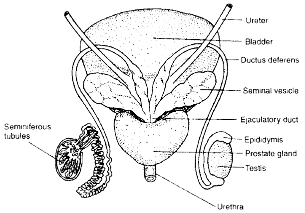
Before entering prostate, the ductus deferens dilates to form ampulla. The final portion of ampulla passes through the prostate to open into the urethra shortly after its origin from the urinary bladder.
The urethra receives the ducts of the prostate and cowper's glands, passes through the penis and opens to the outside.
MALE EXTERNAL GENITAL ORGAN
PENIS
This is the copulatory organ of man. It is a cylindrical and erectile, pendulous organ suspended from pubic region in front of scrotum.
It remains small and limp (= flaccid) but on sexual arousal, it becomes long, hard and erect, ready for copulation (=coitus or intercourse). Erect human penis, on an average is about 15 cm long.
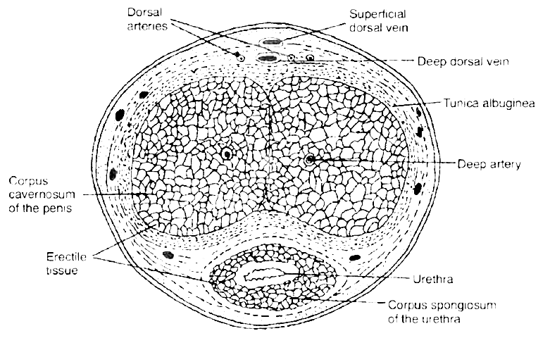
 T.S. of penis
The penial mass is itself encased in a fibrous sheath, called tunica albuginea. The interior of penis is mostly formed of three cylindrical cords of spongy, erectile (= cavernous) tissue.
Two of these cords are thicker and situated parallely on right and left sides, forming the thick part of penis that remains in front when penis is limp, but becomes superio-posterior when penis is erect.
These two cords are called corpora cavernosa.
The fibres of tunica albuginea surround both cords jointly and form a separate sheath around each cord. Some fibres form a partition, called septum penis, between these cords.
The third, smaller cord form that part of penis which remains inferio-anterior in erect penis. Urethra runs through this cord. Hence, this cord is called corpus urethrae or spongiosum.
The extended part of corpus spongiosum is enlarged, forming a bulged, conical structure called glans penis. The surface of glans is formed of a thin, smooth and shiny, hairless skin.
The base line of glans is referred to as neck of the penis. The loose skin of penis folds over and is retractile on glans. It is called foreskin or prepuce.
At the tip of glans penis is the slit like external urethral orifice or meatus by which urethra opens out and discharges urine or semen.
Prepucial glands, present in the skin of penis neck, secrete a white sebaceous substance, called smegma. Microbial infection in smegma causes irritation.
GLANDS
Seminal vesicles - These are paired, tubular, coiled glands situated behind the urinary bladder. They secrete viscous fluid which constitute the main part of the ejaculate. Seminal fluid contains fructose, citric acid, inositol and postaglandins.
Prostate gland - It is a chestnut shaped gland and is a collection of 30-40 tubulo-alveolar glands which lies at the base of the bladder and surrounds the base of the urethra. It contributes an alkaline substance to the seminal fluid.
The substance of prostate help the sperms to become active and counteract any adverse effects of urine on sperms. The prostatic fluid provides a characteristic odour to the seminal fluid. Prostate gland secretes -citrate ion, calcium, phosphate ion, and profibrolysin.
Bulbourethral glands or Cowper's glands - The two bulbourethral glands are pea-sized structures lying adjacent to the urethra at the base of penis. These secrete a viscous lubricant.
The duct system, accessory glands and penis are secondary male sex organs. Their growth, maintenance and functions are promoted by testosterone secreted by Leydig cells.
On the other hand, the growth, maintenance and functions of seminiferous tubules and Leydig cells are regulated, respectively by FSH and ICSH of anterior pituitary.
SEMEN
Semen is a mixture of sperms and the secretions of the seminiferous tubules, seminal vesicles, prostate gland, and bulbourethral glands.
The average volume of semen in an ejaculation is 2.5-5 ml, with a sperm count (concentration) of 200 to 300 million. For normal fertility, atleast 60 percent sperms must have normal shape, size and atleast 40 percent of them must show vigorous motility. When the sperm count falls below 20 million/ml, the male is likely to be infertile.
Semen has a slightly alkaline pH of 7.2-7.7. The prostatic secretion gives semen a milky appearance, whereas fluids from the seminal vesicles and bulbourethral glands give it a sticky consistency.
Semen provides transportation medium and nutrients to sperms. It neutralizes the hostile acidic environment of the male urethra and the female vagina.
(Prostatitis - Inflammation of prostate gland.)
female reproductive system
- Books Name
- A TEXT OF BIOLOGY - CLASS XII
- Publication
- ACME SMART PUBLICATION
- Course
- CBSE Class 12
- Subject
- Biology
FEMALE REPRODUCTIVE SYSTEM
The female reproductive system consists of a pair of ovaries, a duct system, consisting of a pair of fallopian tubes (oviduct), an uterus, cervix and vagina. A pair of mammary glands are accessory genital glands.
OVARIES
The ovary is the primary female sex organ. It produces ova and secretes the female sex hormones, estrogen and progesterone that are responsible for the development of secondary female sex characters and regulate cyclic changes in the uterine endometrium.
The human ovaries are small, almond-like flat bodies of about 3 cm in diameter.
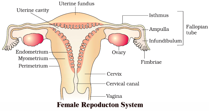
1. Location. Ovaries are located near kidneys and remain attached to the lower abdominal cavity through mesovarium.
2. Structure. The free surface of the ovary is covered by a germinal epithelium made of a single layer of cubical cells. This epithelium is continuous with the mesothelium, called peritoneum. The epithelium encloses the ovarian stroma. The stroma is divided into two zones-a peripheral cortex and an inner medulla. Cortex is covered by a connective tissue, called tunica albuginea.
The cortex contains numerous spherical or oval, sac-like masses of cells, known as ovarian follicles. The medulla consists of loose connective tissue, elastic fibres, blood vessels and smooth muscle fibres.
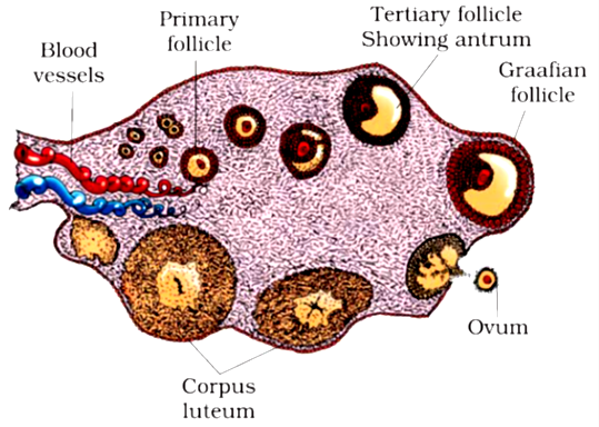
 A section of human ovary
OVARIAN FOLLICLE
The ovarian follicle carries a large, centrally placed ovum, surrounded by several layers of granular cells (follicular granulosa or discus proligerus or cumulus oophorus). It is suspended in small cavity the antrum.
Antrum is filled with liquid folliculi. The secondary oocyte in the tertiary folicle forms a new membrane called zona pellucida. The follicle bulges on the surface of the ovary.
Such a follicle is called the mature Graafian follicle (after De Graaf, who reported them in 1672).
CORPUS LUTEUM
The ovum is shed from the ovary by rupturing the follicle. The release of ovum is called ovulation and occurs nearly 14 days before the onset of the next menstrual cycle.
After the extrusion of the ovum, Graafian follicle transforms into corpus luteum. Corpus luteum is filled with a yellow pigment, called lutein.
Corpus luteum grows for a few days and if the ovum is fertilized and implantation occurs, then it continues to grow. But if the ovum is not fertilized, then corpus luteum persists only for about 14 days.
It secretes progesterone. At the end of its functional life, the corpus luteum degenerates,and becomes converted to a mass of fibrous tissue, called corpus-albicans (white body) that remains as a scar in the ovary throughout the life of female.
FALLOPIAN TUBES (OVIDUCTS)
There are one pair, long (about 10 cm), ciliated, muscular and tubular structures that extend from ovaries to uterus. Each one is suspended by mesosalpinx. Each fallopian tube is differentiated into four parts :
(i) Infundibulum. The part of oviduct closer to the ovary is the funnel shaped infundibulum. The edges of infundibulum possess finger like projections called fimbriae. Fimbriae help in collection of the ovum after ovulation. Infundibulum opens in abdominal cavity by an aperture called osteum.
(ii) Ampulla. The infundibulum leads to a wider part of the oviduct called ampulla.
(iii) Isthmus. It is middle, narrow and ciliated part of the oviduct.
(iv) Uterine part. It is inner and narrow part which opens in the upper part of uterus.
It is involved in the conduction of ovum or zygote towards the uterus by peristalsis and ciliary action. (Fertilization occurs at the junction of ampulla and isthmus).
Uterus. It is a large hollow, muscular, highly vascular and pear shaped structure present in the pelvis between the bladder and rectum. It is suspended by a mesentery, mesometrium. It is formed of three parts.
(i) Fundus. It is upper dome shaped part above the opening of fallopian tubes.
(ii) Body. It is middle and main part of uterus.
(iii) Cervix. It is lower, narrow part which opens in the body of uterus by internal os and in vagina below by external os. It is formed of most powerful sphincter muscle in the body.
Its wall is formed of outer peritoneal layer called perimetrium; middle muscular myometrium made of smooth muscle fibres, and inner highly vascular and glandular endometrium.
It is the site of foetal growth during pregnancy. It also takes part in placenta formation and helps in pushing the baby out during parturition.
VAGINA
It is a long (7.5 cm), fibro-muscular tube. It extends backward in front of rectum and cervix to the vestibule. It is vascular tube internally lined by mucus membrane and is raised into transverse folds called vaginal rugae.
In the virgin female, vaginal orifice is closed by a membranous diaphragm called hymen which becomes centrally perforated at puberty for the discharge of menstrual flow (or menses).
Vagina acts both as copulation canal as it receives the sperms from penis during copulation and birth canal during parturition.
FEMALE EXTERNAL GENITAL ORGAN
Vulva. It is external genitalia of female. It has a depression, the vestibule, in front of anus. Vestibule has two apertures-upper external urethral orifice of urethra and lower vaginal orifice of vagina.
Mons pubis : Is a fleshy and fatty tissue covered by skin and pubic hair.
Labia majora : It is a pair of fleshy folds which extend from the mons pubis and surround the vaginal opening.
Labia minora : Is another pair of tissue folds below the labia majora.
Both labia majora and labia minora are provided with sebaceous glands.
Hymen : Is a membrane that partially covers the vaginal opening. It gets torn during the first coitus.
Clitoris : Is a tiny, erectile, finger-like structure present at the upper junction of the two labia minora above the urethral opening. The fold of skin that covers clitoris is called prepuce. Clitoris is homologous to penis (as both are supported by corpora cavernosa).
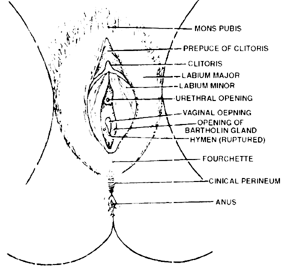
 The external genitalia in the female
GLANDS
Vestibular Glands: These are of two types-greater and lesser. Greater vestibular or Bartholin's glands are a pair of small reddish yellow glands on each side of vaginal orifice and secrete alkaline secretion for lubrication and neutralising urinary acidity.
Lesser vestibular glands or paraurethral or skene's glands are small mucus glands present between urethral and vaginal orifices.
MAMMARY GLAND :
Each mammary gland consists of 15-25 lobules of the compound tubuloalveolar type. These lobules secrete milk to nourish the newborn babies.
Each lobe is separated from the others by dense connective and adipose tissue and represents a gland. From each lobe, excretory lactiferous ducts emerge independently in the nipple, which has 15-25 openings, each about 0.5 mm in diameter.
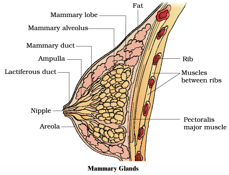
However, the histological structure of mammary glands vary, depending on sex, age and physiological state.
Path of milk ejection :
Mammary alveolus ® Mammary duct ® Ampulla ® Lactiferous duct ® Nipple
Homology Between Male and Female Reproductive System
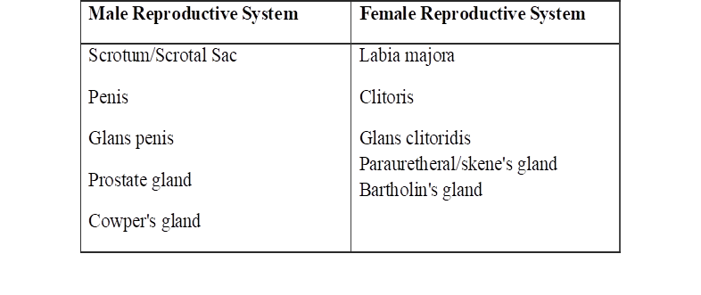
gametogenesis
- Books Name
- A TEXT OF BIOLOGY - CLASS XII
- Publication
- ACME SMART PUBLICATION
- Course
- CBSE Class 12
- Subject
- Biology
THE EVENTS IN HUMAN REPRODUCTION
Gametogenesis Insemination Fertilization Implantation Gestation Parturition
FORMATION OF GAMETES
Sexual reproduction requires the fusion of two haploid gametes to form a diploid individual. These haploid cells are produced through gametogenesis.
As there are two types of gametes, the spermatozoa and ova, gametogenesis can be studied under two broad headings: spermatogenesis and oogenesis.
Spermatogenesis is the formation of spermatozoa, whereas oogenesis is the formation of ova. Both spermatozoa and ova originate from primordial germ cells or PGCs, which are extra-gonadal in origin.
In humans, the PGCs originate during early embryonic development from the extra-embryonic mesoderm. Eventually, they migrate to the yolk sac endoderm, and ultimately, to the gonads of the developing embryo, where they undergo further development.
SPERMATOGENESIS
Spermatozoa are produced in the seminiferous tubules of the testes. Spermatogenesis is the process of maturation of reproductive cells in the testes.
Spermatogenesis includes two stages (i) formation of spermatids and (ii) metamorphosis of spermatids. Spermatids are formed by three phases namely phase of multiplication (mitosis), phase of growth and phase of maturation (meiosis).
During phase of multiplication, the primordial germ cells divide repeatedly by mitosis to form diploid spermatogonia.
During phase of growth, the spermatogonium enlarges in size to form primary spermatocyte and prepares to undergo maturation division.
During phase of maturation, the primary spermatocyte undergoes meiosis I giving rise to two haploid (n) secondary spermatocytes. The secondary spermatocytes undergo meiosis II resulting in the formation of four spermatids.
Transformation of spermatid to sperms is termed spermiogenesis. A spermatid is non-motile. It has organelles like mitochondria, Golgi bodies, centrioles, nucleus etc.
During spermiogenesis, the weight of gamete is reduced along with development of locomotory structures. Nucleus becomes compact forming the major part of head of spermatozoa,
Golgi complex of spermatid give rise to acrosome. Acrosome forms a cap in front of nucleus containing lytic agent which dissolves egg membranes during fertilization.
Acrosome of mammalian sperm produces sperm lysins. The two centrioles of the spermatids become arranged one after the other behind the nucleus. The anterior one is known as the proximal centriole.
The proximal centriole is usually located on the neck of spermatozoan. During fertilization, it is introduced in to the egg and is required for the first cleavage.
The posterior centriole is known as the distal centriole and gives rise to the axial filament of the sperm. Mitochondria from different parts of spermatid to get arranged in the middle piece around the axial filament.
Mitochondria in the middle piece provide energy to the sperm for locomotion.
A typical mammalian sperm is flagellated, consisting of four parts namely head, neck, middle piece and tail.
The human sperm was first seen by Hamm and Leeuwenhoek. Tail-less, (non-flagellate) 'amoeboid' sperm is found in the roundworm Ascaris.
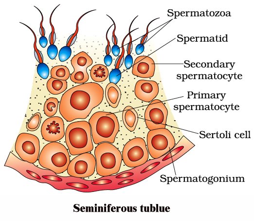
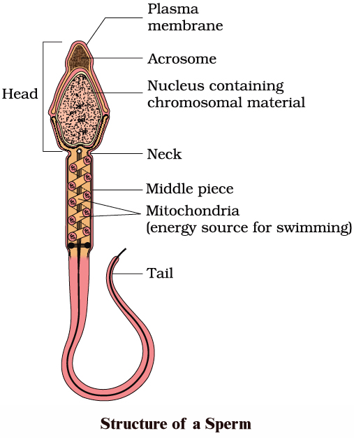
Hormonal control of spermatogenesis
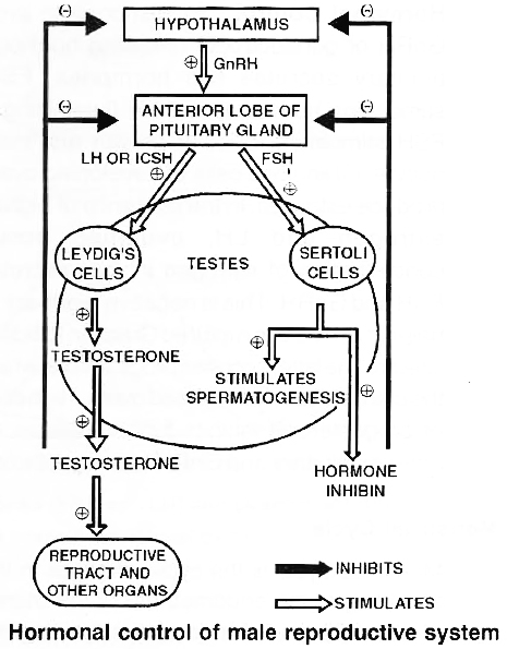
Spermatogenesis is under the control of endocrine hormones. Hypothalamus produces gonadotropin releasing hormone or GnRH.
It acts on anterior pituitary to produce gonadotropins, ICSH and FSH, ICSH interstitial cell stimulating hormone acts on interstitial or Leydig cells which produce testosterone.
Testosterone is essential for formation of sperms, atleast spermiogenesis part by Sertoli cells. Under the influence of FSH, Sertoli cells develop androgen binding protein (ASP).
The latter helps in concentrating testosterone in the seminiferous tubules.
Excess of testosterone inhibits LH/ICSH by anterior pituitary and GnRH production by hypothalamus.
Sertoli cells also produce a glycoprotein called inhibin. Inhibin suppresses FSH synthesis by anterior pituitary and GnRH synthesis by hypothalamus.
Thus normal release of testosterone is under negative feed back control.
OOGENESIS
Oogenesis is the process of maturation of reproductive cells in ovary. Oogenesis starts before birth. In 25 weeks old female foetus, all the oogonia are produced.
Oogenesis is basically similar to spermatogenesis. It includes phase of multiplication, phase of growth and phase of maturation.
During the phase of multiplication, the primordial cells in the ovary divide mitotically to form oogonia (egg mother cell). Each oogonium divides mitotically to form two primary oocytes.
Primary oocytes undergo growth, the growth phase during oogenesis is comparatively longer.
Primary oocytes, begin the first step of Meiosis-I and proceed up to diakinesis.
These oocytes resume their development at puberty. The primary oocyte (2n) completes meiosis-I producing two haploid cells (n), the larger one is secondary oocyte and the smaller one is first polar body.
Secondary oocyte starts Meiosis-II and proceeds upto metaphase-II only. Further development will start only after arrival of spermatozoa.
Entry of sperm restarts the cell cycle by breaking down M PF (M-phase promoting factor) and turning on APC (Anaphase promoting complex). Completion of meiosis II results in the formation of functional egg or ovum and a second polar body.
Compare the structure of Mammalian sperm and Mammalian ovum
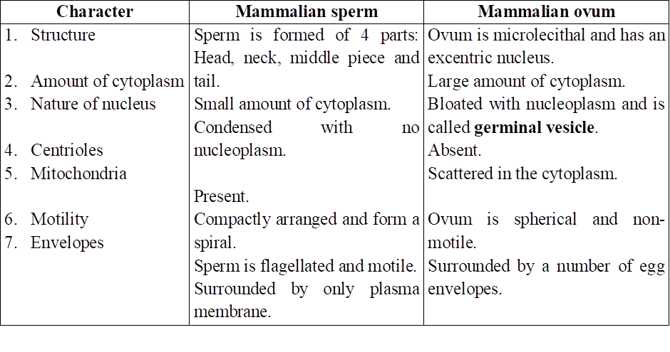
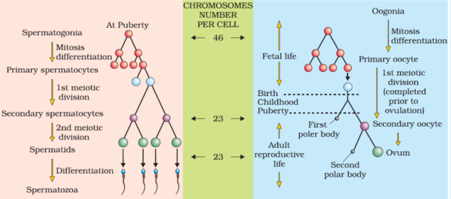
Differences between Spermatogenesis and Oogenesis
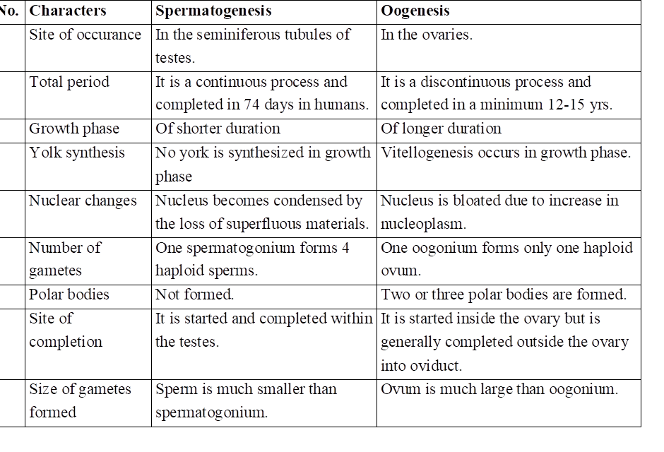
Hormonal control of Oogenesis
In response to production of GnRH or gonadotropin releasing hormone, anterior pituitary secretes two hormones, FSH (follicle stimulating hormone) and LH (luteinizing hormone).
FSH stimulates follicular growth and maturation of oocyte. Granulosa cells of developing ovarian follicle produce estrogen.
In presence of high titre of both estrogen and LH, ovulation occurs. High concentration of estrogen inhibits secretion of both FSH and GnRH.
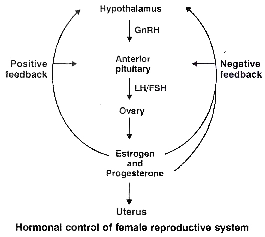
This is negative feedback control LH helps in converting ruptured Graafian follicle into corpus luteum.
The latter secretes progesterone which prepares the uterus to receive fertilised ovum. High concentration of progesterone inhibits further release of LH from anterior pituitary and GnRH from hypothalamus.
menstrual cycle
- Books Name
- A TEXT OF BIOLOGY - CLASS XII
- Publication
- ACME SMART PUBLICATION
- Course
- CBSE Class 12
- Subject
- Biology
MENSTRUAL CYCLE
Menstrual cycle is the cyclic changes in the reproductive tract of primate (Man, Monkey and Apes) females. Menstruation is the periodic shedding of the endometrium of uterus with bleeding. In healthy women, menstruation occurs at intervals of about 28 to 29 days.
Menarche is the starting of menstruation in girls that occurs at about 13 years of her age. Menstrual cycle consists of menstrual phase, proliferative phase (follicular phase) and secretory phase (luteal phase).
Proliferative phase (5th to 14th day of menstrual cycle) consists of growth of endometrium of uterus, fallopian tube and vagina. In ovary, a Graafian follicle secretes estrogen during this phase.
Estrogen is the hormone active during proliferative phase. The ovum is released from the follicle near the end of proliferative phase, i.e., 14th day or midway during menstrual cycle.
Ovulation occurs under the influence of LH from pituitary. The subsequent 14 days in which corpus luteum is active is referred to as the secretory phase.
Progesterone secreted by corpus luteum is active during secretory phase. The uterine endometrium and glands grow further during secretory phase.
At the end of secretory phase, corpus luteum degenerates into corpus albicans in the ovary, progesterone secretion falls, the overgrown uterine endometrium breaks down and mensturation takes place.
Menstrual cycle is controlled by FSH, LH, estrogen and progesterone. The menstrual cycle and menstruation remain suspended during pregnancy and lactation. Menopause (climacteric) is the period of life when menstruation naturally stops.
Menopause occurs in females at the age of around 45-50 years. Ability to reproduce is lost in the female after menopause.
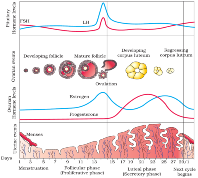
Various Phases of the Menstrual Cycle
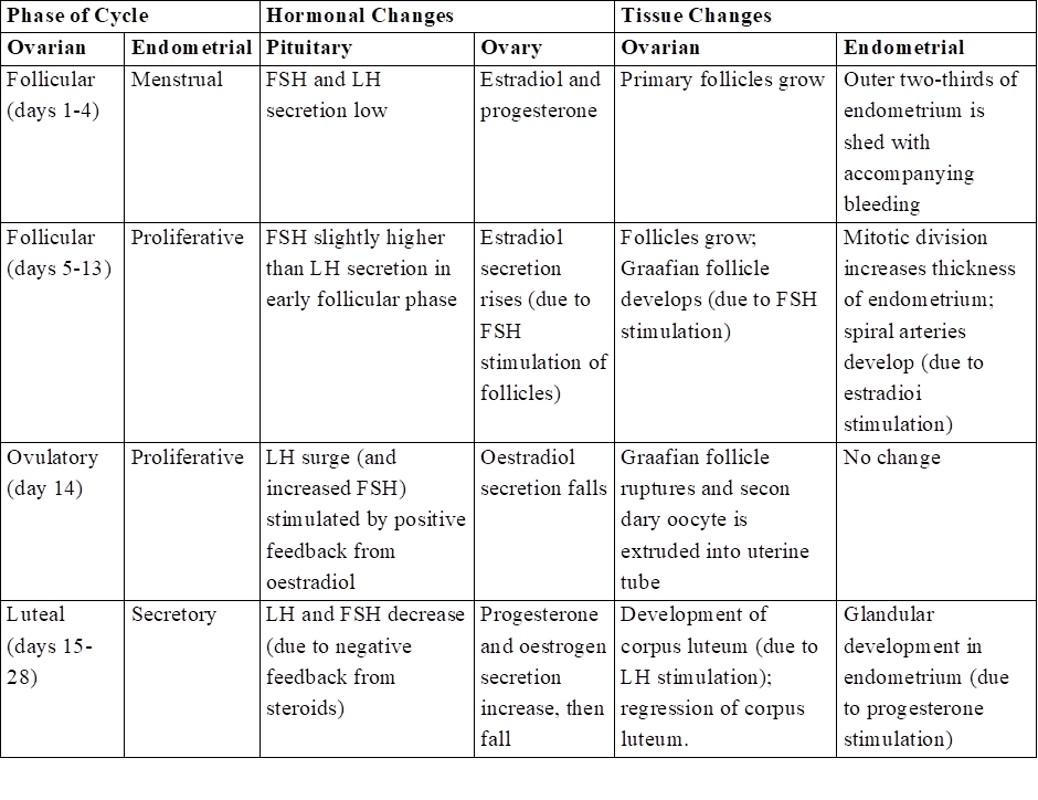
ESTROUS CYCLE
The estrous cycle consists of cyclic changes in the female reproductive system of non-primate mammals. There is no menstruation at the end of estrous cycle.
The estrogen level in blood increases resulting strong sex urge in the female. This is called "period of heat".
The estrous cycles run only during breeding season. The estrous cycles remain suspended in female during non-breeding season. The suspension of estrous cycles is called the state of anestrum.
Those animals that have only a single estrous during the breeding season are called monoestrous, e.g., dog, fox, deer, bat, etc.
The animals that have a recurrence of estrous during breeding season are called polyestrous, e.g., Mouse, squirrel, cow, sheep, pig, horse etc.
Concept Builder
Types of Egg
The eggs are classified on the basis of amount and distribution of yolk in them. Yolk is the reserve food material in the cytoplasm of egg. It may be formed of phospholipids, proteins, or lipid and carbohydrates. The process of formation of yolk and its deposition in egg. is called vitellogenesis.
1. Based on amount of yolk.
(i) Alecithal -Eggs without yolk e.g. human egg.
(ii) Microlecithal -The eggs with very little yolk, e.g. sea urchin, starfish.
(iii) Mesolecithal -The eggs containing moderate amount of yolk, e.g. frog.
(iv) Megalecithal (or Macrolecithal) -The eggs containing very large amount of yolk, e.g. reptiles, birds.
2. Based on distribution of yolk.
On the basis of distribution of yolk, the eggs are classified as follows
(i) Isolecithal -The yolk is uniformly distributed throughout the cytoplasm of egg, e.g., Branchiostoma, Herdmania.
(ii) Telolecithal-The eggs in which the yolk is concentrated towards one pole i.e., vegetal pole & nucleus along with major part of cytoplasm is displaced is animal pole e.g. , amphibians.
(iii) Centrolecithal -Yolk concentrated in centre of the egg with cytoplasm surrounding it, e.g. insects.
Cleidoic Eggs
Eggs of reptiles and birds are insulated by albumen, membranes and shell. The calcareous shell present around the eggs of bird is mainly made up of calcium carbonate (94%) and is secreted by uterus (shell glands). Chalazae are the suspensory ligaments of the yolk in birds's egg.
Egg Membranes
There are three types of egg membrane which can be present around the egg
(i) Primary egg membrane: It is formed around the plasma membrane of egg secreted by ovum itself e.g, Jelly envelope (Echinoderms). Vitelline membrane (Mollusca, amphibians & birds), zona radiata (Shark, some amphibians).
(ii) Secondary egg membrane: It is formed around an egg by the follicle cells of the ovary e.g., chorion around egg of insect; corona radiata, granulosa & theca layers in human.
(iii) Tertiary egg membranes: These are formed by the oviducts & other accessory parts of maternal genital tract while the egg is passing from the ovary to the exterior. e.g., the albumin, shell membrane & outermost calcareous shell of reptiles & birds.
Types of Cleavage
The mode of cleavage is determined by the amount of yolk and its distribution.
(a) Holoblastic: The cleavage in which the segmentation lines pass through the entire egg , dividing it completely. It occurs in alecithal, microlecithal and mesolecithal eggs; e.g. frog, human egg etc. It is of two types equal holoblastic example Amphioxus and Marsupials; unequal holoblastic e.g., lower fishes and amphibians.
(b) Meroblastic: The lines of segmentation do not completely pass through the egg and remain confined to a part of the egg. Such type of cleavage is found in megalecithal eggs as the yolk provides resistance to the cleavage, e.g. insects, birds, reptiles. Meroblastic cleavage may be (i) Superficial which occurs in centrolecithal eggs, e.g . insects, or (ii) discoidal, found in eggs of birds.
Patterns of Cleavage
Cleavage is the successive mitotic cell divisions of the egg & can be in following pattern
(i) Radial Cleavage: Successive clevage plane cuts straight through the egg e.g. Synapta paracen rotus.
(ii) Biradial Cleavage: When the first three division planes do not stand at right angles to each other: Ctenophora
(iii) Spiral Cleavage: There is a rotational movement of cell parts around the egg axis leading to displacement of mitotic spindle with respect to symmetrically disposed radii e.g. turbellarians, nematoda, rotifera, annelida, all mollusc except cephalopods.
(iv) Bilateral Cleavage: Mitotic spindles and cleavage planes remain bilaterally arranged with reference to the plane of symmetery e.g. tunicates, Amphioxous, amphibia and higher mammals.
(v) Meridional Cleavage: When cleavage furrow bisects both the poles of egg passing through animal vegetal axis, the plane of cleavage is called meridional cleavage.
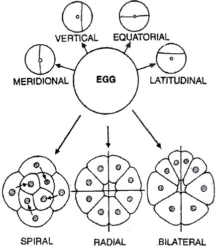
According to the concept of potency, cleavage can be
(i) Indeterminate/Regulative eggs, the fate of different parts of egg or its blastomeres is not predetermined or blastomeres have no characterstic position & alterable fate e.g. Echinoderms and vertebrates.
(ii) Determinate/Mosaic eggs. Every part of fertilized egg has a definite fate, so that fate of every blastomere is determined from beginning i.e. , produces the complete embryo only if all the blastomeres remain together. e.g., Nematoda, Mollusca (Dentalium).
Morula
Cleavage results in the formation of solid mass of cells called Morula as it looks like a mulberry.
Blastula
Blastula is the embryonic stage next to morula.
It contains a fluid-filled cavity called blastocoel surrounded by one or more layers of cells, the blastomeres.
The blastula with blastocoel is called coeloblastula, e.g. frog.
In certain animals, the blastula is solid and is termed stereoblastula e.g., Cnidaria, Nereis & some molluscs.
The blastula formed as a result of superficial cleavage is called superficial blastula, e.g. insects.
Discoblastula is disc shaped blastula formed as a result of discoidal cleavage, e.g. birds.
Gastrula
Gastrula is formed by gastrulation. Gastrulation is the process of the formation of gastrula from the blastula. Gastrula is the embryonic stage of development in which the germinal layers are formed. Gastrulation involves movement of cells.
The movements are called morphogenic movements. They include epiboly (descending of dividing cells to cover other cells) , emboly (upward movement of dividing cells underneath the other cells), involution (inward migration of blastomeres to go into the blastocoel), invagination (tucking in of blastula wall) , and delamination (separation of a sheet of cells from blastula) .
Archenteron is the cavity which occurs inside the gastrula, and is the future alimentary canal. It opens to the outside through blastopore which closes later.
Foetal Membranes (Extra embryonic membranes)
The developing foetus is enclosed by three membranes, amnion, chorion and allantois.
(i) Amnion is formed from mesoderm outside and ectoderm inside. Space between amnion and foetus is called amniotic cavity and it contains amniotic fluid.
(ii) Chorion is formed from ectoderm externally and mesoderm inside. Alongwith the allantois it participates in the formation of placenta. Space between amnion and chorion is embryonic coelom .
(iii) Allantois consists of mesoderm outside, and endoderm inside. It extends to fuse with chorion and forms allanto-chorion which gives rise to foetal part of placenta.
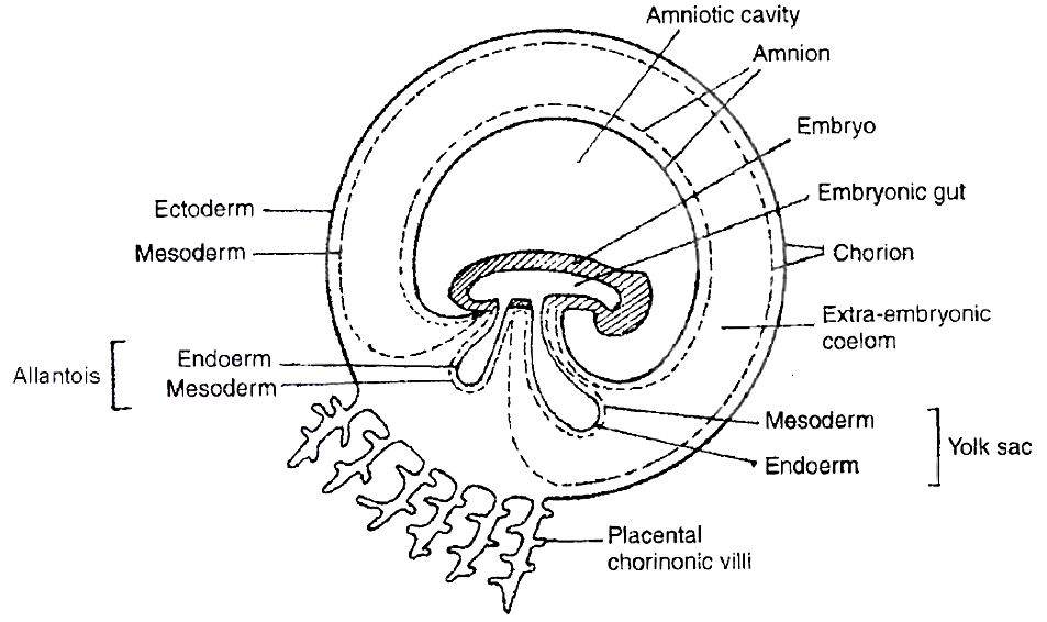
fertilisation and implantation
- Books Name
- A TEXT OF BIOLOGY - CLASS XII
- Publication
- ACME SMART PUBLICATION
- Course
- CBSE Class 12
- Subject
- Biology
THE EVENTS IN MAMMALIAN REPRODUCTION
FERTILISATION
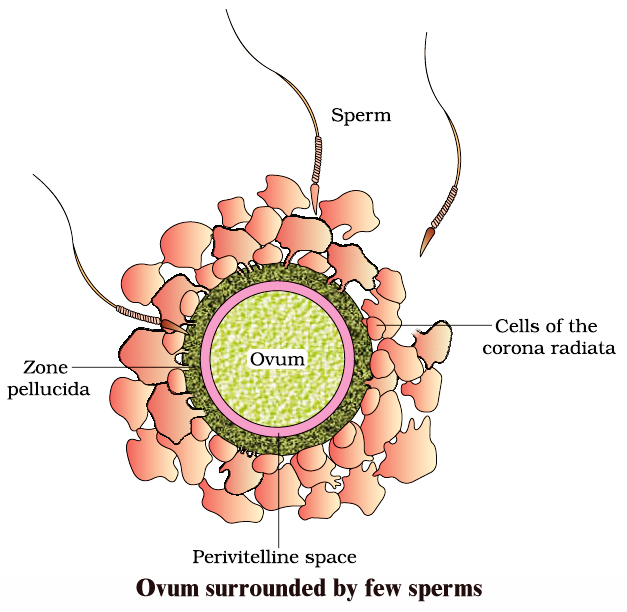
Ovum is released in the secondary oocyte stage (arrested in metaphase-II). Due to ciliary current produced by fimbriae of oviduct, ovum is drawn in through ostium.
It reaches ampulla, the site of fertilisation, by the ciliary action of ciliated columnar epithelium lining of oviduct.
A human sperm can live for many weeks in male genitial duct. Once ejaculated sperm can live alive only for 24 to 48 hours outside the body. Sperms move in the liquid medium secreted by female genital tract (1.5-3.0 mm/minute).
Prostaglandins of semen help in movement of spermatozoa and finally reach ampulla portion of the oviduct.
Capacitation of sperm occurs in the female genital system due to
(1) Removal of membrane cholesterol present over acrosome, weakening the membrane cover.
(2) Dilution of decapacitation factors.
(3) Entry of Ca2+ into sperms causing rapid whiplash motion of the tail.
Fusion of gametes/Syngamy: The various steps involve :
Acrosomal reaction: Number of sperms adhare to the surface of egg covers (Agglutination). The acrosome starts releasing its hydrolytic enzymes (sperm lysins).
It includes,
(a) Hyaluronidase: Dissolves the hyaluronic acid responsible for cementing of follicle cells or granulosa cells.
(b) Corona digesting enzyme (CDE) : Dissolves corona radiata.
(c) Zona lysin/Acrosin : Digests the zona pellucida. It involves zona pellucida compatibility reaction determined by 'fertilizin' protein over zona pellucida and 'antifertilizin' in case of sperm.
Contact of acrosome stimulates development of an outgrowth by the oocyte called fertilisation cone or cone of reception.
As the sperm head gets in contact with the fertilization cone, it cause opening up of Na+ channel to cause depolarisation of membrane (fast block to check polyspermy) and Ca2+ wave inside the egg.
Sperm and egg membranes dissolve. Male pronucleus & proximal centriole of sperm enter the cytoplasm of egg & rest part is left out.
Ca2+ wave causes extrusion of cortical granules (cortical reaction) and zona reaction makes the zona pellucida impervious to second sperm by destroying sperm receptors.
Cortical reaction and zona reaction constitute slow block to check polyspermy.
Entry of sperm causing breakdown of metaphase promoting factor (MPF) and turning on anaphase promoting complex (APC). This results in oocyte completing its meiosis -II.
Male and female pronuclei approach each other and finally mixing up of paternal and maternal chromosomes (Amphimixis) occur resulting in the formation of zygote/ synkaryon.
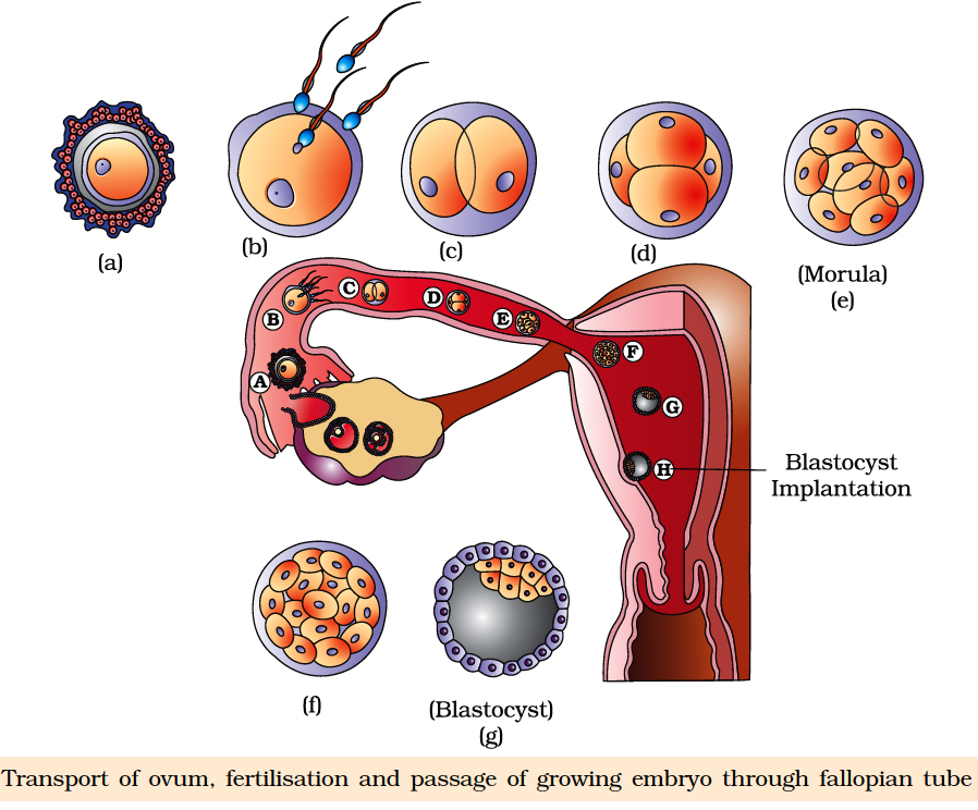
embryonic development
- Books Name
- A TEXT OF BIOLOGY - CLASS XII
- Publication
- ACME SMART PUBLICATION
- Course
- CBSE Class 12
- Subject
- Biology
EMBRYONIC DEVELOPMENT
It includes cleavage, blastulation, implantation, gastrulation and organogenesis.
Cleavage:
First cleavage is completed after 30 hours of fertilization. Cleavage furrow passes from animal-vegetal axis as well as centre of zygote (Meridional cleavage).
It divides the zygote into two blastomeres (Holoblastic cleavage). Second cleavage is completed after 60 hours of fertilization.
It is also meridional but at right angle to the first one. It is completed earlier in one of the two blastomeres resulting in transient 3-celled stage.
Third cleavage is horizontal forming 8 blastomeres. lt is slightly unequal. Thereafterthe rate and pattern of cleavage is not specific.
Morula:
Cleavage results in solid ball of celled Morula with 16 cells (occasionally 32 cells). Zona pellucida is still present as the outer cover. Morula undergoes compaction.
The outer/peripheral cells are small/flat with tight junction while the inner cell mass slightly large round and with gap junction.
Morula descends slowly towards uterus in 4-6 days and corona radiata detaches during this period.
BLASTULATION OR BLASTOCYST FORMATION
Endometrium secretes a nutrient fluid and its mucosal cells become enlarged with stored nutrients. As the morula enters uterus, it obtains enriched supply of nutrients.
Outer peripheral cells enlarge and flatten further. They form trophoblast or trophoectoderm. Trophoblast secretes a fluid into the interior. It creates a cavity called blastocoel.
The inner cell mass now comes to lie on one side as embryonal knob.
With the formation of blastocoel, morula is converted into blastula which is called blastocyst in mammals because of different nature of surface layer and eccentric inner cell mass.
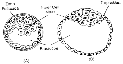
Due to pressure of growing blastocyst a slit is produced in zona pellucida. The growing blastocyst comes out. At times it gets broken into two parts which then give rise to identical twins.
Trophoblast cells in contact with embryonal knob are called cells of Rauber. Area of embryonal knob represents animal pole.
The opposite side is embryonal pole. Soon embryonal knob shows rearrangement to form embryonal disc. Cells of trophoblast layer divide periclinally.
This makes trophoblast two layered, outer syncytotrophoblast and inner cytotrophoblast. The two layers later form chorion, amnion and foetal part of placenta.
IMPLANTATION
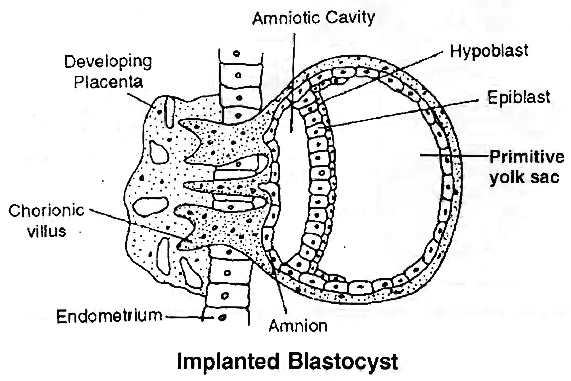
It is embedding of the blastocyst into endometrium of uterus.
Blastocyst comes in contact with the endometrium in the region of embryonal knob or embryonic disc. It adheres to the same.
The surface cells of trophoblast secrete lytic enzymes which cause corrosion of endometrial lining.
They also give rise to finger-like outgrowths called villi. Villi not only help in fixation but also absorption of nourishment.
Implantation causes nutrient enrichment, enlargement of cells and formation of uterine part of placenta called decidua (L. deciduus-falling off).
Decidua has three regions (i) Decidua Basalis (Basal Decidua, Tunica Serotina). Part of decidua underlying the chorionic villi and overlying the myometrium. (ii) Decidua Capsularis (Decidua Reflexa). It lies between embryo and lumen of uterus. (iii) Decidua Parietalis (Decidua Vera).
It is part of decidua that lines the uterus at a place other than the site of attachment of embryo.
Trophoblast secretes hormone called human chorionic gonadotropin (hCG). Detection of hCG in the urine is the basis of pregnancy/Gravidex test.
hCG maintains the corpus luteum beyond its normal life. It continues to secrete progesterone which prevents menstruation and maintains the uterine lining in nutrient rich state.
Progesterone induces the cervical glands to secrete viscous mucus for filling the cervical canal to form a protective plug.
Progesterone is also called pregnancy hormone as it is essential for maintenance of pregnancy. The hormone is secreted by placenta as well.
GASTRULATION
It is characterised by movement of cells in small masses or sheets so as to form primary germinal layers. There are three primary germinal layers -endoderm, ectoderm and mesoderm.
The cell movements that occur during gastrulation are called morphogenetic movements since they lead to initiation of morphogenesis. The product of gastrulation is called gastrula.
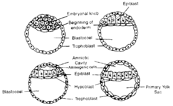
 Formation of endoderm, and amniotic cavity
FORMATION OF PRIMARY GERMINAL LAYERS
Cells of the inner cell mass or embryonal knob get rearranged to form a flat embryonic or germinal disc. The latter differentiate into two layers, outer epiblast of larger columnar cells and inner hypoblast of smaller cuboidal cells.
Gastrulation begins with the formation of the primitive streak on the surface of the epiblast.
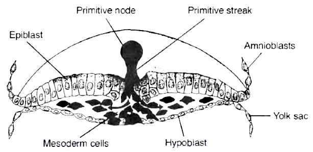
 1. Cross section through the cranial, region of the streak at 15 days showing movement of epiblast cells. The first cells to move inward displace the hypoblast to create the definitive endoderm.
2. Once definitive endoderm is established, inwardly moving epiblast forms mesoderm.
3. Cells remaining in the epiblast form ectoderm. Thus the epiblast is the source of all the germ layers in the embryo.
FATE OF GERM LAYERS
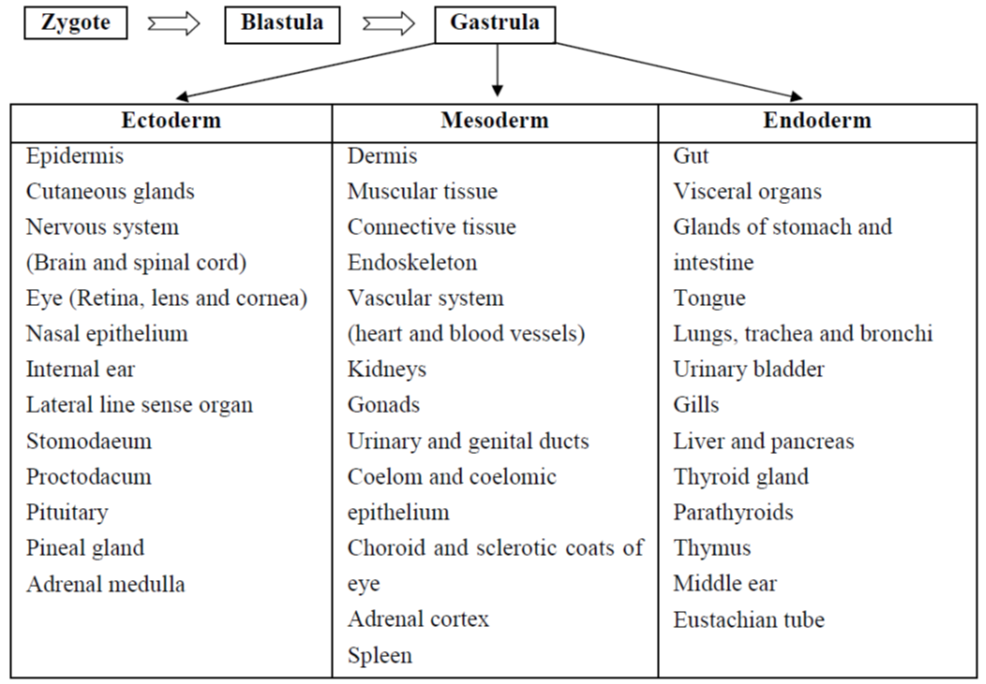
PLACENTA
Placenta is an organ which connects the foetus and uterine wall.
It is contributed by both-maternal as well as foetal part although there is no blending of the maternal and foetal blood supplies, the placenta acts as an ultrafilter, soluble inorganic and organic materials nutrients, hormones, antibodies against diphtheria, small pox, scarlet fever, measles, etc. can pass from the mother to the foetus.
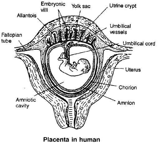
Placenta acts as an endocrine gland and synthesises large quantities of proteins and some hormones, such as human chorionic gonadotropin (hCG), chorionic thyrotropin, chorionic corticotropin, chorionic somatomammotropin, estrogens and progesterone.
The hCG stimulates the corpus luteum to secrete progesterone until the end of pregnancy. In addition, it secretes relaxin that facilitates parturition by softening the connective tissue of the symphysis pubica.
The metabolic activity of the placenta is almost as great as that of the foetus itself. The umbilical cord connects the foetus to the placenta.
During the first trimester (first 3 months) of pregnancy, the basic structure of the baby is formed.
This involves cell division, cell migration, and the differentiation of cells into many types found in the baby. During this period, the developing baby-called foetus is very sensitive to anything that interferes with the steps involved.
Virus infection of the mother e.g., by Rubella (German measles) virus or exposure to certain chemicals, may cause malformations in the developing embryo. Such agents are called teratogens (monster forming).
By 3 months, all the systems of the baby have been formed, at least in a rudimentary form.
From then, development of the foetus is primarily a matter of growth and minor structural modifications.
The foetus is less susceptible to teratogens.
Table-I: Important Developmental Events in the Human Embryo
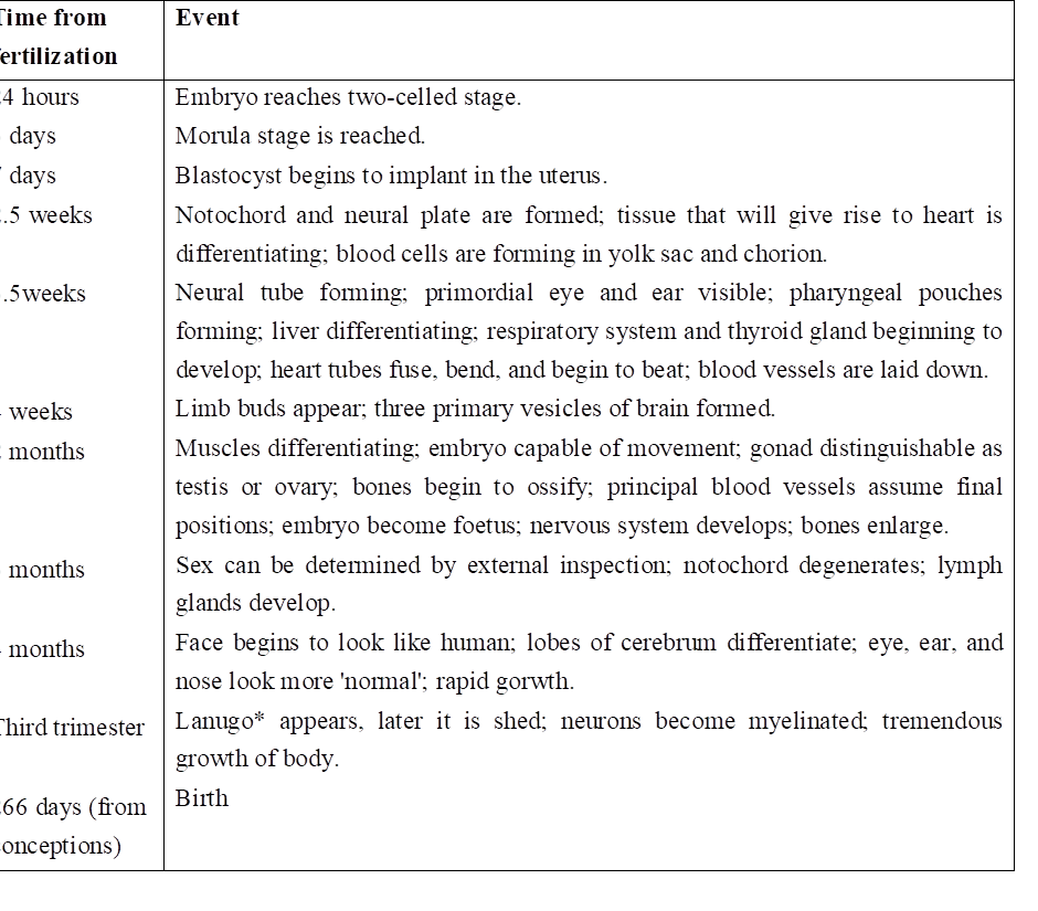
* Lanugo is the soft hairy covering of the foetus which begins to be shed before birth.
parturition and lactation
- Books Name
- A TEXT OF BIOLOGY - CLASS XII
- Publication
- ACME SMART PUBLICATION
- Course
- CBSE Class 12
- Subject
- Biology
PARTURITION
The gestation period of the human is about 38 weeks/266 days followed by birth. The process of giving birth to a baby is called parturition.
It starts with rise in estrogen/progesterone ratio, increase in the level of oxytocin secretion by both, mother and foetus.
It includes 3 stages.
1. Dilation Stage:
The uterine contraction starts from top and occur at long intervals (once every thirty minutes). This forces the baby to push its head against cervix.
As a result, cervix gets dilated with vagina also showing similar dilation. Dilation of cervix increases the stimulus for oxytocin secretion, further increasing the strength and frequency of contractions (1-3 every minute).
With continued powerful contractions, the amnion ruptures and the amniotic fluid flows out through vagina.
2. Expulsion Stage:
With further increase in the intensity of uterine and abdominal contraction, the baby comes out through cervix and vagina with head coming out first.
It may take 20 -60 min. Umbilical cord is cut. The infant's lungs expand and it begins breathing. This requires a major switch over in the circulatory system.
Blood flow through the umbilical cord ductus arteriosus and foramen ovale ceases; the adult pattern of blood flow through the heart, aorta and pulmonary arteries begins.
In some infants, the switch over is incomplete, and blood flow through the pulmonary arteries is inadequate. Failure to synthesise enough nitric oxide (NO) is one cause.
3. After Birth:
Within 10-15 minutes after delivery, the placenta and the remains of the umbilical cord which is called 'after birth' is expelled out.
LACTATION
Although estrogen and progesterone are essential for the physical development of the breasts during pregnancy, a specific effect of both these hormones is to inhibit the actual secretion of milk. Conversely, the hormone prolactin has exactly the opposite effect on secretion: promotion of milk secretion.
This hormone is secreted by the mother's anterior pituitary gland and its concentration in her blood rises steadily from the fifth week of pregnancy until birth of the baby, at that time it has risen to 10 to 20 times the normal nonpregnant level.
In addition, the placenta secretes large quantities of human chorionic somatomammotropin, which probably also has lactogenic' properties, thus supporting the prolactin from the mother's pituitary during pregnancy.
The fluid that is secreted in the last few days before and first few days after parturition is called colostrum, it contains essentially the same concentrations of proteins and lactose as milk but almost no fat.
Ejection (or "Let-Down") Process in Milk Secretion
Milk is secreted continuously into the alveoli of the breasts, but milk does not flow easily from the alveoli into the duct system and, therefore, does not continually leak from the breast nipples.
Instead, the milk must be ejected from the alveoli into the ducts before the baby can obtain it. This is caused by a combined neurogenic and hormonal reflex that involves the posterior pituitary hormone oxytocin.
When the baby suckles, sensory impulses are transmitted through somatic nerves from the nipples to the mother's spinal cord and then to her hypothalamus, there causing nerve signals that promote oxytocin secretion at the same time they cause prolactin secretion.
The oxytocin is carried in the blood to the breasts, where it causes myoepithelial cells (that surround the outer walls of the alveoli) to contract, thereby expelling the milk from the alveoli into the ducts.
Summary of Human Pregnancy from Fertilization to Birth of the Baby
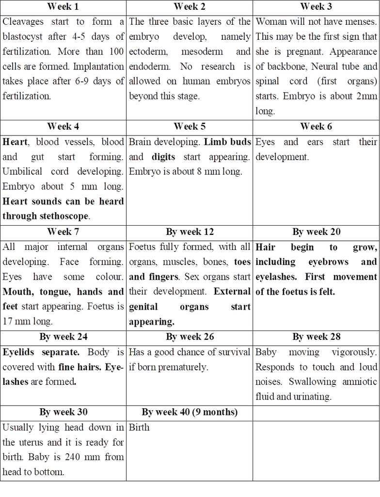
Concept Builder
Types of Placenta
(a) On the basis of structure, the placentae are of following types:
(i) Epitheliochorial - Placenta with all the six barriers between foetal and maternal blood; e.g. Horse, Ass.
(ii) Syndesmochorial - Uterine epithelium breaks down; only five barriers left; e.g. Cow, Buffalo, Sheep, Goat, Camel.
(iii) Endotheliochorial - Uterine epithelium and connective tissue eroded; only four barriers left; e.g. Tiger, Lion, Cat, Dog.
(iv) Haemochorial - Placenta with only three barriers, the maternal part of placenta eroded; e.g. Human, Ape, Lemurs.
(v) Haemo-endothelial-All barriers except endothelium of foetal part of placenta get eroded e.g. Rat, Rabbit.
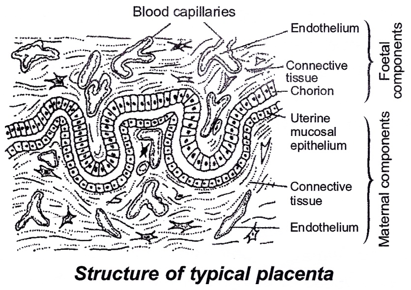
(b) On the basis of nature of uterine wall after parturition, the placenta may be :
(i) Non-deciduous-no part of uterine portion of placenta is broken off, e.g. Horse, Ass.
(ii) Deciduous-a portion of uterine tissue called decidua is detached and passed out at birth, e.g. most of the mammals.
(iii) Contra deciduous – even the foetal part of placenta is retained and gets absorbed to provide nourishment, e.g. Talpa, Parameles.
(c) On the basis of distribution of villi on the surface, the placentas are categorised into six types :
(i) Diffuse placenta-Villi distributed uniformly all over the surface. e.g. Horse, Pig .
(ii) Cotyledonary-the villi form tufts which fit into corresponding areas, the caruncles in uterine part of placenta, e.g.' Cow, Buffalo, Sheep.
(iii) Intermediate-villi occur singly as well as in tufts, e.g. Camel, Giraffe
(iv) Zonary-villi arranged in two transverse bands, e.g. Tiger, Lion, Cat, Dog, Elephant.
(v) Discoidal-When the villi are confined to a disc-like area, e.g. Rat, Rabbit, Bat.
(vi) Metadiscoidal-The placenta in which the villi are initially distributed uniformly all over the surface but later on get confined to a disc-like area fitting into a corresponding depression on the uterine wall. That is, the placenta is diffuse at first but later on becomes discoidal, e.g. Human beings and Apes.
Concept Builder
1. Amenorrhoea: Non occurrence of menses.
2. Inguinal hernia: Protrusion of intestinal loop into scrotum
3. Spontaneous ovulator : Ovulation with any external induction.
4. Orchidectomy: Removal of testes. It produces eunuchs.
5. Induced ovulator : Ovulation after copulation e.g., Rabbit.
6. Hysterectomy: Surgical removal of uterus.
7. Unejaculated sperms: Sperm production is a continuous process. unejaculated sperms are absorbed in vas deferens.
8. Corpus luteum : Persists for two weeks in case of non-pregnancy and four months when pregnancy has taken place.
9. Cryptorchidism: Failure of testes to descend in to scrotum.
10. Spermatogenesis takes about 74 days.
11. Androgenesis: Development in which the embryo has only paternal chromosomes, male parthenogenesis.
12. Gynogenesis : Development in which the embryo has only female chromosomes, female parthenogenesis.
13. Mictic females produce: haploid eggs which can be fertilized by males. Amictic females produce diploid eggs which develop parthenogenetically. A diploid egg may be produced by -autofertilization (polar body fuses with secondary oocyte) or Restitution: Chromosomes of both secondary oocyte and polar bodies arrange at equator during 1st cleavage e.g. hymenopterans (bees and wasps)
14. Paras: Fertile, can have pregnancy
15. Nulliparous: Women with no children
16. The most common method by which a baby comes out at parturition is left anterior oblique with head coming out first.
17. Fetal hemoglobin differs from adult hemoglobin because it has and chains. It has higher affinity for O2 and shifts HbO2 dissociation curve to left.
18. 1st Menses is called menarche while the onset of menses and development of secondary sexual characters is called puberty. Dysmenorrhoea - painful menses.
19. In the three trimesters of preganancy. (0-12 weeks 12-22 weeks and 22-28 weeks) if the baby dies from 0-22 weeks by natural or man-made causes it is called abortion because embryo is not called viable.
20. After 20-22 weeks abortion is banned in India.
21. The number of eggs at the time of birth in a young female undergoes drastic reduction by a process called atresia.
22. Gynandromorphs: An individual possessing both male and female parts.
23. Menopause: Stopping of menses, average age 51 years. Post menopausal effects includes increased risk to heart attack due to decreased estrogens, hot flushes, irritability, fatigue, anxiety decreased strength of bones.
24. Implantation takes place in 7 days. Conceptus remains in fallopian tube for 3-4 days and then by 7-9 days implantation occurs.
25. Vasectomy : Cutting of vas deferens.
26. Gynaecomastia: Males developing breasts due to hormonal imbalances.
Gestation: The period of development of the young in the womb of the mother is called gestation. Maximum weight gain and maximum growth rate of foetus takes place between 7-9 months of pregnancy.
27. Human Egg 0.1 mm in diameter or 100 µm.
28. Human sperm 0.06 mm or 60 µm long
29. Largest sperm 5.8 cm (20 times longer than the body of fly) in Drosophila bifurca.
30. Ectopic Pregnancy : It is the growth of the foetus outside the uterus, commonly inside the fallopian tube. Hence also called as tubal pregnancy. By 6th week as growing foetus may cause rupturing of fallopian tube hence its immediate surgical removal.
31. Animal cloning: First performed by Willmut and Campbell (1997). The first cloned animal was sheep Dolly (Feb. 13, 1997). Egg cell was taken out when maturation promoting factors (MPF) were maximum. It was denucleated. A nucleus from udder cell arrested in non-dividing growth phase (GP) by serum starvation was inserted in the denucleated egg.
32. Spermatozoa of turbellarians and Ascaris (nematode) and sporozoans lack acrosome.
33. Non flagellate sperms are found in Ascaris, Procambarus (Crayfish) Callinectes (blue crab).
34. Biflagellate spermatozoa are found in Opsanus (toad fish)
35. Ascaris megalocephola univalens shows the process of chromosome dimunition. In these spiral clevage of egg takes place and each cell except the one giving rise to gametes looses a part of chromosome set. Other e.g. -Gall midges (Mayetiola) and fungus grats (Wachtiella)
36. Compact ovaries are found in -cyclostomes, elasmobranchs, osteichthyes fishes, reptiles, birds and mammals.
37. Saccular ovaries are found in amphibians.
38. Lampbrush chromosomes : Found in oocytes of amphibians. They resemble the brushes used to clean the chimneys in London. They store extra information for survival of egg. They consist of two homologous chromosomes held together at chiasmata present in (diplotene) prophase cell stage
39. Polytene chromosomes : Found in the interphase stage of cells of salivary glands of chromosomes of Drosophila larvae. They exhibit gene amplification.
40. Fecundity: Animals potential capability to produce off springs.
41. Fertility -Animal's actual capability to conceive or induce conception.
42. Precocial young ones: ungulates and jackrabits -hair and open eyes, ability to move around.
43. Altricial: young ones are naked blind and helpless.
44. Estrous cycle: When females are most receptive to male (not in man) -phases -Estrus -Metaestrus -Diestrus -Proestrus. It occurs in non-primates.
45. Rut cycle: Testicular activity in the majority of mammals, is a seasonal event as is the associated period of sexual excitement.
46. F.R. Lillie gave the fertilization theory. Fertilizin of egg of one species locks with antifertilizin of same species (1914).
47. Sperms are decapacitated in epididymis where they mature. They are recapacitated in vaginal fluid / uterus.
48. Spermetogensis : Is the process of formation of haploid sperm from the diploid primary spermatocyte in the testis.
49. Spermiogenesis : Is the process of transformation of haploid spermatid to the functional sperm.
50. Spermiation : Is the process of releasing the mature sperms from the semniferons tubuels.
51. Insemination : Is the process of introduction of sperms (or semen) in the female reproductive system.
52. Semination : Is the process of ejaculation of semen from the male reproductive system.
53. Menarche : Is the process of start of menstrual cycle in the life of a female human at puberty.
54. Follicular phase : Primary follicles in the ovary grow to become mature follicle (Graafian follicle).
55. Luteal phase : The process of transformation of ruptured Graafian follicle into corpus luteum.
56. Capacitation : Is the process of activation of sperms in the female reproductive system after their ejaculation into it.
introduction to reproductive health
- Books Name
- A TEXT OF BIOLOGY - CLASS XII
- Publication
- ACME SMART PUBLICATION
- Course
- CBSE Class 12
- Subject
- Biology
REPRODUCTIVE HEALTH
According to World Health Organisation (WHO), reproductive health means a total well-being in all aspects of reproduction, i.e., physical, emotional, social and behavioural.
Thus, a society with people who have physically and functionally normal reproductive organs and normal emotional and behavioural interactions among them in all sex-related aspects might be called reproductively healthy.
REPRODUCTIVE HEALTH -PROBLEMS AND STRATEGIES
Problems and strategies of reproductive health in human beings are explained as follows.
(i) Over population :
Main problem of India is its excess population which is directly connected with reproductive health.
To achieve total reproductive health, some plans and programmes were started.
Family planning programme was initiated in 1951 and was periodically assessed.
These programmes were popularly named Reproductive and Child Health-care (RCH).
The major tasks carried out under these programmes are to provide facilities and support for building up a reproductive healthy society.
(ii) Awareness about reproduction :
Audio-visual and print media, governmental and non-governmental agencies are doing good job to create awareness among people about reproduction in humans.
Parents, close relatives, friends and teachers also have a major role in giving the above information.
(iii) Sex Education :
Sex education in schools should also be introduced and encouraged to provide right information about myths and misconceptions about sex-related aspects.
(iv) Knowledge of growth of reproductive organs and STDs :
Proper information about reproductive organs, adolescence (period of rapid growth between childhood and adulthood), safe and hygienic sexual practices, sexually transmitted diseases (STDs), e.g., AIDS etc., would help to lead a reproductive healthy life.
(v) Birth control devices and care of mother and child (Prenatal, Natal and Post-natal Care) :
Fertile couples and people of marriageable age group should know about available birth control devices, care of pregnant mothers, postnatal (after birth) care of the mother and child, importance of breast feeding, equal importance for the male and female child, etc.
(vi) Prevention of sex abuse and sex related crime:
Awareness of problems due to uncontrolled population growth, social evils like sex abuse and sex-related crimes, etc. need to be created so that people should think and take up necessary steps to prevent them and thereby build up a reproductively healthy society.
(vii) lnformation about reproduction related problems:
For successful action plans to attain reproductive health, requires good infrastructural facilities, professional expert knowledge and material support.
These are necessary to provide medical help and care for reproduction related problems like menstrual problems, infertility, pregnancy, delivery, contraception, abortions, sexually transmitted diseases (STDs).
Implementation of better techniques and new strategies are also required to provide better care and help to people for reproductive health.
(viii) Amniocentesis -Meaning and Use:
 Amniocentesis is a foetal sex determination and disorder test based on the chromosomal pattern in the amniotic fluid surrounding the developing embryo.
Procedure:
Amniotic fluid contains cells from the skin of the foetus and other sources.
These cells can be used to determine the sex of the infant, to identify some abnormalities in the number of chromosomes and to detect certain biochemicals and enzymatic abnormalities.
If it is established that the child is likely to suffer from a serious incurable congenital defect, the mother should get the foetus aborted.
Misuse of Amniocentesis: It is being used to kill the normal female foetus. Female foeticide is illegal.
(ix) Research in reproductive health area:
It should be encouraged and supported to find out new methods.
"Saheli" a new oral contraceptive for the females was developed by our scientists at Central Drug Research Institute (CDRI) in Lucknow, India.
(x) Medical facilities :
Better awareness about sex related problems, prenatal care of mother, medically assisted deliveries and post-natal care of mother and infant, decrease maternal and infant mortality. Small families, better detection and cure of sexually transmitted diseases (STDs) and increased medical facilities for sex-related problems, etc. indicate improved reproductive health of male and female individuals and children.
Measures to control Over Population
1. Education:
People, particularly those in the reproductive age group, should be educated about the advantages of a small family.
Mass media and educational institutions can play an important role in this campaign.
Posters showing a happy couple and two children with a slogan "Hum Do Humare Do" should be displayed.
Many couples even adopted "one child norm".
2. Marriageable Age :
Raising of the age of marriage is more effective means to control the population (now marriageable age of female is 18 years and that of male is 21 years).
3. Incentives: Couples with small families should be given incentives.
4. Family planning: There are many birth control measures which can check birth rate.
methods of birth control
- Books Name
- A TEXT OF BIOLOGY - CLASS XII
- Publication
- ACME SMART PUBLICATION
- Course
- CBSE Class 12
- Subject
- Biology
POPULATION EXPLOSION AND BIRTH CONTROL
Rapid increase in population over relatively short period is called population explosion.
Census gives information about the number of individuals present in a given region at a given time.
World Population 1900 2 billion
2000 6 billion
Indian Population 1947 350 million
2000 1 billion
It means every sixth person in the world is an Indian.
The time required for a population to double itself is called the doubling time.
The present growth rate of approximately 1.6 percent per year for India is smaller than the peak of about 2.1 percent per year during 1965-1970.
Population growth rate is indicated by (i) the annual average growth rate and (ii) the doubling time.
Growth rate depends on birth (fertility) rate, death (mortality) rate migration and age sex ratio.
The major reasons for this growth are :
(i) A rapid decline in death rate
(ii) A decline in maternal mortality rate (MMR)
(iii) A decline in infant mortality rate (IMR)
(iv) Increase in the number of people reaching reproducible age
BIRTH CONTROL
Is the process to prevent conception or pregnancy without interfering the reproductive health of the individuals.
Characteristics of an ideal contraceptive are: (i) User friendly, i.e., comfortable and easy to use. (ii) Without side effects. (iii) Reversible. (iv) Completely effective against pregnancy.
There are several methods of contraception-natural or traditional, barriers, IUDs, oral contraceptives, injectables, implants and surgical methods.
Couple protection is the process of bringing eligible couples under family planning measures. In India, it is over 55% at present and is voluntary in nature.
In 2004, there were 60·79 lakh IUD insertions, 48·74 lakh sterilisations, 249·9 lakh condom users and 87·54 lakh oral pill users.
METHODS OF BIRTH CONTROL
1. Natural Methods:
These are the methods which do not require any device or medicine so no side effects but chances of failure are very high.
Natural methods are of three kinds-safe period, withdrawal and breast feeding.
(i) Safe period (Rhythm Method) :
Ovulation occurs roughly about the middle of menstrual cycle.
Fertility period is upto 48 hours after ovulation, when fertilization can occur.
Avoiding sex during the fertility period will naturally prevent conception.
Ovulation period can be known by the rise in body temperature by about 1º F, cervical mucus is slippery and can be drawn into a thread (Spinnbarkeit test) when stretched between two fingers.
Period prior to ovulation is safe.
Period after fourth day of rise in body temperature (or last positive Spinnbarkeit test) is also considered safe.
It is however, always better to avoid sex from day 10-17 of the menstrual cycle.
(ii) Withdrawal Method (Coitus Intruptus) :
The method is based on withdrawal of penis from the vagina before ejaculation.
This method has high failure rate due to pre-ejaculatory release of sperms or failure to withdraw penis from the vagina before ejaculation.
(iii) Lactational Amenorrhoea :
Just after parturition, there is a phase of amenorrhoea or absence of menstruation.
It is also the phase of intense lactation.
Breast feeding the child fully prevents conception.
The method is, however, effective only upto a maximum period of six months.
Concept Map
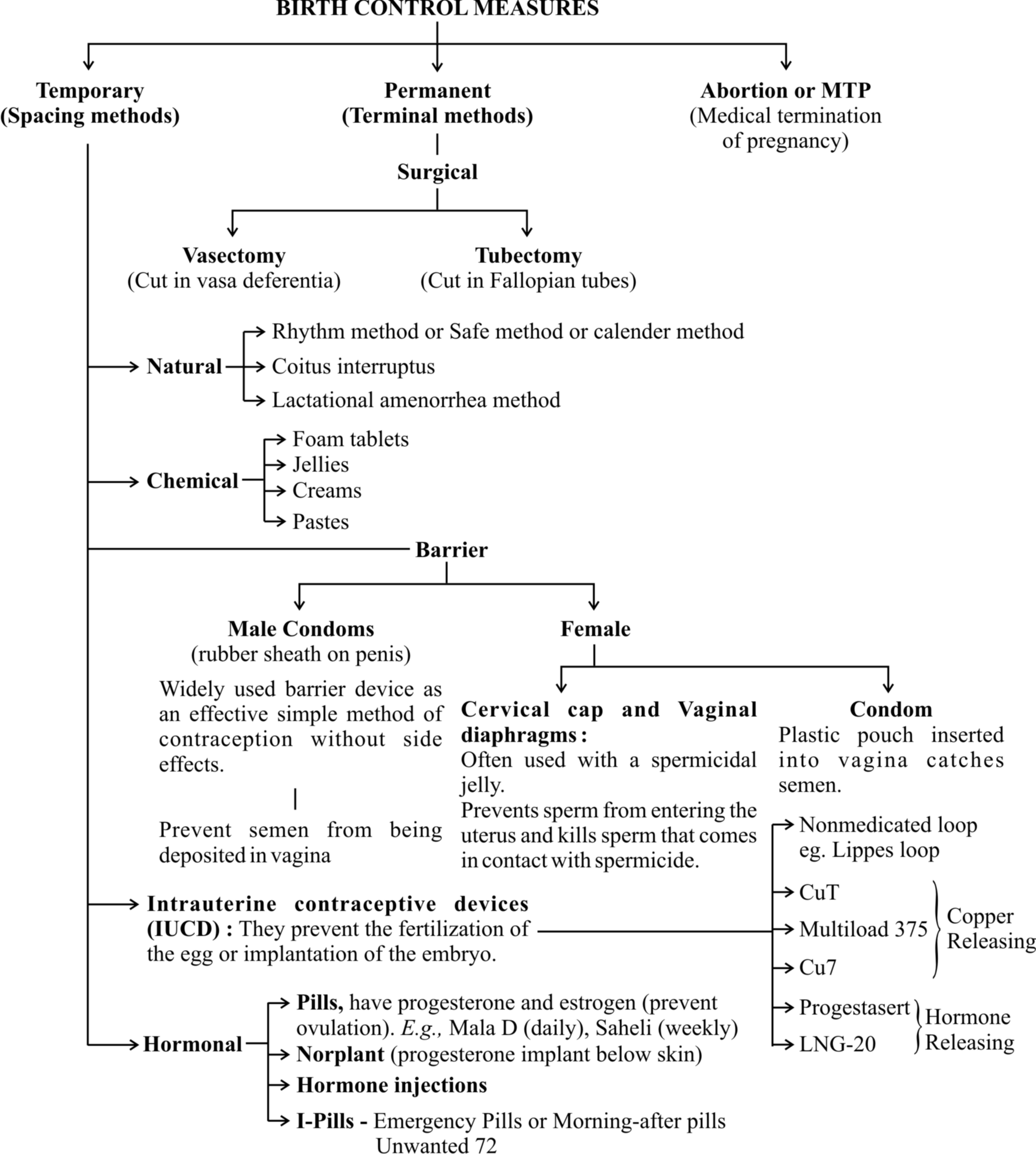
2. Barrier Methods :
These are mechanical devices which prevent the deposition of sperms into vagina and their passage into the uterus.
Further, they can be self inserted by the user in complete privacy.
The common barrier methods are condoms, diaphragm, fem shield and cervical cap.
(i) Condom:
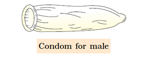
It is tubular latex sheath which is rolled over the male copulatory organ during sex.
The common brand provided by family welfare services is Nirodh.
The device also provides protection against sexually transmitted diseases including AIDS.
(ii) Fem Shield (Female Condom) :
The device is polyurethane pouch with a ring at either end.
The inner ring is smaller and present at the inner closed end.
The device covers the external genitalia as well as lines the vagina.
Fem shield provides protection from sexually transmitted diseases also.
(iii) Diaphragm:
It is a tubular rubber sheath with a flexible metal or spring ring at the margin which is fitted inside the vagina.
 (iv) Cervical Cap:
It is rubber nipple which is fitted over the cervix and is designed to remain there by suction.
The device prevents the entry of sperms into the uterus.
 (v) Vault Cap :
It is hemispheric dome like rubber or plastic cap with a thick rim which is meant for fitting over the vaginal vault over the cervix.
3. Chemical Methods:
These are contraceptives which contain spermicidal chemicals.
The chemical contraceptives are available in the form of creams (e.g., delfen), jelly (perceptin, volpar paste), foam tablets (e.g., aerosol foam, chlorimin T or contab).... etc.
These commonly contain lactic acid, boric acid, citric acid, zinc sulphate and potassium permanganate.
The contraceptives are introduced in vagina prior to sex.
Sponge (Today) is a foam suppository or tablet containing nonoxynol-9 as spermicide. It kills the sperm by disrupting the membrane. It is moistened before use to activate the spermicide. The device also absorbs the male ejaculate.
4. Intrauterine Devices (IUDs) (Intrauterine Contraceptive Devices or IUCDs) :
 These devices are inserted by doctors or expert nurses in the uterus through vagina. IUDs affect motility of sperms within the uterus. These IUDs can be –
(a) Non-medicated (e.g., Lippes loop)
(b) Copper releasing (e.g., CuT, Cu7, Multiload 375) – The copper ions suppress the motility and fertilization capacity of sperms.
(c) Hormone releasing (e.g., Progestasert, LNG-20)- The hormone releasing IUDs, in addition, make the uterus unsuitable for implantation and the cervix hostile to the sperms.
IUDs are ideal contraceptives for the females who want to delay pregnancy and / or space children. It is one of the most widely accepted methods of contraception in India.
5. Oral Contraceptives (Oral Pills) :
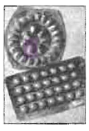
 Are preparations containing either progestin (= progestogen = progesterone) alone or a combination of progestogen and oestrogen (= estrogen).
The pills are taken orally for 21 days in a menstrual cycle starting from 5th day and ending on 25th day.
However, it is advisable to restart the course after a gap of 7 days irrespective of the onset or nonset of menstruation during the pill free days.
When a pill is missed, it should be taken whenever one remembers, sometimes two at a time.
This helps in keeping the hormonal level optimum for contraception.
Hormonal pills act by four ways :
(a) Inhibition of ovulation.
(b) Alternation in uterine endometrium to make it unsuitable for implantation.
(c) Changes in cervical mucus impairing its ability to allow passage and transport of sperms.
(d) Inhibition of motility and secretory activity of fallopian tubes.
Oral pills are of two types, combined and minipills.
Combined pills contain both oestrogen and progestin.
They are synthetic products.
Oestrogen is an ovulatory, that inhibits FSH production. Progestin is anovulatory that inhibits LH production.
It protects the endometrial lining from adverse effect of oestrogen.
The hormone also changes cervical mucus.
The most commonly used progestin is levonorgestrel or desogestrel.
The most common oestrogen is ethinyl oestradiol or menstranol.
In monophasic combined pill, both oestrogen and progestin are present in nearly the same amount, e.g., Mala D, Mala L.
In multiphasic combined pills, oestrogen is maintained at the same level throughout the 21 day course (0.03 mg). But the amount of progestin is increased (0.05 mg for first six days, 0.075 for next 5 days and 0.125 mg for last ten days), e.g., triquilar, orthonovum.
Minipills are progestin pills only (with no estrogen). They are taken daily without break.
Saheli, a nonsteroidal preparation, is taken once a week after an initial intake of twice a week dose for 3 months.
6. Injectable Contraceptives (Depo-Provara) :
Two types of progestin preparations are used singly.
They are depot-medroxy progesterone acetate (DMPA) 150 mg every three months or 300 mg every six months and norethisterone enanthate (NET EN) 200 mg every two months.
Cyclofem and mesigna and combined injectable contraceptives which are given once every month.
They contain progestin preparation as well as oestradiol.
7. Implants:
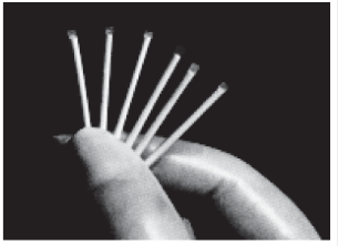
 They are hormone containing devices which are implanted subdermally for providing long term contraception.
Norplant is progestin only. The device is with six small permeable capsules (34 mm × 2.4 mm) each with about 36 mg levonorgestrel.
They are inserted under the skin in a fan shaped manner inside upper arm or fore arm through a small incision.
Suturing is not required. Norplant remains effective for about 5 years.
Implanon is a single rod-like device which is implanted through a wide bored needle. It contains 3-keto desogestrel. It remains functional for three years.
Average Failure Rate of Various Contraceptive Techniques
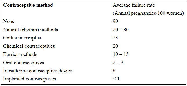
8. Emergency Contraception :
It is treatment for unprotected sex, sexual assault, missed pills and other reasons which have risk of pregnancy.
The drugs used for treating emergency contraceptions are called morning-after pills.
They are also available in India under Family Welfare Programme since 2002-2003.
Two ovral tablets to start and two tablets after 12 hours provide relief.
Other morning-after pills are noral, norgynon and ovidon.
An antiprogesterone pill (mifepristone) is a single pill treatment.
Insertion of IUD within five days of unprotected sex prevents implantation.
9. Surgical Methods of Family Planning :
They are also called terminal methods of family planning.
Surgical methods are permanent methods of family planning where there is no need of replacement or augmentation but the reversibility is poor.
The methods are operative procedures which block the passage of semen in males and ova in females.
The techniques are also called sterilisation procedures.
They are called vasectomy in males and tubectomy in females.
Vasectomy (L. vas-vessel, ektome-excision) :
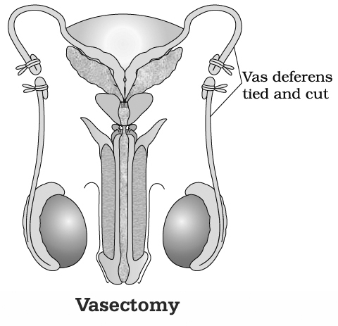
It is a surgical method of sterilisation of males.
Vasa deferentia are blocked by cutting and occluding them so that sperms are unable to pass down the male reproductive system.
(i) Conventional Vasectomy (Scalpel Surgery) :
Under local anaesthesia, transverse 1cm incision is made through the skin of the scrotum with the help of the scalpel over the area of vasa deferentia.
Each vas deferense is exposed and cut.
The two ends are separated and tied.
A gap of 1-4cm is must between the two ends otherwise reunion can occur.
(ii) No-scalpel Vasectomy:
Here instead of scalpel, a dissecting forceps and a ringed forceps are required.
The skin is punctured and the vas deferense is taken out.
It is occluded by removal of 1-2 cm followed by ligation of ends.
Occlusion can also be achieved by heat and clips.
Vasectomy is a reversible procedure as the cut ends can be joined together to open to sperm passage.
Tubectomy (L. tubes-pipe, ektome-excision):
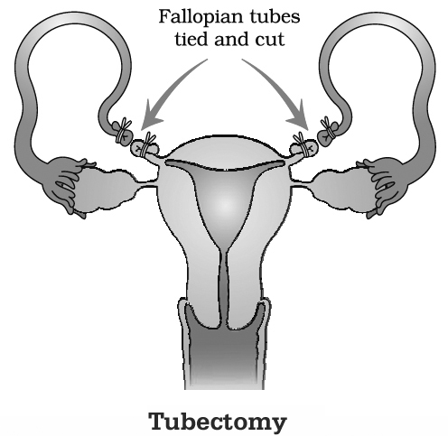
It is a surgical procedure of female sterilisation where a portion of both the fallopian tubes is excised or ligated to block the passage of ovum through them.
Tubectomy is performed by conventional transabdominal surgery, conventional laparotomy and minilaparotomy.
In surgical procedures, the fallopian tubes are cut and the cut ends are tied to prevent reunion.
The procedure is reversible as the cut ends can be rejoined.
In laparoscopic procedure, sterlisation is achieved by loop development and constricting the basal region of loop with the help of elastic ring either through a small incision in the abdomen or through vagina.
Difference between Vasectomy and Tubectomy
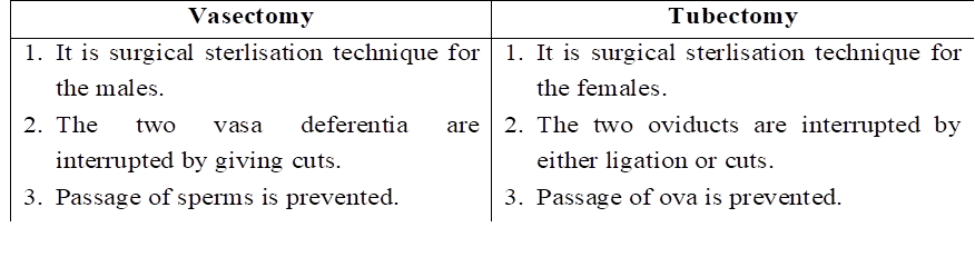
Medical Termination of Pregnancy (MTP)
It is voluntary or intentional abortion induced and performed to end pregnancy before the completion of full term.
Worldwide, nearly 20% of the total pregnancies get aborted.
The number of MTPs is 40-50 million/yr.
Therefore, MTPs have a significant role in containment of population though they are not performed for this purpose.
They are mainly meant for removing unsustainable pregnancies.
Many countries do not have a law about MTPs because the latter involve emotional, ethical, religious and social issues.
However, in India there is a proper act, Medical Termination of pregnancy Act, 1971.
It is mainly meant for preventing unnatural maternal deaths due to unsafe abortions (8.9% of the total maternal deaths).
The act has been amended in 2002.
Under this act termination of pregnancy can be done upto 20 weeks, if pregnancy is likely to produce a congenitally malformed child, is a result of rape and contraceptive failure or is likely to harm the mother.
MTP is safe if it is performed upto 12 weeks (first trimester) of pregnancy.
Misoprostol (prostaglandin) alongwith mifepristone (antiprogesterone) is an effective combination.
Vacuum aspiration and surgical procedures are adopted thereafter.
Second trimester abortions are risky.
They are generally performed after testing the sex of the baby through amniocentesis or sonography.
It has resulted in large scale female foeticide and complications due to unsafe abortions in the hands of untrained persons.
To prevent such happening, the government has enacted a law, Pre-natal diagnostic techniques (Regulation and Prevention of Misuse) Act, 1994 with amendments in 2003.
It prohibits preconception and prenatal sex determination.
sexually transmitted diseases
- Books Name
- A TEXT OF BIOLOGY - CLASS XII
- Publication
- ACME SMART PUBLICATION
- Course
- CBSE Class 12
- Subject
- Biology
SEXUALLY TRANSMITTED DISEASES
The general term sexually transmitted disease (STD) is applied to any of the large group of diseases that can be spread by sexual contact.
The group includes conditions traditionally specified as venereal diseases (VD), such as chlamydia, gonorrhea, syphilis, and genital herpes.
AIDS and hepatitis, which are sexually transmitted diseases and are that also can be contracted in other ways also.
Some common sexually transmitted diseases (STDs)
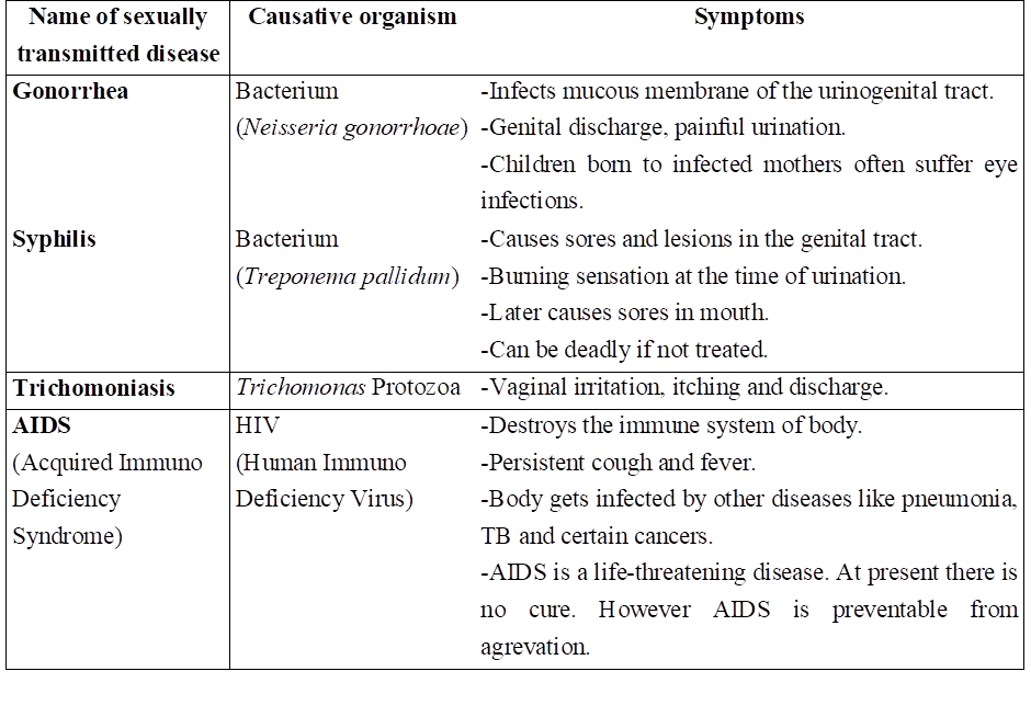
infertility
- Books Name
- A TEXT OF BIOLOGY - CLASS XII
- Publication
- ACME SMART PUBLICATION
- Course
- CBSE Class 12
- Subject
- Biology
INFERTILITY
Infertility (L. in-not, fertilis-fruitful) is failure to conceive even after 1-2 years of regular unprotected sex.
The term is not synonym of sterility which means complete inability to produce offspring.
Infertility can best be defined as relative sterility.
It is of two types, primary and secondary.
Primary infertility is the infertility found in patients who have never conceived.
Secondary infertility is found in patients who have previously connceived.
Infertility is caused by defects found in males, females as well as both.
Infertility in Males
Semen of a fertile male is 2.5 to 5 ml per ejaculation with a sperm count of over 200-300 million, mostly motile, having proper fructose content and fluidity which is deposited high in the vagina.
Any defect in sperm count, sperm structure, sperm motility of seminal fluid leads of infertility.
Low sperm count is called oligospermia while near absence of sperms is known as azoospermia.
Low sperm motility is called asthenozoospermia while defective sperm morphology is termed as teratozoospermia.
Infertility in Females
A fertile woman is the one who regularly ovulates once every cycle, passes the egg down the reproductive tract which develops conditions for smooth passage of sperms and implantation of fertilised egg.
The various causes of infertility in females are as follows :
1. Anovulation (nonovulation) and oligoovulation (deficient ovulation).
2. Inadequate growth and functioning of corpus luteum.
3. The ovum is not liberated but remains trapped inside the follicle due to hyperprolactinaemia.
4. Fallopian tube may fail to pick up ovum.
5. Noncanalisation of uterus.
6. Defective uterine endometrium.
7. Fibroid uterus.
8. Defects in cervix.
9. Defective vaginal growth.
ASSISTED REPRODUCTIVE TECHNOLOGIES (ART)
More than two decades ago, in an experimental procedure called invitro fertilization (IVF), doctors joined a woman's egg and a man's sperm in a glass dish on the laboratory table.
For the first time, fertilization happened outside a woman's body. Nine months later, the first test-tube baby was born.
Today, assisted reproductive technology (ART) refers not only to IVF but also to several variations tailored to patient's unique conditions.
These procedures are usually paired with more conventional therapies, such as fertility drugs, to increase success rates.
Almost one out of every three cycles of ART results in the birth of a baby.
But ART procedures are invasive and expensive.
Though no long-term health effects have been linked to children born using ART procedures, most doctors recommend reserving ART as a last resort for having a baby.
Following is the list of important techniques which could benefit such infertile couples.
IVF & ET
In Vitro Fertilization (IVF) is the fertilization outside the body, in almost similar conditions as that in the body, which is followed by embryo transfer (ET). In this method, popularly known as, Test-Tube baby method, the ova from the wife/donor (female) and sperms from the husband/donor (male) are collected and are induced to form zygote under simulated conditions in the laboratory. The zygote or early embryos are then transferred into fallopian tube or uterus to complete its further development.
1. ZIFT
In ZIFT (Zygote Intra Fallopian Transfer) the zygote, formed in vitro, or early embryo up to 8-blastomere, is transferred into fallopian tube.
2. IUT
In IUT (Intra Uterine Transfer) the embryos with more than 8 blastomeres are transferred into the uterus for further development. The embryos formed by in-vivo fertilization can also be used for transfer to assist those females who cannot conceive.
3. GIFT
The GIFT (Gamete Intra Fallopian Transfer) is the transfer of an ovum collected from a donor into the fallopian tube of the recipient who can provide suitable environment for fertilization and further development.
4. ICSI
In ICSI (Intra Cytoplasmic Sperm Injection) is another specialized procedure to form an embryo in the laboratory in which a sperm is directly injected into the ovum.
5. AI
The AI (Artificial Insemination) is used for the cases of infertility which is either due to inability of the male partner to inseminate the female, or due to very low sperm counts in the ejaculate.
In this technique, the semen collected either from the husband or a healthy donor is artificially introduced either into the vagina or into the uterus (IUI -intra-uterine insemination) of the female.
ART requires extremely high precision handling by specialized professionals and expensive instrumentation. The infertility facilities are presently available only in very few centres in the country.
Obviously their benefits are affordable to only a limited number of people. Emotional, religious and social factors are also involved in the adoption of these methods.
Adoption
Our law also permits legal adoption. Adoption can benefit not only the people who are looking for parenthood but also to many orphaned and destitute children in India, who would probably not survive till maturity, unless taken care of.
Surrogacy or use of a gestational carrier: Another woman carries embryo or a donor embryo to term.
Concept Builder
Conventional Methods of Contraception: Rhythm method, spermicides, vaginal diaphragms and condoms.
Gossypol : It is a yellow pigment (C30H30O8) present in cotton seed taken orally (10-20 mg) daily for 3 months and thereafter twice weekly (20 mg) inhibits spermatogenesis.
Saheli : (Centchroman) is nonsteroidal drug taken orally twice a week for three months and then once a week is a potent contraceptive which prevents implantation but does not inhibit ovulation. It is a research product of Central Drug Research Institute, Lucknow.
IVE-ET — In vitro fertilisation and embryo transfer
GIFT — Gamete intra-fallopian transfer
ZIFT — Zygote intra-fallopian transfer
lUI — Intrauterine insemination
POST — Peritoneal oocyte and sperm transfer
SUZI — Subzonal insemination
ICSI — Intra-cytoplasmic sperm injection
TESE — Testicular sperm extraction
MESA — Microsurgical epididymal sperm aspiration
