 ACME SMART PUBLICATION
- Books Name
- A TEXT OF BIOLOGY - CLASS XII
- Publication
- ACME SMART PUBLICATION
- Course
- CBSE Class 12
- Subject
- Biology
PRE-FERTILIZATION : Structures and Events
Much before the actual flower is seen on a plant, the decision that the plant is going to flower has taken place. A number of hormonal and structural transformations occur prior to initiation of flowering.
Shoot apical meristem is transformed into reproductive meristem.
Reproductive meristem grows to form inflorescence axis over which floral primordia develop.
The primordia grow into floral buds and then flowers. In the flower, the androecium and gynoecium differentiate and develop.
Stamen, Microsporangium and Pollen grain
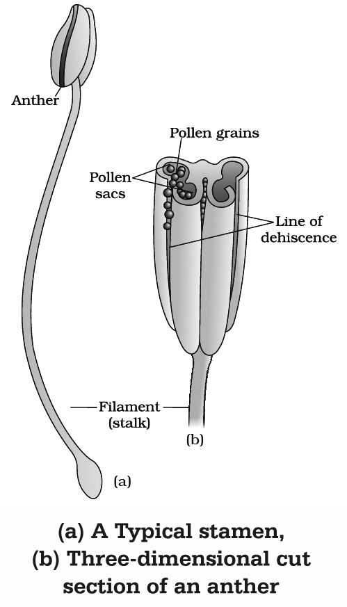
(a) Stamen or Microsporophyll (Male sex organ)
It consists of two parts :
(i) Anther
It is broader knob-like fertile part.
A typical angiospermic anther has two lobes and such anther is called dithecous.
Dithecous anther is a four sided (tetragonal) structure consisting of four microsporangia located at the corners, two in each lobe.
(ii) Filament
It is sterile, long and slender stalk.
The proximal end of the filament is attached to the thalamus, petal or tepal.
Concept Builder
(i) Floriculture is the science of cultivation, breeding, marketing and arrangement of flowers.
(ii) In members of Malvaceae, anther consists of one lobe and two microsporangia. Such anthers are monothecous and bisporangiate.
(iii) In Arceuthobium, the smallest dicot parasite, anther consists of one microsporangium, i.e., monothecous and monosporangiate
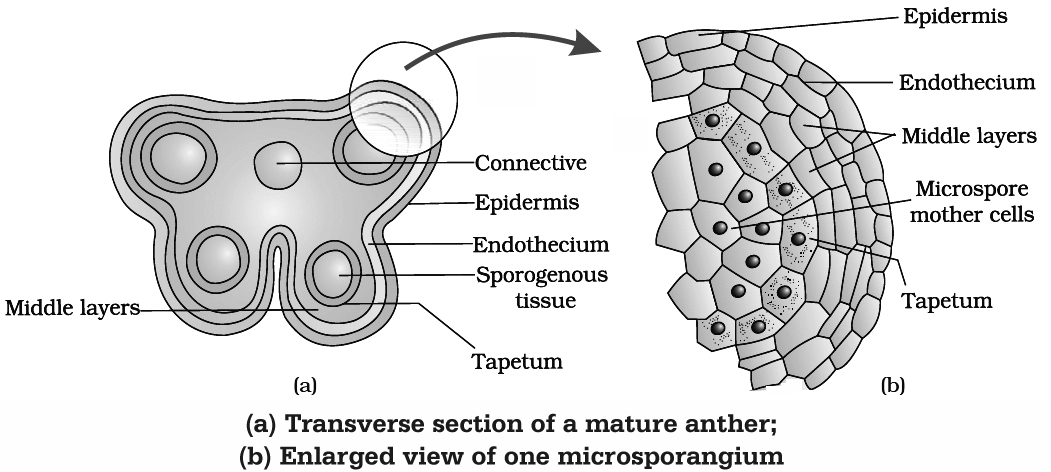
(b) Structure (T.S.) of Anther
A young anther consists of homogenous mass of meristematic cells called primary sporogenous cells surrounded by anther wall. Primary sporogenous cells form microspore mother cells (2n) inside the microsporangium.
Anther Wall Layers : Anther wall consists of following layers :
(1) Epidermis : Outermost single layered and protective in function. The epidermis of Arceuthobium develops some fibrous thickenings and is called exothecium.
(2) Endothecium : Cells of this layer have -cellulosic fibrous bands arising from inner tangential wall which help in dehiscence of anther due to their hygroscopic nature. Fibrous bands are absent in hydrophytes, e.g., Hydrocharitaceae.
(3) Middle layer : Cells of this layer are ephemeral and are 1-3 layered. It degenerates at maturity.
(4) Tapetum : This is the innermost layer of anther wall which surrounds the sporogenous tissue. Tapetal cells nourishes the developing pollen grains. Cells of the tapetum possess dense cytoplasm and generally have more than one nucleus. They are polyploid. The tapetal cells show increase in their DNA content.
Concept Builder
Increase in DNA content of tapetum may be 'achieved by the following ways:
(i) Endomitosis : It involves DNA replication and splitting of chromosomes through endoprophase, endometaphase, endoanaphase and endotelophase.
(ii) Formation of restitution nuclei : It involves normal mitosis upto anaphase but the chromosomes at two poles get surrounded by a common nuclear membrane so as to form a restitution nucleus.
(iii) Polyteny : If DNA replication is not accompanied by splitting of chromosomes, polytenic chromosomes are formed.
The tapetum is of two types :
(a) Secretory or glandular tapetum : These cells secrete sporopollenin, pollenkitt and compatibility proteins. These cells provide Ubisch bodies which help in the ornamentation of exine, as they have a chemical called sporopollenin which is deposited on them.
(b) Amoeboid or plasmodial or invasive tapetum : Cells undergo breakdown and their entire protoplasts move in the centre to nourish microspores.
(c) Microsporogenesis
The process of formation of microspores from a pollen mother cell (PMC) or microspore mother cell (MMC) through meiosis is called microsporogenesis. As each cell of the sporogenous tissue is capable of giving rise to a microspore tetrad, therefore each one is a potential pollen mother cell.
Cytokinesis, after the meiotic divisions in PMCs, is of two types:
(i) Successive : In this type, cytokinesis occurs after each meiotic division, thus isobilateral tetrad of microspores is formed, e.g., monocots. Successive type of cytokinesis is advanced type.
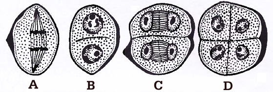
during microsporogenesis, (A-B) Dividing mother cell, (C) Dividing dyad, (D) Tetrad
(ii) Simultaneous : It occurs after complete meiotic (I and II) division, thus tetrahedral tetrad of microspores is formed, e.g., dicots.
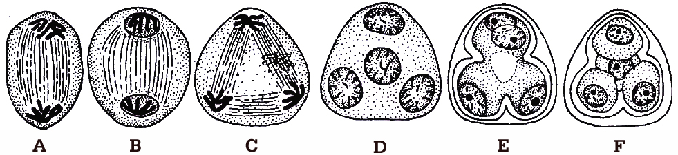
The microspores, as they are formed, are arranged in a cluster of four cells, called microspore tetrad. Usually the arrangement of microspores in a tetrad is tetrahedral or isobilateral.
However, T-shaped, linear and decussate tetrads are also found. In Aristolochia elagans, all the five types of tetrads are present.
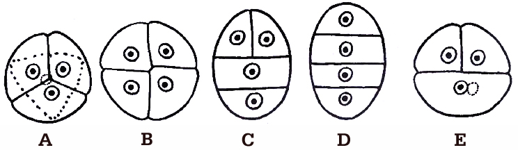
A. Tetrahedral, B. Isobilateral, C. T-shaped, D. Linear, E. Decussate
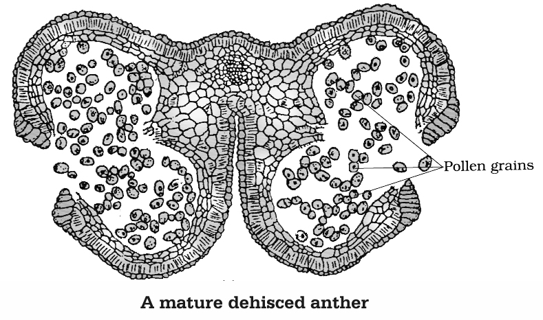
As the anthers mature and dehydrate, the microspores dissociate from each other and develop into pollen grains. These are released with the dehiscence of anther.
Concept Builder
(i) R. Camerarius described sexual reproduction for the first time in plants.
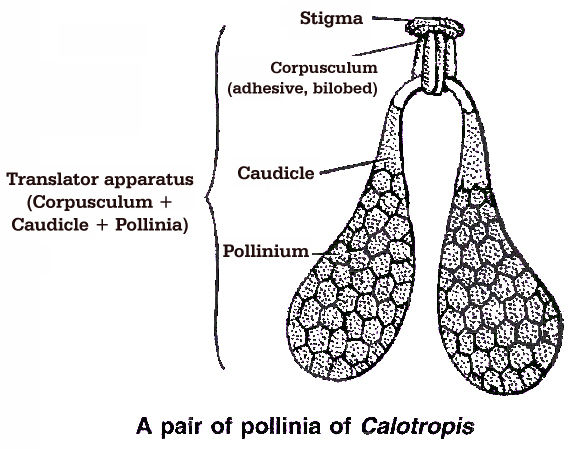
(ii) 'In family Asclepiadaceae (Calotropis) and Orchidaceae, all the microspores in a sporangium adhere together in a single mass known as pollinium.
(iii) In Calotropis, the pollinia of adjacent anthers of different stamens are attached by thread like caudicles (retinaculi) to a sticky disc called corpusculum. The whole structure is called translator.
(iv) After their formation, the microspores are separated from tetrad, but in Elodea, Drosera, Typha, the microspores do not separate from each other, thus developing into compound pollen grains.
(v) In family Cyperaceae, out of 4 microspores formed, 3 degenerate, so ultimately one MMC (2n) produces only one microspore or pollen grain.
(vi) Sometimes more than four microspores are produced from one microspore mother cell. It is called polyspory, e.g., Cuscuta reflexa.
(d) Pollen Grain
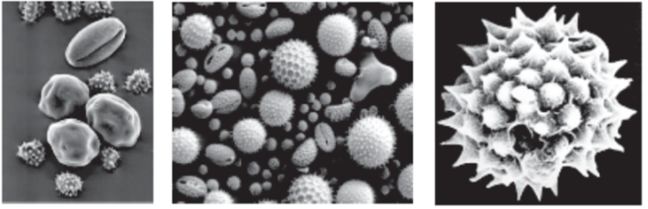
(i) These are generally spherical measuring about 25-50 µm in diameter. The cell wall of pollen grain is called sporoderm which consists of two layers. The hard outer layer called the exine and inner thin layer is called the intine.
(ii) Intine : It is made of cellulose and pectin.
(iii) Exine : The exine is made up of sporopollenin is a fatty substance and one of the most resistant organic material known. It is not affected by high temperature, strong acids or alkali. No enzyme is known to degrade it. Because of the presence of sporopollenin, pollen grains of the past plants are well preserved as fossils. Exine is made of two layers :
(a) Ektexine : It is highly sculptured and is differentiated into outer tectum, middle baculum and innermost foot layer. Tectum provides a characteristic sculpturing or designs over the surface of pollen grain. The design helps the experts to identify the pollen grain as to its class, family, genus or species.
(b) Endexine: It is not sculptured.
(iv) Pollen grain exine has prominent apertures called germ pores where sporopollenin is absent. Pollen grains can be monocolpate (having one germ pore called germinal furrow, e.g., monocots), bicolpate (2 germ pores) and tricolpate (3 germ pores, e.g., dicots).
(v) The pollen kitt is a sticky layer found on the outer side of exine of mature pollen grains of many insect pollinated species, it is made of carotenoids and lipids. Pollenkitt material is contributed by the tapetal cells. Pollen kitt acts as an insect attractant and can help against UV.
(vi) Pollen grains of many species (especially anemophilous plants) cause severe allergies and bronchial affictions in some people. Weed Parthenium hysterophorus (carrot grass) came to India as a contaminant with improved wheat. The weed has become a major cause of pollen allergy. Hay fever is an allergic reaction due to the presence of pollen in the air. Plants commonly causing hay fever are Amaranthus, Chenopodium and Parthenium.
(vii) Pollen grains are rich in nutrients. They are taken as tablets and syrups to improve health. Pollen consumption has been claimed to enhance the performance of athletes and race horses.
(viii) The period for which pollen grains retain the ability to germinate is called Pollen Viability. It is highly variable and to some extent depends upon the environmental factors like temperature, humidity.
In cereals, like rice, wheat etc, pollen viability is minimum upto 30 minutes, while in Rosaceae, Leguminosae and Solanaceae it is upto several months.
(ix) Pollen grains can be cryopreserved in liquid nitrogen (-196°C) and used as pollen banks.
Concept Builder
(i) The study of pollen grain is called Palynology. (Term given by Hyde and Williams).
(ii) In Hyacinthus, Nemec observed 8-nucleated embryo sac type organisation in pollen grain. This is called Nemec phenomenon.
(iii) Smallest pollen grain: Myosotis (2.5 -3.5 µ).
(iv) Largest pollen grain : Mirabilis (250 µ).
(v) Longest or Filamentous pollen grain : Zostera (2500 µ).
(e) Development of Male Gametophyte
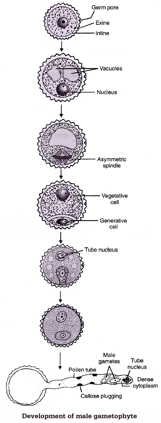
The microspore is the first cell of male gametophyte. The germination of microspore starts in situ (in the mother place). Microspore may be best defined as partially developed male gametophyte.
Microspores divide mitotically into large tube cell and small generative cell Pollination takes place at two celled stage in 60% of angiosperms (in some cases at three-celled stage). The further development of these male gametophyte takes place on the stigma.
Pollen grain expands by absorbing the liquid from the moist surface of stigma. Stigma provides, boron, sugar, amino acids etc. The intine comes out in the form of pollen tube, from germ pores.
The pollen grains are either monosiphonous (with one pollen tube) or polysiphonous (with more than one pollen tubes), e.g., members of Cucurbitaceae and Malvaceae.
The generative nucleus divides mitotically to form two male gametes called sperm. The male gametes are non-motile and amoeboid. They are slightly unequal in size, such a pollen will be called three celled pollen or mature male gametophyte.
The function of pollen tube is to carry aflagellated sperm. Tube nucleus enters first in the pollen tube and is a vestigial structure and soon disintegrates. Growth of the pollen tube is chemotropic apicle and entire cytoplasm of pollen grain is confined to tip of the pollen tube.
Concept Builder
(i) The pollen tube was first observed by G.B. Amici (1824) in Portulaca.
(ii) Longest pollen tube occurs in Zea mays.
(iii) B-Ca-inositol sugar complex act as chemotropic agent for pollen tube growth.
The Pistil, Ovule and Embryo sac
(a) Pistil (Female Sex Organ)
The gynoecium may consist of a single pistil (monocarpellary) or may have more than one pistil (multicarpellary).
If there are more than one, the Pistils may be fused together (Syncarpous) or may be free (apocarpous).
There are three parts of each pistil -the stigma, style and ovary. The stigma is upper broader region which is specialised for receiving pollen grains.
The style is long stalk-like structure and ovary is basal swollen ovule containing region.
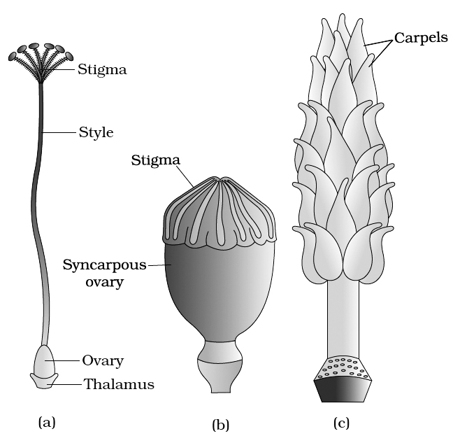
(c) A multicarpellary, apocarpous gynoecium of Michelia
(b) Ovule (Integumented Megasporangium)
(i) Ovary has an ovarian cavity with one or more chambers (lobules). The placenta is located inside the ovarian cavity. One or many ovules are present inside the ovary.
Plants with one ovule in an ovary: (i) Wheat, (ii) Paddy, (iii) Mango
Plants with many ovules in an ovary: (i) Papaya, (ii) Water melon, (iii) Orchids
(ii) The ovule is a small structure attached to the placenta by means of a stalk called funicle. The point of attachment of the funicle with the main body of the ovule is called hilum. Thus, hilum represents the junction between ovule and funicle. Sometimes funicle gets fused with the body of the ovule along one side and forms a ridge known as raphe. The basal region of the ovule is known as Chalaza.
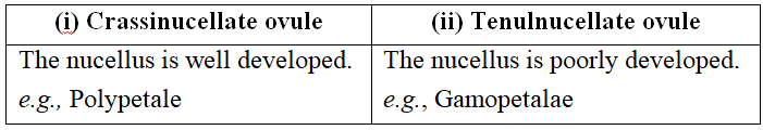
(iii) Main body of an ovule is called nucellus (megasporangium) which consists of a mass of parenchymatous tissue. Cells of nucellus have abundant reserve food materials. Depending upon the development of nucellus, ovules are of two types:
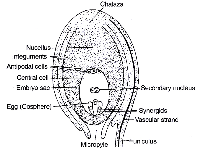
containing developed embryo sac (female gametophyte)
(iv) The nucellus is invested all around by one or two layered protective covering called integuments, except apex where a small passage is formed known as micropyle. On the basis of number of integuments, ovules are of following types:
(a) Unitegmic: Ovules with one integument, e.g., members of gamopetalae and gymnosperms.
(b) Bitegmic : Ovules with two integuments, e.g., members of polypetalae and monocots.
(c) Ategmic : Ovules are without integument, e.g., Santalum, Loranthus (Parasites) and Liriosoma.
(v) On the basis of the relative position of micropyle, body of the ovule and funicle, i.e., the degree of curvature, there are six types of ovules:
(a) Orthotropous : The micropyle, chalaza and funicle are in a straight line. This is the most primitive type of ovule, e.g., Piper, Polygonum, Cycas.
(b) Anatropous : The ovule turns 180º angle. Thus it is inverted ovule. Micropyle lies close to hilum or at side of hilum, e.g., found in 82% of angiosperm families.
(c) Hemianatropous: Ovule turns at 90º angle upon the funicle or body of ovule is at right angle to the funicle, e.g., Ranunculus.
(d) Campylotropous: Ovule is curved more or less at right angle to funicle. Micropylar end is bend down slightly, e.g., in members of Leguminosae, Cruciferae.
(e) Amphitropous: Ovule as well as embryo sac is curved like horse shoe, e.g., Lemna, Poppy, Alisma.
(f) Circinotropous: The ovule turns at more than 360º angle, so funicle becomes coiled around the ovule, e.g., Opuntia (Cactaceae), Plumbaginaceae.
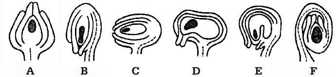
A. Orthotropous, B. Anatropous, C. Hemianatropous,
D. Campylotropous, E. Amphitropous, F. Circinotropous
Concept Builder
(i) Third integument in the form of aril develops from base of ovule or funicle in many plants, e.g., Litchi, Asphodelus and Inga dulce. In litchi and Inga dulce, aril is fleshy and edible.
(ii) In some ovules, e.g., Zostera, a group of thickened cells is found in the chalazal region just below the embryo sac, called hypostase. When a similar group of specialized cells is found in the nucellar region just above the embryo sac, it is called epistase.
(iii) In castor (Ricinus), proliferation of the outer integumentary cells at micropylar region is called caruncle or strophiole. It performs two functions :
a. It acts as water absorbing pad.
b. It is made up of sugary substance that attract and helps in the seed dispersal by ants (myrmecochory).
(iv) The placental or funicular outgrowth present at, the micropylar end is called obturator. It directs the passage of pollen tube towards the ovule.
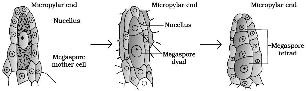
(c) Megasporogenesis
(i) Any of the cells of nucellus hypodermis towards the micropylar end gets differentiated from the other cells. This cell is called archesporial cell. In crassinucellate ovules, it undergoes periclinal division to form an outer primary parietal cell and inner primary sporogenous cell. The later behaves as megaspore mother cell (MMC). The archesporial cell directly behaves as megaspore mother cell in tenuinucellate ovules.
(ii) The megaspore mother cell is large sized containing dense cytoplasm and a prominent nucleus. Some carbohydrate storing bodies appear between cell wall and plasma membrane of megaspore mother cell in Lilium, called paramular bodies.
(iii) The MMC (2n) undergoes meiosis and forms a linear tetrad of 4 haploid megaspores. The process of formation of megaspores from the MMC is called megasporogenesis.
(d) Female Gametophyte or Embryo sac
(i) P. Maheshwari classified the embryo sac on the basis of number of megaspore nuclei participating in embryo sac formation into following types :
Monosporic embryo sac : Only one megaspore nucleus forms embryo sac, e.g., Polygonum, Oenothera.
Bisporic embryo sac: Two megaspore nuclei take part in development of embryo sac, e.g., Allium, Endymion.
Tetrasporic embryo sac: All the four megaspore nuclei take part in development of embryo sac, e.g., Adoxa, Plumbago, Drusa, Fritillaria, Paenaea, Plumbagella, Peperomia.
(ii) Development of Monosporic Embryo sac (Polygonum type)
(A) In majority of angiosperms, one of the megaspore is functional while the other three degenrate. Only the functional megaspore (n) develops into the female gametophyte. This process of embryo sac formation from a single megaspore is termed monosporic development.
(B) Polygonum type of embryo sac is found in 80% flowering plants. This development has been studied in (Polygonum by Strasburger). The nucleus of chalazal functional megaspore (4th from micropyle) divides by three mitotic divisions to form 8 nuclei, four towards each pole. One nucleus from each pole moves to the middle and they form polar nuclei. These mitotic divisions are strictly free nuclear, i.e., nuclear divisions are not followed immediately by cell wall formation.
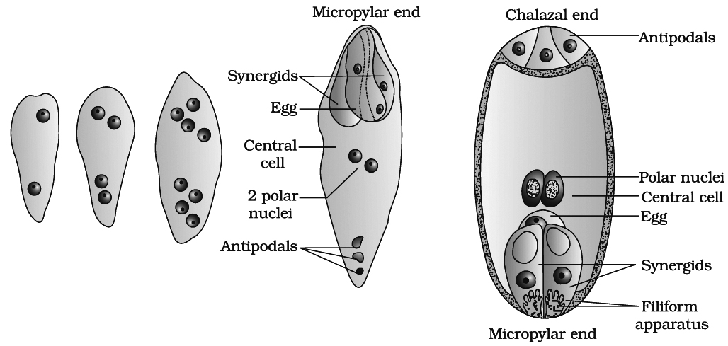
At this stage, following changes occur:
(a) Three of the nuclei (n) get organised as cells at micropylar end forming egg apparatus. One is the egg cell (n) and two are synergids (n).
(b) Three nuclei get organised as antipodal cells (n) at chalazal end
(c) Two nuclei in the centre are called polar nuclei (n)
This constitutes a 7-celled and 8-nucleated embryo sac.
Organization of Embryo Sac
(a) Synergids or helper cells or co-operative cells: These cells generally possess a micropylar nucleus and a chalazal vacuole. The electron microscopic studies have revealed that the synergids lack a cell wall on their chalazal side at maturity. They are characterised by the presence of a 'filiform apparatus at the' micropylar tip. It is in the form of finger like projections, each projection comprising a core of microfibrils enclosed in a sheath. Usually one synergid starts to degenerate just with pollination. The synergids perhaps secrete some chemotropic substance and thus, direct the pollen tube growth inside embryo sac.
(b) Egg : The egg shows cytoplasmic polarity opposite to synergid and its wall is thicker at the micropylar end. Usually the egg has a micropylar vacuole and a chalazal nucleus. Plasmodesmata connection is present in between egg and synergids.
(c) Antipodals or vegetative cells : These are vegetative cells of embryo sac. In most of the plants there are three antipodal cells.
(d) Central Cell: It is the largest cell of the embryo sac. It initially contains two polar nuclei which fuse just before, fertilization to form a secondary nucleus or definitive nucleus (2n).
Concept Builder
(i) Synergids are absent in Plumbago and Plumbagella. Number of synergid is one in Peperomia.
(ii) In Zea mays 20 antipodal cells are present, in Sasa paniculata they are 300 in number. This is perhaps the highest number recorded for any plant. These are absent in Oenothera type.
