 ACME SMART PUBLICATION
- Books Name
- A TEXT OF BIOLOGY - CLASS XII
- Publication
- ACME SMART PUBLICATION
- Course
- CBSE Class 12
- Subject
- Biology
HUMAN REPRODUCTION
Human beings are unisexual.
The growth, maintenance and function of the gonads are regulated by gonadotropins secreted from anterior lobe of pituitary gland.
The organs which neither produce gametes nor secrete sex hormones but perform important functions in reproduction are called secondary sex organs.
The latter include the prostate, seminal vesicles, vas deferentia and penis in males, and the fallopian tubes, uterus, vagina and mammary glands in females.
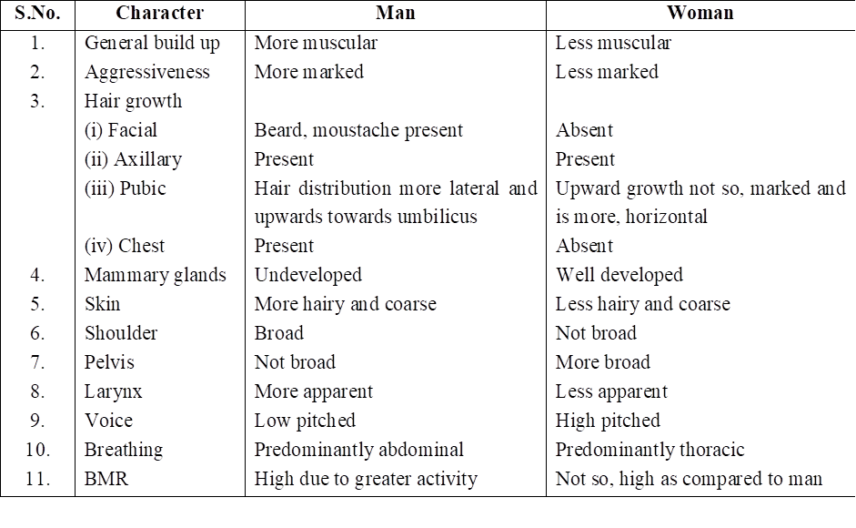
The characters which distinguish the male from the female externally are called accessory or external sex character. They are also called secondary sex characters.
Secondary Sexual Features in Man and Woman
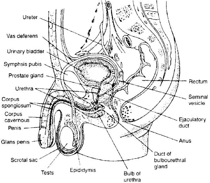
MALE REPRODUCTIVE SYSTEM
The male reproductive system of mammals consists of a pair of testes, several accessory glands, a duct system and a mating organ called the penis. Testis is the primary male sex organ. It produces spermatozoa and secretes the male sex hormone testosterone.
The human testis measures about 5 cm, 3 cm and 2.5 cm, respectively, in length, thickness and width. It is covered by thick, fibrous, connective tissue called tunica albuginea.
In man, both testes normally remain suspended in a pouch called scrotum outside the abdominal cavity. This keeps the testes at a low temperature than the body temperature (about 2°C below)-this is essential for the maintenance and normal functioning of the spermatogenic tissue of the testes.
Testes descend in the scrotal sac when foetus is about 7 months old and this occurs under the influence of FSH and testosterone.
If testes fail to descend, than the condition is called cryptorchidism that leads to sterility. Scrotum remains connected with the abdomen or pelvic cavity by the inguinal canal. Blood vessels, nerves and conducting tubes pass through inguinal canal.
Cremaster muscles and connective tissue form spermatic cord and surround all structures passing through inguinal canal. Cremaster muscles and dartos muscles of the scrotal sac help in the positioning of testes.
Whenever the outside temperature is low, these contract to move the testes close to the abdominal cavity/pelvic cavity. When outside temperature is high, these relax moving the testes away from the body.
In some seasonally breeding mammals, testes descend into the scrotum during breeding season but ascend back into the abdomen in the non-breeding season. e.g. rats and bats.
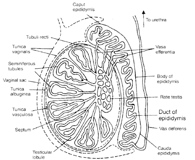
ANATOMY OF TESTIS
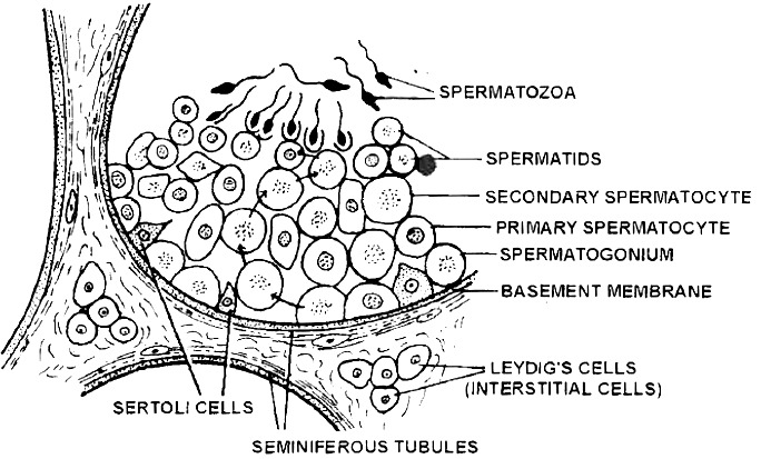
Each testis contains numerous tiny, highly convoluted tubules, called seminiferous tubules. They constitute the spermatogenic tissue of the testis.
Cells lining these tubules give rise to spermatozoa which are released into the lumen of the tubule.
In between spermatogenic cells, Sertoli or sustentacular or nurse cells are present which provide nourishment to the developing spermatozoa and regulate spermatogenesis by releasing inhibin to check FSH overactivity.
The other functions of sertoli cells include
(i) Provide nourishment to the developing spermatozoa,
(ii) Absorb the parts being shed by developing spermatozoa
(iii) Release anti mullerian factor (AMF) to prevent development of mullerian duct/oviduct
(iv) Release of androgen binding protein (ABP).
Groups of polyhedral cells called Interstitial cells or Leydig cells, are located in the connective tissue around the seminiferous tubules.
They constitute the endocrine tissue of the testis. Leydig cells secrete testosterone into the blood.
Seminiferous tubules unite to form several straight tubules called tubuli recti which open into irregular cavities in the posterior part of the testis.
It is highly anastomosing labyrinth of cuboidal epithelium lined channels called rete testis.
Several tubes called vasa efferentia arise from it and conduct spermatozoa out from the testis (Seminiferous tubule to vasa efferentia form intratesticular genital duct system).
The extratesticular duct system consists of tubes which conduct sperms from the testes to the outside.
It starts with vasa efferentia which arise from each testis and becomes confluent to form a folded and coiled tube called epididymis behind each testis.
The epididymis consist of 3 parts: (i) Caput, (ii) Corpus, (iii) Cauda. The epididymis stores the sperms temporarily.
From cauda epididymis, a partially coiled tube called vas deferens ascends into the abdomen through inguinal canal, passes over the urinary bladder, receives the duct from the seminal vesicle behind the urinary bladder to from an ejaculatory duct.
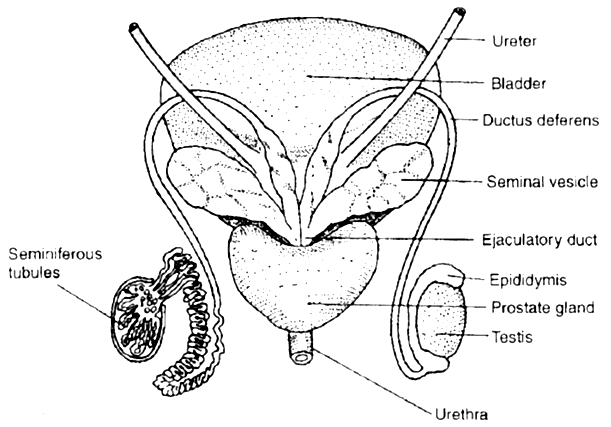
Before entering prostate, the ductus deferens dilates to form ampulla. The final portion of ampulla passes through the prostate to open into the urethra shortly after its origin from the urinary bladder.
The urethra receives the ducts of the prostate and cowper's glands, passes through the penis and opens to the outside.
MALE EXTERNAL GENITAL ORGAN
PENIS
This is the copulatory organ of man. It is a cylindrical and erectile, pendulous organ suspended from pubic region in front of scrotum.
It remains small and limp (= flaccid) but on sexual arousal, it becomes long, hard and erect, ready for copulation (=coitus or intercourse). Erect human penis, on an average is about 15 cm long.
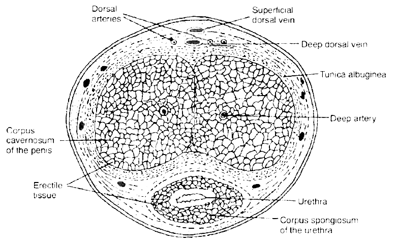
 T.S. of penis
The penial mass is itself encased in a fibrous sheath, called tunica albuginea. The interior of penis is mostly formed of three cylindrical cords of spongy, erectile (= cavernous) tissue.
Two of these cords are thicker and situated parallely on right and left sides, forming the thick part of penis that remains in front when penis is limp, but becomes superio-posterior when penis is erect.
These two cords are called corpora cavernosa.
The fibres of tunica albuginea surround both cords jointly and form a separate sheath around each cord. Some fibres form a partition, called septum penis, between these cords.
The third, smaller cord form that part of penis which remains inferio-anterior in erect penis. Urethra runs through this cord. Hence, this cord is called corpus urethrae or spongiosum.
The extended part of corpus spongiosum is enlarged, forming a bulged, conical structure called glans penis. The surface of glans is formed of a thin, smooth and shiny, hairless skin.
The base line of glans is referred to as neck of the penis. The loose skin of penis folds over and is retractile on glans. It is called foreskin or prepuce.
At the tip of glans penis is the slit like external urethral orifice or meatus by which urethra opens out and discharges urine or semen.
Prepucial glands, present in the skin of penis neck, secrete a white sebaceous substance, called smegma. Microbial infection in smegma causes irritation.
GLANDS
Seminal vesicles - These are paired, tubular, coiled glands situated behind the urinary bladder. They secrete viscous fluid which constitute the main part of the ejaculate. Seminal fluid contains fructose, citric acid, inositol and postaglandins.
Prostate gland - It is a chestnut shaped gland and is a collection of 30-40 tubulo-alveolar glands which lies at the base of the bladder and surrounds the base of the urethra. It contributes an alkaline substance to the seminal fluid.
The substance of prostate help the sperms to become active and counteract any adverse effects of urine on sperms. The prostatic fluid provides a characteristic odour to the seminal fluid. Prostate gland secretes -citrate ion, calcium, phosphate ion, and profibrolysin.
Bulbourethral glands or Cowper's glands - The two bulbourethral glands are pea-sized structures lying adjacent to the urethra at the base of penis. These secrete a viscous lubricant.
The duct system, accessory glands and penis are secondary male sex organs. Their growth, maintenance and functions are promoted by testosterone secreted by Leydig cells.
On the other hand, the growth, maintenance and functions of seminiferous tubules and Leydig cells are regulated, respectively by FSH and ICSH of anterior pituitary.
SEMEN
Semen is a mixture of sperms and the secretions of the seminiferous tubules, seminal vesicles, prostate gland, and bulbourethral glands.
The average volume of semen in an ejaculation is 2.5-5 ml, with a sperm count (concentration) of 200 to 300 million. For normal fertility, atleast 60 percent sperms must have normal shape, size and atleast 40 percent of them must show vigorous motility. When the sperm count falls below 20 million/ml, the male is likely to be infertile.
Semen has a slightly alkaline pH of 7.2-7.7. The prostatic secretion gives semen a milky appearance, whereas fluids from the seminal vesicles and bulbourethral glands give it a sticky consistency.
Semen provides transportation medium and nutrients to sperms. It neutralizes the hostile acidic environment of the male urethra and the female vagina.
(Prostatitis - Inflammation of prostate gland.)
