ACME SMART PUBLICATION
- Books Name
- A TEXT OF BIOLOGY - CLASS XII
- Publication
- ACME SMART PUBLICATION
- Course
- CBSE Class 12
- Subject
- Biology
FEMALE REPRODUCTIVE SYSTEM
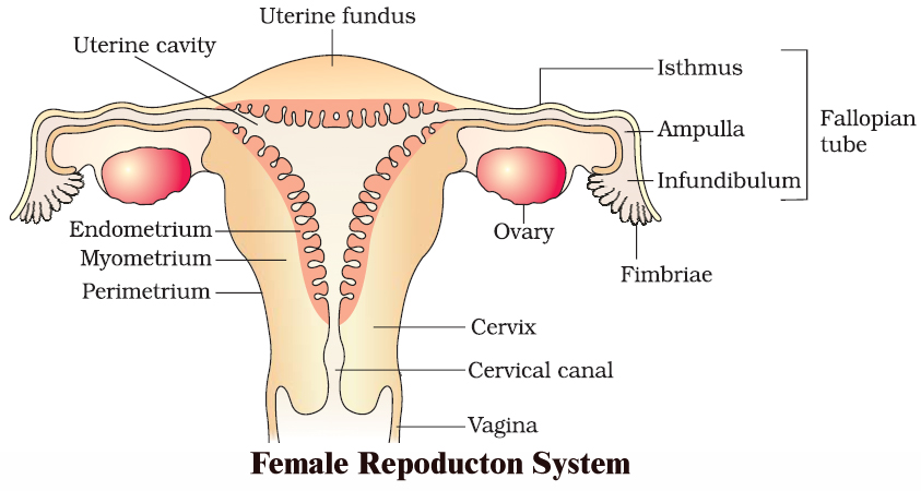
The female reproductive system consists of a pair of ovaries, a duct system, consisting of a pair of fallopian tubes (oviduct), an uterus, cervix and vagina. A pair of mammary glands are accessory genital glands.
OVARIES
The ovary is the primary female sex organ. It produces ova and secretes the female sex hormones, estrogen and progesterone that are responsible for the development of secondary female sex characters and regulate cyclic changes in the uterine endometrium.
The human ovaries are small, almond-like flat bodies of about 3 cm in diameter.
1. Location. Ovaries are located near kidneys and remain attached to the lower abdominal cavity through mesovarium.
2. Structure. The free surface of the ovary is covered by a germinal epithelium made of a single layer of cubical cells. This epithelium is continuous with the mesothelium, called peritoneum. The epithelium encloses the ovarian stroma. The stroma is divided into two zones-a peripheral cortex and an inner medulla. Cortex is covered by a connective tissue, called tunica albuginea.
The cortex contains numerous spherical or oval, sac-like masses of cells, known as ovarian follicles. The medulla consists of loose connective tissue, elastic fibres, blood vessels and smooth muscle fibres.
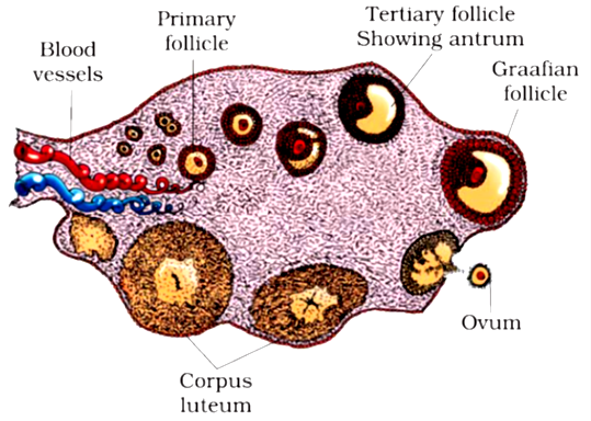
 A section of human ovary
OVARIAN FOLLICLE
The ovarian follicle carries a large, centrally placed ovum, surrounded by several layers of granular cells (follicular granulosa or discus proligerus or cumulus oophorus). It is suspended in small cavity the antrum.
Antrum is filled with liquid folliculi. The secondary oocyte in the tertiary folicle forms a new membrane called zona pellucida. The follicle bulges on the surface of the ovary.
Such a follicle is called the mature Graafian follicle (after De Graaf, who reported them in 1672).
CORPUS LUTEUM
The ovum is shed from the ovary by rupturing the follicle. The release of ovum is called ovulation and occurs nearly 14 days before the onset of the next menstrual cycle.
After the extrusion of the ovum, Graafian follicle transforms into corpus luteum. Corpus luteum is filled with a yellow pigment, called lutein.
Corpus luteum grows for a few days and if the ovum is fertilized and implantation occurs, then it continues to grow. But if the ovum is not fertilized, then corpus luteum persists only for about 14 days.
It secretes progesterone. At the end of its functional life, the corpus luteum degenerates,and becomes converted to a mass of fibrous tissue, called corpus-albicans (white body) that remains as a scar in the ovary throughout the life of female.
FALLOPIAN TUBES (OVIDUCTS)
There are one pair, long (about 10 cm), ciliated, muscular and tubular structures that extend from ovaries to uterus. Each one is suspended by mesosalpinx. Each fallopian tube is differentiated into four parts :
(i) Infundibulum. The part of oviduct closer to the ovary is the funnel shaped infundibulum. The edges of infundibulum possess finger like projections called fimbriae. Fimbriae help in collection of the ovum after ovulation. Infundibulum opens in abdominal cavity by an aperture called osteum.
(ii) Ampulla. The infundibulum leads to a wider part of the oviduct called ampulla.
(iii) Isthmus. It is middle, narrow and ciliated part of the oviduct.
(iv) Uterine part. It is inner and narrow part which opens in the upper part of uterus.
It is involved in the conduction of ovum or zygote towards the uterus by peristalsis and ciliary action. (Fertilization occurs at the junction of ampulla and isthmus).
Uterus. It is a large hollow, muscular, highly vascular and pear shaped structure present in the pelvis between the bladder and rectum. It is suspended by a mesentery, mesometrium. It is formed of three parts.
(i) Fundus. It is upper dome shaped part above the opening of fallopian tubes.
(ii) Body. It is middle and main part of uterus.
(iii) Cervix. It is lower, narrow part which opens in the body of uterus by internal os and in vagina below by external os. It is formed of most powerful sphincter muscle in the body.
Its wall is formed of outer peritoneal layer called perimetrium; middle muscular myometrium made of smooth muscle fibres, and inner highly vascular and glandular endometrium.
It is the site of foetal growth during pregnancy. It also takes part in placenta formation and helps in pushing the baby out during parturition.
VAGINA
It is a long (7.5 cm), fibro-muscular tube. It extends backward in front of rectum and cervix to the vestibule. It is vascular tube internally lined by mucus membrane and is raised into transverse folds called vaginal rugae.
In the virgin female, vaginal orifice is closed by a membranous diaphragm called hymen which becomes centrally perforated at puberty for the discharge of menstrual flow (or menses).
Vagina acts both as copulation canal as it receives the sperms from penis during copulation and birth canal during parturition.
FEMALE EXTERNAL GENITAL ORGAN
Vulva. It is external genitalia of female. It has a depression, the vestibule, in front of anus. Vestibule has two apertures-upper external urethral orifice of urethra and lower vaginal orifice of vagina.
Mons pubis : Is a fleshy and fatty tissue covered by skin and pubic hair.
Labia majora : It is a pair of fleshy folds which extend from the mons pubis and surround the vaginal opening.
Labia minora : Is another pair of tissue folds below the labia majora.
Both labia majora and labia minora are provided with sebaceous glands.
Hymen : Is a membrane that partially covers the vaginal opening. It gets torn during the first coitus.
Clitoris : Is a tiny, erectile, finger-like structure present at the upper junction of the two labia minora above the urethral opening. The fold of skin that covers clitoris is called prepuce. Clitoris is homologous to penis (as both are supported by corpora cavernosa).
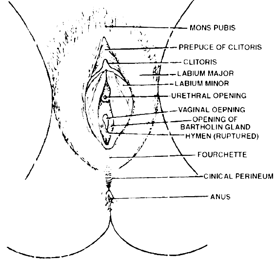
 The external genitalia in the female
GLANDS
Vestibular Glands: These are of two types-greater and lesser. Greater vestibular or Bartholin's glands are a pair of small reddish yellow glands on each side of vaginal orifice and secrete alkaline secretion for lubrication and neutralising urinary acidity.
Lesser vestibular glands or paraurethral or skene's glands are small mucus glands present between urethral and vaginal orifices.
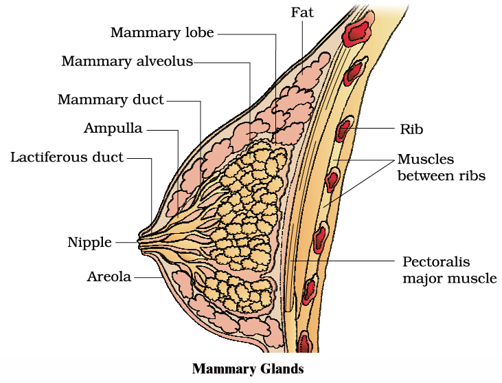
MAMMARY GLAND :
Each mammary gland consists of 15-25 lobules of the compound tubuloalveolar type. These lobules secrete milk to nourish the newborn babies.
Each lobe is separated from the others by dense connective and adipose tissue and represents a gland. From each lobe, excretory lactiferous ducts emerge independently in the nipple, which has 15-25 openings, each about 0.5 mm in diameter.
However, the histological structure of mammary glands vary, depending on sex, age and physiological state.
Path of milk ejection :
Mammary alveolus ® Mammary duct ® Ampulla ® Lactiferous duct ® Nipple
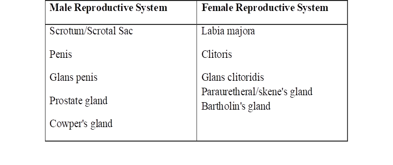
Homology Between Male and Female Reproductive System