 ACME SMART PUBLICATION
- Books Name
- A TEXT OF BIOLOGY - CLASS XII
- Publication
- ACME SMART PUBLICATION
- Course
- CBSE Class 12
- Subject
- Biology
BIOTECHNOLOGICAL APPLICATION IN MEDICINE
1. Therapeutic agents
Proteins with potential as pharmaceutical agents are produced by using genetically engineered organisms.
Enzymes have also been used for this purpose, e.g., DNase I and alginate lyase have been used in aerosols.
Some known examples are given below:
1. Human growth hormone obtained from E. coli is used for treatment of dwarfness.
2. Chorionic gonadotropin hormone produced by genetic engineering is used for treatment of infertility.
3. Interferons produced by E. coli are commercially used for treatment of viral infections and cancer. Interferons were first obtained through DNA recombinant technique by Charles Weisman in 1980. He inserted the gene for interferon production in E. coli.
4. Interleukins produced by E. coli are used for stimulating immunity system.
5. Tissue Plasminogen Activator (TPA) -an enzyme is used for dissolving blood clot after heart attack and stroke.
6. Antihemophilic human factor VIII is used by people with hemophilia to prevent and control bleeding or to prepare them for surgery.
7. Platelet derived growth factor produced by recombinant DNA technology is useful for stimulating wound healing.
8. Penicillin G acylase is also produced by genetic engineering. This enzyme is used for converting penicillin into 6-amino penicilline acid for the formation of new antibiotics.
2. Genetically engineered insulin
Since the discovery of insulin by Banting and Best (1921), and its use for the treatment of diabetes, it was derived from pancreatic glands of abattoir animals.
This hormone, produced and secreted by the beta cells of the pancreas islets of Langerhans, regulates the use and storage of food, particularly carbohydrates.
Although bovine and porcine insulin is similar to human insulin, their composition is slightly different.
It, therefore, causes adverse effects due to regular injection, this being a foreign substance.
This observation led to the synthesis of human insulin which is chemically identical to its naturally produced component.
Insulin consists of 51 amino acids forming two short polypeptide chains-chain A having 21 amino acids and chain B with 30 amino acids.
The two chains are linked by disulfide bond. In animals, including humans, insulin occurs as proinsulin.
It is made of chain A, chain B and chain C (30 amino acids). As the insulin matures, chain C is removed.
The genetic engineering of insulin begins with identification and separation of DNA sequences coding for chain A and chain B.
This was found to be present at the top of the short arm of the eleventh chromosome.
It contains 153 nucleotides-63 nucleotides for chain A and 90 nucleotides for chain B.
These sequences were introduced into plasmid (pBR322) of Escherichia coli -common human colon bacterium.
It is said to be the factory used in genetic engineering of insulin.
In E. coli, -galactosidase controls the transcription of these genes, therefore, insulin gene needs to be tied to this enzyme.
The protein formed by E. coli consists partly of -galactosidase joined to either A or B chain of insulin.
These are then extracted from -galactosidase fragment and purified.
The two chains are mixed and reconnected in a reaction that forms disulfide bridges resulting in pure humulin-the synthetic human insulin.
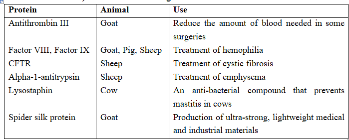
Proteins with Therapeutic and Industrial Value that have been Produced (but not Commercialized) in the Milk of Transgenic Animals
GENE THERAPY
Much attention has been focussed on the so-called genetic metabolic diseases in which a defective gene causes an enzyme to be either absent or ineffective in catalyzing a particular metabolic reaction effectively.
A potential approach to the treatment of genetic disorders in man is gene therapy.
This is a technique whereby the absent or faulty gene is replaced by a working gene, so that the body can make the correct enzyme or protein and consequently eliminate the root cause of the disease.
The first clinical gene therapy was given in 1990 to a 4-year old girl with adenosine deaminase (ADA) deficiency.
This enzyme is crucial for the immune system to function.
The disorder is caused due to the deletion of the gene for adenosine deaminase.
In some children, ADA deficiency can be cured by bone marrow transplantation; in others, it can be treated by enzyme replacement therapy, in which functional ADA is given to the patient by injection.
But the problem with both of these approaches is that they are not completely curative.
As a first step towards gene therapy, lymphocytes from the blood of the patient are grown in a culture outside the body.
A functional ADA cDNA (using a retroviral vector) is then introduced into these lymphocytes, which are subsequently returned to the patient.
However, as these cells are not immortal, the patient requires periodic infusion of such genetically engineered lymphocytes.
However, if the gene isolated from bone marrow cells producing ADA is introduced into cells at early embryonic stages, it could be a permanent cure.
Before treatment for a genetic disease can begin, an accurate diagnosis of the genetic defect needs to be made.
It is here that biotechnology is also likely to have a great impact in the near future.
Genetic engineering research has produced a powerful tool for pinpointing specific diseases rapidly and accurately.
Short pieces of DNA called DNA probes can be designed to stick very specifically to certain other pieces of DNA.
The technique relies upon the fact that complementary pieces of DNA stick together.
DNA probes are more specific and have the potential to be more sensitive than conventional diagnostic methods, and it should be possible in the near future to distinguish between defective genes and their normal counterparts, an important development.
Molecular Diagnosis
For effective treatment of a disease, early diagnosis and understanding its pathophysiology is very important.
Using conventional methods of diagnosis (serum and urine analysis, etc.), early detection is not possible.
Recombinant DNA Technology, Polymerase Chain Reaction (PCR) and Enzyme Linked Immuno-Sorbent Assay (ELISA) are some of the techniques that serve the purpose of early diagnosis.
Presence of a pathogen (bacteria, viruses, etc.) is normally suspected only when the pathogen has produced a disease symptom.
By this time the concentration of pathogen is already very high in the body.
However, very low concentration of a bacteria or virus (at a time when the symptoms of the disease are not yet visible) can be detected by amplification of their nucleic acid by PCR, which is now routinely used to detect HIV in suspected AIDS patients.
It is being used to detect mutations in genes in suspected cancer patients too.
It is a powerful technique to identify many other genetic disorders.
DNA is usually isolated from White blood cells & has to be cut into smaller pieces to be analysed.
This is accomplished by restriction enzymes. Eco RI (a restriction enzyme from E. coli) will cut DNA wherever the sequence GAATTC appears.
Exposure to this enzyme results in the DNA being chopped into millions of fragments of varying size, called restriction fragments.
Once cut, the DNA is loaded into a well on one end of a slab of gel.
The fragments are then separated according to size by electrophoresis.
As electric current passes through the gel, the fragments move according to size.
The bigger fragments stay close to the origin, and the smaller fragments move farther down the length of the gel.
The DNA is then denatured (by exposure to alkaline solutions) to render the DNA single stranded (instead of the natural double-stranded form).
Since the gel is difficult to handle, the DNA is transferred to a nitro cellulose paper to create a Southern blot (named after the researcher who developed the procedure).
The DNA probe which is radioactively labeled (or fluorescently labeled) is then applied to the Southern blot.
Since the probe is also single-stranded, it will seek the single-stranded DNA fragments that are complementary, and undergo hybridization.
The excess probe is washed out and only the bound probe will remain on the Southern blot paper.
This is then laid on an X-ray film.
The radioactive probe will leave bands on the X-ray film.
Depending on the type of probe used, there could be hundreds of bands (much like bar codes) or only a few bands present on the X-ray film.
By having several wells on the end of the gel, several samples can be loaded, and DNA fragments in corresponding lanes can be analyzed concurrently.
By running control samples, with known DNA fragment sizes, on the same gel with patient samples, it is possible to identify changes in the size of a DNA fragment and, therefore, a change in a specific gene.
Since each step takes about a day and since samples are batched, the procedure ordinarily takes one to two weeks to complete.
ELISA is based on the principle of antigen-antibody interaction. Infection by pathogen can be detected by the presence of antigens (proteins, glycoproteins, etc.) or by detecting the antibodies synthesised against the pathogen.
