ACME SMART PUBLICATION
- Books Name
- ACME SMART COACHING Biology Book
- Publication
- ACME SMART PUBLICATION
- Course
- CBSE Class 11
- Subject
- Biology
KINGDOM : FUNGI
This kingdom contains achlorophyllous, eukaryotic, heterotrophic, spore producing, thalloid organisms. The study of fungi is called mycology.
Pier Antonio Micheli is considered as father or founder of mycology.
Mycologist H.A. de Bary is the father of modern mycology.
The father of Indian mycology is E.J. Butler.
General Characters
They are cosmopolitan and occur in air, water, soil and on animals and plants. They are mostly terrestrial. They prefer to grow in warm and humid places.
They may grow on tree bark, dung, wood, burnt wood and keratinous material (e.g., hair, horns) and are called corticolous (bark), coprophilous (cow dung), epixylic (wood), xylophilous (burnt wood) and keratinophilous (keratin) respectively.
The body is haploid (n) and thalloid, i.e., not differentiated into root, stem and leaves. They are multicellular (except Yeast and Synchytrium).
The fungal body is made up of thread like elongated tubular structures, called hyphae. These cris-cross with one another to form a network known as mycelium.
The hyphae may be aseptate and multinucleate. Such a hypha is termed coenocytic. In most of the fungi, the mycelium is septate.
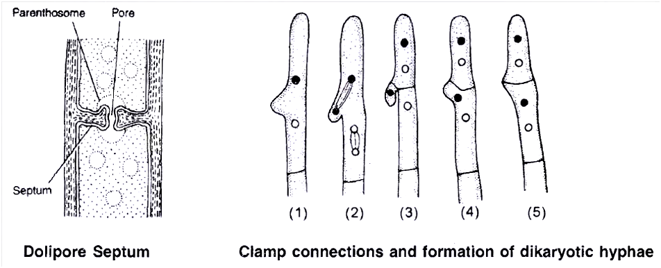
The septum, however, is not complete, but has a pore through which continuity of the cytoplasm of the adjoining cells is maintained.
The septum may have simple central pore as in ascomycetes, but in higher fungi (class basidiomycetes), the septum is dolipore septum, in which central pore possesses a barrel shaped inflation.
In septate mycelium, individual cell may contain single nucleus (monokaryotic -feature of primary mycelium) or an intermediate phase of two nuclei (dikaryotic -feature of secondary mycelium).
The cell wall of the hyphae is made up of chitin or fungal cellulose, which is a polysaccharide containing nitrogenous compound and it is basically made up of acetylglucosamine.
In some fungi, the cell wall is made up of cellulose (e.g., Phytophthora, Pythium and other oomycetes). Reserve food material is stored in the form of oil and glycogen.
Cells have unicisternal golgi bodies.
Mitosis in somatic cells is Karyochorisis type (mitosis with intranuclear spindle formation).
Nutrition is heterotrophic which includes saprophytes, parasites and symbionts.
In most of the fungi, there are two distinct phases in the life cycle, the vegetative or assimilative phase and the reproductive phase.
In vegetative phase, fungus is microscopic hidden in the substratum and is hardly visible to the naked eyes.
The fungus enters into reproductive phase after attaining maturity in the vegetative phase.
In unicellular yeasts, the same cell performs both assimilative and reproductive functions.
Such type of fungal bodies in which entire cell gets transformed into reproductive structures are known as holocarpic.
Fungal body is termed eucarpic in which a part of mycelium is used up in the development of reproductive structures.
Concept Builder
Modification of mycelium:
(a) Plectenchyma :
When hyphae of a mycelium grow together like plates and intertwine with one another forming a thick woven structure, it is called plectenchyma.
Plectenchyma may have :
(i) Prosenchyma : Loosely interwoven structure whose hyphal components lie more or less parallel to each other and are recognizable.
(ii) Pseudo parenchyma : Hyphae are compactly arranged and hyphal components have lost their identity and appear isodiametric and continuous in section resembling parenchyma of higher plants.

(b) Sclerotia (Singular Sclerotium) :
In some fungi like Claviceps the mycelium may pass into a dormant or resting stage by the formation of hard resting bodies resistant to unfavourable conditions.
Each sclerotium is composed of central prosenchymatous and peripheral pseudoparenchymatous arrangement which are again surrounded by a ring of pigmented hyphae.
(c) Rhizomorph:
- When the fungal hyphae aggregate together below surface they behave as an organized unit to form a root like strand with a thick hard cortex.
- It also develops a growing tip somewhat resembling that of a root tip, e.g., Agaricus.
(d) Appressorium: Terminal swollen structure of germ tube for attachment, and penetration.
(e) Haustoria: Terminal swollen structure for absorption of food, e.g., Albugo.
(f) Snares/hyphal traps: Helps in capturing nematodes in predaceous fungi, e.g., Arthrobotrys, Dactylaria
Reproduction in Fungi
- Fungi reproduce by all the three modes, i.e., vegetative, asexual and sexual.
1. Vegetative reproduction: It occurs by the following methods:
(a) Fragmentation : The mycelium breaks up into two or more fragments due to mechanical injury, decay or some other reasons. Each fragment grows into independent mycelium.
(b) Fission: Here, simple splitting of vegetative cells into two daughter cells takes place by simple constriction.
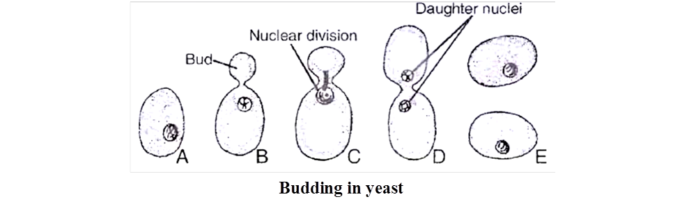
(c) Budding: Some fungi like yeast produce small outgrowths, i.e., buds from their vegetative body. Eventually, the buds are cut off from parent cell and mature to form new individuals.
2. Asexual reproduction:
It occurs through spores.
These are single celled specialized structures which separate from the organism, get dispersed and germinate to produce new mycelium after falling on suitable substrate.
The spores produced during asexual reproduction in fungi are formed by mitotic division and are thus termed, mitospores.
The various means of asexual reproduction are as follows :
(a) Zoospore :
Many fungi, especially aquatic members produce these types of spores.
Zoospore may be uniflagellate, e.g., Synchytrium or biflagellate, e.g., Saprolegnia, Pythium and are naked uninucleate structures formed in zoosporangia.
They germinate to give rise to new mycelium.
Biflagellate zoospores are of two kinds (e.g., Saprolegnia) pear shaped or pyriform with 2 flagella placed at anterior end (primary zoospore) and kidney shaped or bean shaped, bearing two laterally inserted flagella (secondary zoospore).
This phenomenon of having two types of zoospores is called diplanetism.
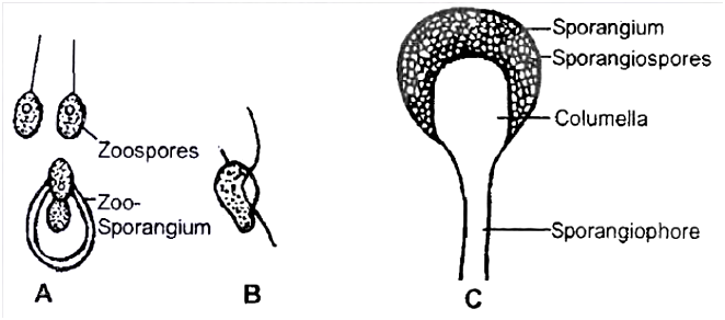
(b) Sporangiospore :
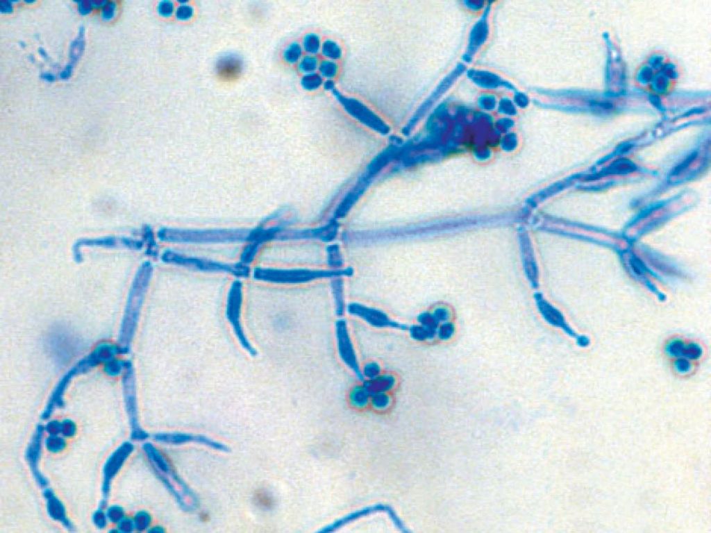
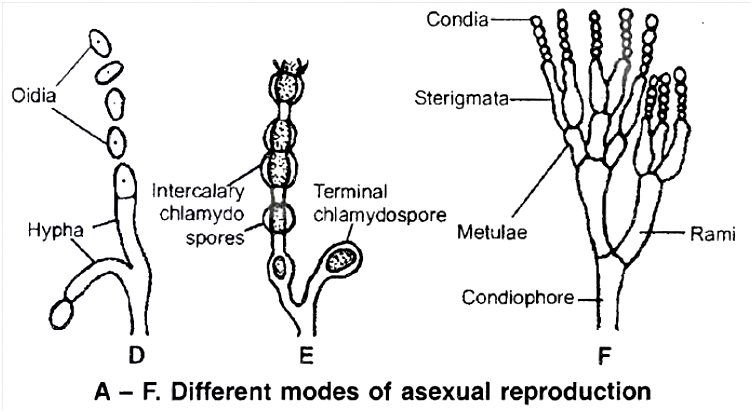
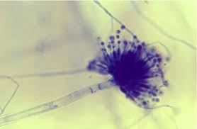
(c) Conidia:
Conidia are non motile, thin walled exogenous spores produced at the tips of erect hyphae called conidiophore.
They are arranged in chains upon the conidiophore, e.g., Aspergillus and Penicillium
(d) Chlamydospore:
In some fungi the hyphae under unfavourable conditions, forms thick walled resting resistant spores which later get separated from each other.
They may be terminal or intercalary.
They may remain viable for several years.
On return to favourable conditions they germinate to give rise to new individuals.
Thus, chlamydospores are structures for perennation also, e.g., Rhizopus.
(e) Oidia:
Non-motile thin walled spores developing under sugar rich conditions in medium.
Their budding condition is called torula stage.
3. Sexual reproduction:
Sexual reproduction is reduced in fungi and takes place by two fusing gametes.
It includes 3 stages :
(a) Plasmogamy:
There is union between two haploid protoplasts which results in bringing the fusing nuclei of different parents close together.
In some fungi, plasmogamy is immediately followed by karyogamy.
However, in Ascomycetes and Basidiomycetes, an intermediate dikaryotic (n + n) condition occurs. This phase is called dikaryophase.
(b) Karyogamy: The two haploid nuclei which come together in plasmogamy fuse and thus, a diploid zygote is produced.
(c) Meiosis: Reduction division takes place in the zygote thus, reducing the number of chromosomes to half.
Plasmogamy occurs by the following methods :
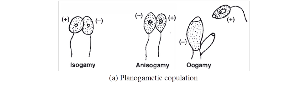
(a) Planogametic copulation / Gametic fusion:
This is the simplest form of sexual reproduction.
In this process, fusion of two gametes of opposite sex or strains takes place.
One or both of the fusing gametes are motile.
It results in the formation of a diploid zygote, e.g., Allomyces.
This process is usually of three types: Isogamy, Anisogamy, Oogamy.
(b) Gametangial contact :
In this process two gametangia come in contact with one another.
A fertilization tube is developed to facilitate the migration of entire contents of male gametangium into the female gametangium.
Both the gametangia never fuse together losing their identity, e.g., Pythium, Albugo (Oomycetes).
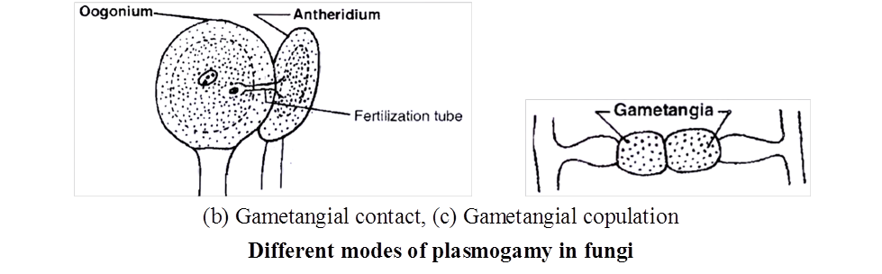
(c) Gametangial copulation :
In this process, direct fusion of entire contents of two gametangia is accomplished by dissolution of their common walls resulting in the formation of a single cell, in which protoplasts of two gametangia fuse, e.g., Mucor, Rhizopus (Zygomycetes).
(d) Spermatization :
Some fungi produce many minute, spore like, single-celled structures called spermatia (non motile male gametes) on spermatiophores (hyphae).
These structures are transferred through agencies like water, wind and insects to special female receptive, hyphae (Basidiomycetes).
The contents migrate into receptive structure.
Thus, dikaryotic condition is established, e.g., Puccinia.
(e) Somatogamy :
This takes place in most of the higher true fungi, where formation of gametes is absent.
In such fungi, direct fusion of somatic hyphal cells occur to establish dikaryophase, e.g., Agaricus
Classification of Fungi
A number of criteria are used for classifying fungi.
Morphology of mycelium, mode of spore formation and fruiting bodies form the basis for the classification of this kingdom.
A common systematic presentation is given below
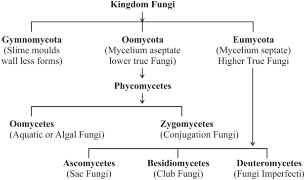
I. Oomycetes : The algal fungi
Hyphal wall contains cellulose and other glucans in many members.
The mycelium is coenocytic (multinucleate and aseptate).
Asexual reproduction involves the formation of spore containing sacs or sporangia. In aquatic forms, the sporangia produce zoospores.
Zoospores generally have two laterally inserted flagella with heterokont condition, in which one flagellum is smooth (whiplash) while the other is of tinsel type (having fine surface outgrowths called mastigonemes).
Sexual reproduction is by planogametic fusion or gametangial contact.
The product of sexual reproduction and site of meiosis is oospore.
Concept Builder
(i) Phytophthora infestans causes late blight of potato and occasionally of tomato as well. Great Irish famine of 1845 -1847 was due to late blight of potato.
(ii) Albugo candida (Cystopus candidus) causes white rust of crucifers and is characterised by the appearance of irregular white blisters on the leaves and stems.
(iii) Pythium debaryanum causes damping off disease in seedlings of tomato, chillies, castor, mustard.
(iv) Sclerospora graminicola causes downy mildew in cereals particularly, Pennisetum typhoides (vern. Bajra) green ear disease.
(v) Peronospora parasitica causes downy mildew in a number of plants, such as mustard, spinach, onion etc.
(vi) Saprolegnia causes salmon disease of gills in fishes.
II. Zygomycetes: The conjugation fungi
It is class of terrestrial fungi which are mostly saprotrophic and rarely parasitic.
Hyphal wall contains chitin or fungal cellulose.
The mycelium is coenocytic (multinucleate, aseptate) like the one found in Oomycetes.
Motile cells (zoospores or planogametes) are absent.
Mitospores are non motile. They are called sporangiospores as the spores are formed inside sporangia that are borne at the tips of special hyphae called sporangiophores.
Sexual reproduction occurs through gametangial copulation or conjugation. Because of it, zygomycetes are also called conjugation fungi.
The gametes are commonly multinucleate and are called coenogametes.
Sexual reproduction produces a resting diploid spore called zygospore. Because of the presence of zygospore, the group of fungi is called zygomycetes. Zygospore differs from oospore in that, for its formation a distinct food laden, non motile, large female gamete is not produced.
Zygospore is the site of meiosis and does not give rise to new mycelium directly. Instead it produces a new sporangium called germ sporangium (previously called zygosporangium). Germ sporangium forms meiospores called germ spores.
Sometimes, gametangia fail to fuse. Gametangia become surrounded by a thick wall resulting in formation of azygospore (parthenogenetically produced zygospore).
Concept Builder
(i) Rhizopus stolonifer (= R. nigricans) is popularly known as black bread mould. Mucor mucedo is called dung mould or pin mould. Rhizopus and Mucor are the common saprotrophic fungi that attack a variety of food stuffs. Soft rot or leek disease of strawberry, apple, jack fruit, sweet potato etc. is due to Rhizopus. Mucor pusillus causes infection of internal organs in human beings. Absidia corymbifera causes bronchomycosis.
(ii) Ramysin (antibiotic) is obtained from Mucor ramannianus.
Life Cycle of Rhizopus
Rhizopus is a saprophytic fungus, commonly found on dead organic matter rich in carbohydrates.
Mycelium is made up of white narrow thread like hyphae growing on the surface of substratum.
Two types of vegetative hypha (Rhizoidal and Stoloniferous) arise from definite points called hold fast / apparent nodes.
Third hypha is asexual, called Sporangiophore and fourth type is sexual hypha called zygophore.
The absorptive hyphae are called rhizoidal hyphae which penetrate into the substratum.
The hyphae appeared over the surface of substratum are in the form of stolons.
From the nodes of stolons arise branched rhizoidal hyphae on the under surface and a group of aerial structures called sporangiophore (asexual hypha) from the upper surface of 'apparent nodes'.
The apical portion of each sporangiophore ends into a swollen structure called sporangium which is filled with spores.
The hyphae are coenocytic, aseptate and branched. It has many nuclei, oil drops, glycogen bodies and vacuoles in the cytoplasm.
The reproduction in Rhizopus is vegetative, asexual and sexual.
- Vegetative reproduction: This takes place by fragmentation of hyphae.
- Asexual reproduction :
Asexual reproduction takes place by the formation of non-motile spores inside a sporangium.
The tip of aerial hypha swells in which cytoplasm migrates with nuclei.
The tip swells considerably and the nuclei divide repeatedly.
The contents of the young swollen tip differentiate into a central zone called columellaplasm mainly filled with vacuolated cytoplasm surrounded by a peripheral zone called sporangioplasm containing dense cytoplasm and many nuclei.
Vacuoles ultimately form a continuous vacuolated layer by fusing laterally one after the other and ultimately develop into a dome shaped septum known as columella (sterile part).
In the meantime, cleavage of sporangioplasm takes place resulting into innumerable, small 2-10 nucleate portions which round up, become invested with spore membranes, and develop into nonflagellate spores, the sporangiospores.
These are formed under most favourable conditions. Thus, the sporangium is large, globose and contains many spores.
Spores are dispersed by bursting of the thin wall of the sporangium due to pressure that is set up in the columella.
Other two asexual spores are oidia and chlamydospores (formed under unfavourable conditions).
The spores on germination produce a germ tube giving rise to new mycelium.
3. Sexual Reproduction :
Sexual reproduction in Rhizopus takes place by the formation of two multinucleate gametangia.
Both the gametangia are similar externally but are different physiologically i.e. they are of (+) and (-) type.
It is called heterothallism (This was discovered by Blakeslee in Rhizopus stolonifei ).
Another species called Rhizopus sexualis is a homothallic species.
When two mycelia of opposite strains, (+) behaving as male and (-) behaving as female respectively, come near one another, under influence of a chemical called trisporic acid, this stimulates the formation of special sub-aerial hypha called zygophores.
This hypha produces small outgrowths, called progametangia.
Their apical ends are swollen and filled with multinucleate protoplasm.
This apical portion of these progametangia, comes in contact with one another.
A septum is laid down, separating the terminal portion which is now termed gametangium. The remaining part of progametangia is called suspensor.
The multinucleate undifferentiated protoplast of each gametangium is termed as coenogamete.
As the gametangia mature, the separating wall dissolves from the middle to outward and intermingling of the contents of two gametangia takes place (Gametangial copulation).
Nuclear pairing and fusion of one (+) and another (-) nuclei gives rise to a large number of diploid nuclei.
The young zygospore lying within the parent gametangial wall enlarges considerably and secretes several layers of thick wall (5 layered, 2 layered exosporium and 3 layered endosporium) around it.
The zygospore matures it breaks up the original gametangial wall into small pieces that fall apart exposing the outer thick, spiny and black exosporium.
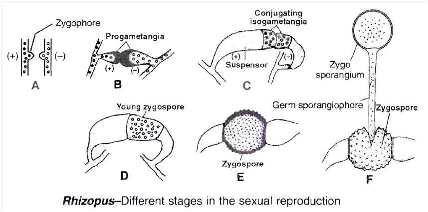
Meiosis occurs at the time of germination of zygospore.
The zygote germinates after rest period.
On germination, the exosporium cracks and endosporium produces a germsporangiophore (promycelium) that terminally develops a germ sporangium (zygosporangium) which bears large number of spores.
The meiosis produces 4 haploid nuclei where only one remain functional.
This divides repeatedly to produce coenocytic mycelium with many haploid nuclei.
Occasionally, failure of gametangial copulation results in parthenogenetic development of zygospore, which are called azygospores (parthenospores
III. Ascomycetes: The sac fungi
The mycelium consists of septate hyphae. (Yeasts are an exception in that they are basically unicellular).
They are saprophytic, decomposers, parasitic or coprophilous (growing on dung).
The septa possess central pores called septal pores. The pores allow communication and transport between adjacent cens.
Cell wall contains chitin.
Motile structures do not occur in the life cycle.
In majority of ascomycetes, the common mode of asexual reproduction is through the formation of conidia . Conidia are borne on branched or unbranched hyphae called conidiophores , e.g., Penicillium, Aspergillus.
Female sex organ is called ascogonium.
Plasmogamy occurs by means of -
- Gametangial contact (e.g., pyronema)
- Conjugation (e.g., Yeast)
- Spermatization (e.g., Ascobolus)
- Somatogamy (e.g., Peziza)
- Autogamy (e.g., Morchella).
Karyogamy is delayed after plasmogamy. A new transitional phase appears in the life cycle. It is called dikaryophase. The cells of dikaryophase are called dikaryotic cells. Each such cell possesses two different nuclei (Dikaryon). This forms a shorter phase of life cycle.
Once a cell becomes dikaryotic, it transfers the nucleus to other cells by the crozier method (method of dikaryotization) to make them dikaryotic.
Some dikaryotic cells function as ascus mother cells. This converts the cells into asci (singular ascus). Ascus is a sporangial sac peculiar to Ascomycetes. Ascus is the site of karyogamy and meiosis. 4 to 8 haploid meiospores named ascospores are produced endogenously in each ascus. In most of the cases, half the number of ascospores belong to one mating type(+) while the other half belong to the second mating type (-).
Ascospores may be arranged linearly (Neurospora) or unorderly (yeast).
The asci may occur freely or get aggregated into specific fructifications called ascocarps. Ascocarps are of many types : cup-like (apothecium, e.g., Peziza), flask-shaped (perithecium, e.g., Neurospora, Claviceps) , elongated with a slit (hysterothecium), closed (cleistothecium , e.g., Penicillium) cushion like, chambered (Ascostroma, e.g., Pleospora). The fructifications of some ascomycetes are edible, e.g., morels, truffles.
Concept Builder
Members of this class are said to be our worst fungal enemies.
(i) Morels are Ascomycetes with edible ascocarps having fleshy sponge-like conical cap or pileus and a stalk like stipe, e.g., Morchella esculenta (vern. Guchhi), M. deliciosia.
(ii) Truffles: They are edible tuber-like subterranean ascocarps, e.g., Tuber aestivum.
(iii) Claviceps purpurea causes ergot of rye and C. microcephala causes ergot of bajra in which ears are filled with sclerotia of the fungus. Eating of infected cereals produces ergotism. (It produces an alkaloid called ergotine which causes abortion, if eaten, unknowingly). This is used as a drug to promote expulsion of foetus.
(iv) Neurospora crassa (Pink bread mould), is often employed in experimental genetics, so is called Drosophila of plant kingdom.
(v) Erisyphe: The fungus produces powdery mildew (fungal disease in which pathogen results in a powdery coating of spores on the sut1ace of the host), e.g., Erysiphe graminicola.
(vi) Penicillium chrysogenum is used in commercial production of the antibiotic penicillin. The later was the first commercial antibiotic. It was discovered from P. notatum. The fungus is employed in ripening of cheese, e.g., P.roqueforti and P. camemberti.
(vii) Aspergillus: It is common green smoky mould which not only contaminates laboratory cultures (hence called weed of laboratory), but also various food stuffs including bread, butter etc. Aspergillus flavus is highly poisonous due to the presence of aflatoxins. A. oryzae is the source of diastase enzyme.
(viii) Brewing Industry: Under anaerobic conditions, sugary solutions inoculated with yeasts are converted into alcoholic beverages, e.g., beer, wine, cider, toddy. The two common yeasts used by brewing industry are Saccharomyces cerevisiae (Beer or Baker's yeast) and S. ellipsoidens (Wine yeast).
(ix) Gibberellins: They were first discovered in the extracts of Gibberella fujikuroi growing on rice (bakanae disease of rice). It is the perfect stage of fungus Fusarium moniliforme (Deuteromycete). Gibberellins are natural plant growth hormones.
Life cycle events of (A) Yeast and (B) Penicillium are described below:
(A) Yeast
Yeasts are a group of non mycelial or pseudomycelial ascomycetes which multiply asexually by budding or fission and where asci are not organised into ascocarps.
These are facultative aerobes.
Depending upon the mode of asexual reproduction, yeasts are of three types -budding yeast (e.g., Saccharomyces), fission yeast (e.g., Schizosaccharomyces) and helobial yeast (both budding and fission, e.g., Saccharomycoides).
Yeasts in which ascus formation is known are named as true yeasts.
Related forms which resemble yeasts in main characterstics, but where ascus formation is not reported, are called false yeasts, e.g., Candida, Mycoderma, Geotrichum, Cryptococcus (false yeasts belong to deuteromycetes).
Life cycle of Yeast
It is a saprophytic fungus found on substratum which is rich in sugars, e.g., sugarcane juice, fruits (banana, plums, grapes), milk etc.
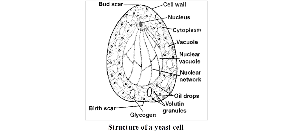
Structure
This is a circular or elliptical, unicellular, colourless fungi not having a typical mycelium.
Their cell wall contains mannans, glucans, lipids, proteins and chitin.
Protoplasm of yeast can be differentiated into two regions.
Outer region called ectoplasm in the form a thin layer and endoplasm which is granular.
The protoplasm has stored foods in the form of glycogen bodies and volutin granules and fats.
Mitochondria and ribosomes are found in the cytoplasm.
1. Vegetative Reproduction : Yeast reproduces vegetatively either by Transverse wall fission or by budding. Depending on this character, they are grouped as fission yeast (Schizosaccharomyces) and budding yeast (Saccharomyces).
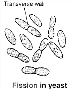
(a) By fission:
During reproduction by fission the parent cell elongates, the nucleus divides into two daughter nuclei and gradually a transverse partition wall is laid down somewhat near the middle, starting from periphery to the centre dividing the mother cell into daughter cells.
The two daughter cells so formed, may remain together for sometime and begin to divide again or they may separate soon and then divide.
(b) By budding:
Budding yeasts are rather common than the fission yeast. At the commencement of budding a small portion of the cell wall, usually near the end, softens.
The protoplast of the mother cell covered by a thin membrane bulges out in the form of a bud which ultimately develops into a daughter cell.
Meanwhile, the nucleus of the mother cell divides mitotically, (according to some, the division is amitotic).
One of the two daughter nuclei migrates into the enlarging bud.
The bud grows until it attains the size of the mother cell.
The daughter cell then becomes separated from the mother cell and the process may be repeated indefinitely.
Under conditions of rapid growth, the daughter cell also starts producing buds before being detached from the mother cell and the process may be repeated several times, giving rise to chains or groups of yeast cells.
This results in the formation of branched or unbranched pseudomycelium. The cells in chains of pseudomycelium are loosely joined together.
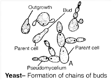
Sooner or 'A later, however, the chains break into their constituent cells.
2. Sexual Reproduction:
It takes place by the union of two cells (more often similar in size).
The copulating pair of cells may be vegetative cells or ascospores.
Yeasts may be homothallic or heterothallic and stages of fusion are extremely variable.
Three life cycle patterns are distinguishable among yeasts. These are :
(a) Haplontic life cycle: This is exhibited by Schizosaccharomyces octosporus (fission yeast).
(b) Diplontic life cycle: This is exhibited by Saccharomycoides ludwigii (Helobial yeast).
(c) Diplohaplontic life cycle: This is exhibited by Saccharomyces cerevisiae (Budding yeast).
Concept Builder
In addition to the above given methods of sexual reproduction of yeast, the following methods are also found:
- (a) Adelphogamy: Copulation between two adjoining sister cells. This is isogamous and the cells which fuse, do not separate after fission but remain united to form short chains.
- (b) Pedogamy: It is copulation between mother and the daughter cell formed by budding. The daughter remains attached to the mother and the nucleus of the bud migrates into the mother.
Saccharomyces cerevisiae = Brewer's Beer yeast or Baker's yeast
S.ellipsoidens = Wine yeast
Torulopsis utilis and Endomyces vermalis are rich in proteins. Rhodotorula is rich in vitamin A and Ashbya gossypii is rich in vitamin B2.
Yeasts are used in curing cocoa beans.
Diseases caused by yeast
(a) Candidiasis/moniliasis – Candida albicans
(b) Blastomycosis – Blastomyces dermatidis
(c) Histoplasmosis – Histoplasma capsulatus
(d) Cryptococcosis – Cryptococcus neoformans
Some yeasts reduce the yield of silk industry by attacking silkworms.
Species of Nematospora attack cotton, tomato and beans.
(B) Penicillium
Important characters
Facultative parasite and saprophytic fungi.
Mycelium is branched septate, with simple septal pore and each cell is uni or multinucleate depending upon the species.
Asexual reproduction occurs by conidia.
Conidiophores are often branched.
The first branch level is called rami and second or ultimate branches are called metulae having bottle shaped sterigmata.
Each sterigmata produces a chain of conidia.
The conidia in chain are arranged in basipetal order.
Each conidium is uninucleate, non motile, two layered, dispersed by air and germinates to form new mycelium.
Sexual reproduction: It exhibits dikaryophase and produces ascocarp.
The ascocarp is cleistothecium type.
Each ascus has 8 ascospores.
Ascospore germinates to form new mycelium.
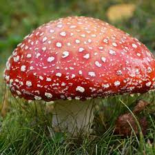
IV. Basidiomycetes: The Club Fungi
They are the most advanced and most commonly seen fungi. Their fructifications are often large and conspicuous, e.g., mushrooms, toadstools, puff balls, bracket fungi etc.
Basidiomycetes are among the best decomposers of wood. They are able to decompose both cellulose and lignin. Lignin is not metabolised by most other fungi and even bacteria. Ganoderma species causes decay of wood even on standing trees.
Motile structures or cells are absent.
Mycelia are of two types, primary and secondary. Primary mycelium contains monokaryotic cells and is short lived.
Monokaryotic phase or primary mycelium may multiply by oidia, conidia-like spores and pycniospores.
Secondary mycelium is long lived and dominant phase of life cycle. It is represented as dikaryophase. It consists of profusely branched septate hyphae.
Septa possess dolipores or central pores with barrel-shaped outgrowths (except rusts and smuts).
Handle like outgrowths are found on the sides of septa. They are called clamp connections. Clamp connections are meant for proper distribution of dikaryons at the time of cell division.
Secondary mycelium can perennate in the soil or wood by means of sclerotia or rhizomorphs.
Dikaryophase or secondary myceium may multiply by different types of spores chlamydo-spores, aecidiospores, uredospores, teleutospores etc.
There is often differentiation of two mating types (+) and (-) in thallus.
Sexual reproduction does not involve sex organs. Instead fusion occurs between basidiospores and other monokaryotic spores, between a spore or spermatium and a hypha or between two hyphal cells of primary mycelia.
Karyogamy and meiosis occur in club-shaped structures known as basidia (singular -basidium). The name of the class is based after them.
A basidium commonly produces four meiospores or basidiospores exogenously at the tips of fine outgrowths called sterigmata or directly on the basidium.
The fungi may or may not produce fructifications called basidiocarps. The basidiocarps vary from microscopic forms to large macroscopic structures. Some puff balls and brackets can be over 50 cm in diameter.
Concept Builder
(i) Smuts : They produce thick-walled, black-coloured resting spores called smut spores. Smuts are of two types, covered and loose. In covered smut, the spore mass remains within the membranous covering of sorus, e.g., Ustilago hordei (covered smut of barley), Ustilago maydis (smut of corn). In loose smut the spores are exposed while attached to the host, e.g., Ustilago nudatritici (loose smut of wheat), U. avenae (loose smut of oat).
(ii) Mushrooms : They are edible and non edible Agaricales which possess umbrella like basidiocarp. Common examples of edible mushrooms are Agaricus campestris, A. bisporous, Volvariella volvacea (Paddy straw mushroom), Pleurotus ostreatus etc.
(iii) Toadstools: Toadstools are poisonous mushrooms which generally have white spores. Amanita caesarea (Caeser's mushroom) was used in poisoning Roman emperor Caesar. The other toadstools are-Amanita phalloides (Death cup), A. muscaria (Fly agaric) and Gynomitra esculenta (heat labile carcinogenic toxin).
(iv) Rusts : They are characterised by the formation of rusty pustules containing the spores.
(a) Puccinia graminis tritici -Black rust of wheat.
(b) Puccinia glumarum or P striiformis -Yellow rust of wheat.
(c) P. recondita -Brown rust of wheat
(v) Hallucinogens: Psilocybe mexicana (Sacred mushroom) has hallucinating properties similar to LSD. It is used by Mexican Indians during certain religious ceremonies.
(vi) Armillaria (largest fungi): A. mellea (Honey mushroom) is a serious root parasite of both hardwoods and conifers. The fungus develop rhizomorphs into the phloem of the host and hence, blocks the food supply.
(vii) Puffballs: The basidiocarp is a stalked rounded structure which, upon ripening, releases out puffs of spores. The fructification may grow above or below the substratum, e.g., Lycoperdon oblongisporum, L. giganteurn.
(viii) Bracket fungi (Shelf fungi) : They are basidiomycetes whose basidiocarps or fructifications appear on tree trunks, logs, lumber etc. just as brackets or shelves, e.g., Fomes applanatus, Polyporus sulphureus, Ganoderma.
(ix) Predator fungi : e.g., Dactylaria, Arthrobotrys.
(x) Stinkhorn. Phallus impudicus (Dead man's finger). Spore mass produces a stinking odour to attract flies.
Life History of a Mushroom
Agaricus campestris is the common field mushroom which has edible basidiocarp. The fungus is saprotrophic.
The vegetative or assimilative part of mycelium is subterranean. It is found in moist humus rich soil of open fields, grassland, piles of straw or within rotting togs.
The mycelium multiplies by fragmentation; occasionally by oidia and chlamydospores.
Life cycle of mushroom contains two types of mycelia, primary and secondary. Sex organs do not differentiate.
Primary mycelium is short lived. It consists of septate hyphae having monokaryotic cells. The mycelia are heterothallic.
The hyphae of two mating types come in contact and show somatogamy. However, only plasmogamy occurs at this time.
It gives rise to a dikaryotic cell that grows, divides and produces a long-lived and extensive dikaryotic or secondary mycelium.
The hyphae of secondary mycelium show clamp connections and dolipore septa. Its celis possess two haploid nuclei instead of single diploid nucleus.
Under favourable conditions, hyphae of secondary mycelium collect at places and give rise to rounded or pyriform compact masses of hyphae called buttons.
The buttons enlarge and produce aerial fructifications or basidiocarps.
The latter are popularly termed as mushrooms. In contrast the secondary mycelium, from which mushrooms develop, is known as spawn.
The basidiocarps or mushrooms often lie in rings. The latter are called as fairy rings, its diameter increases centrifugally every year.
Each basidiocarp or mushroom is cream to pinkish brown in colour and consists of two parts, stipe and pileus.
The stipe or stalk is fleshy. It is slightly swollen at the base.
Pileus is umbrella-like cap of the mushroom. In the button stage, the pileus is connected to stipe by membrane called veil or velum.
It ruptures during growth of pileus. However, its remains can be seen on the upper part of stipe as annulus.
The pileus is circular in outline. Its upper surface is more or less convex. The under surface is flat or concave.
It bears 300-600 radiating rows of vertical plates named gills (lamellae).
The two sides of vertically placed gills are lined by thousands of club-shaped basidia alongwith sterile paraphyses.
The two, together constitute the fertile layer or hymenium of the gill. Hymenium is subtended by compact subhymenium.
The centre consists of interwoven hyphae called trama. Each basidium functions as the site for both karyogamy and meiosis. The two nuclei fuse to form a short-lived diploid synkaryon.
The latter, then divides meiotically giving rise to four haploid nuclei, two of (+) strain and two of (-) strain. The free end of the basidium now develops four peg-like outgrowths called sterigmata.
Each sterigmata bears an ovoid pinkish-purple meiospore termed as basidiospore. A droplet appears at the tip of sterigmata which creates tension and hanging basidiospores are carried away by air currents.
The basidiospores are liberated successively for several days. After falling on a suitable substratum, each basidiospore germinates to produce monokaryotic primary mycelium.
Life Cycle of Puccinia graminis tritici
P. graminis tritici is a macrocyclic (producing many dikaryotic spores), Heteroecious (requires more than one host i.e., primary host -wheat, secondary or alternate host -barberry).
It produces uredospores and teliospores on the wheat plant, basidiospores in soil and pycniospores and aeciospores on barberry.
It causes black or stem rust of wheat. Hence, we describe the various stages of life cycle on the basis of the host.
A. Stage of life cycle on wheat (primary host)
(i) Uredia and uredospores :
The dikaryotic aeciospores germinate on the leaves of wheat on both the surfaces and may also germinate on the stem.
They form the dikaryotic mycelium.
The germ tube protrudes out which swells up to form an elongated appressorium near the stomata.
A peg like outgrowth now arises from the appressorium and penetrates the stomata. It ramifies repeatedly to form a mass of mycelium this mycelium forms the uredospore.
As a result, some pressure is exerted on the epidermis which bursts exposing the uredospore. These clusters of uredospores have been variously referred to as uredosori or urediopustules.
A uredospore is reddish-brown, unicelled, oval or globose, stalked, dikaryotic spore. Its wall is three layered, the outer being somewhat spiny.
The uredial stage multiplies through the uredospores which germinate on fresh wheat plants (due to the red colour of spores this stage is called red rust stage).
(ii) Telia and teliospores (black rust stage) :
Towards the end of the season, the dikaryotic mycelium of the uredosorus begins to produce teliospores (or teleutospores) in the same sorus.
They are dark brown or black, stalked, thick walled, bi-celled spores. The upper cell is pointed, both the cells are binucleate.
The pustules containing teliospores are called as tella or teleutosori.
The teliospores also exert pressure on the epidermis which bursts open, exposing the spores.
It is at this stage the symptoms develop in stem also, so the disease is named black stem rust of wheat.
Karyogamy occurs inside each cell of teliospore and as a result they become diploid.
The teliospores cannot infect fresh wheat plants. They germinate in soil to form the basidiospores.
(iii) Basidia and basidiospores:
The two cells of the teliospores now act as hypobasidium. They germinate in soil and form a tube called promycelium.
The diploid nucleus migrates into epibasidium and then undergoes meiosis to form four haploid nuclei each of them develops as a basidiospore on the sterigmata.
Of these, two basidiospores belong to +ve strain and two to -ve strain.
These spores are not capable to infect a wheat plant. Each spore is unicelled, monokaryotic and unstalked.
These can infect the barberry plant (Berberis vulgaris) which is the secondary or alternate host occurring on the hills in India.
B. Stage of life cycle on barberry
(i) Spermatia or pycniospores
The basidiospores of both the strains (+ and -) can germinate on upper surface of barberry leaf.
They produce haplophase or primary mycelium of the respective strains which grows through the intercellular spaces.
Soon the mycelium forms a palisade like mat which organises like a flask shaped structure near the upper epidermis called as spermogonia or pycnidium.
They open by a single pore called ostiole.
The spermogonium is lined by palisade Ilike uninucleate cells called spermatiophores.
From the tips of spermatiophores are pinched off a large number of uninucleate cells called pycniospores or spermatia.
Besides, some sterile hyphae also arises which grow out through the ostiole.
They are called as periphysis.
In addition to these, some thin walled hyphae are also given out which become more elongated.
They are called as flexuous hyphae or receptive hyphae.
While the spermatia function as the male cells, the flexuous hyphae behave as the female hypha.
The pycniospores or spermatia protrude out of the ostiole in a nectar.
The insects feeding on this nectar transfer the spermatia from one spermogonium to the other.
Spermatization is brought about when spermatia of one strain come in contact with the trichogyne of flexuous hyphae of the other strain.
The nucleus of pycniospore or spermatium passes into the flexuous hyphae, thus bringing
(ii) Aecia and aeciospore :
An aecium or aeciosorus is produced on the lower surface of barberry leaves. It arises just beneath pycnium. The dikaryotized mycelium aggregates sub-epidermally.
This gives rise to a group of elongated dikaryotized cells which function as aecial mother cell. The mother cells differentiate a row of binucleate cells.
The alternate binucleate cells enlarge and they are identified as aeciospores. The alternate cell remaining small is disjunctor or sterile cell.
The sterile wall of the aecial cup called peridium or pseudoperianth presses the lower epidermis which eventually bursts open. Thus, the aeciospores are set free.
The aeciospores are polyhedral or ovate, binucleate, unicelled and double layered. The outer thick wall is called exine and the inner as intine.
They are set free in spring. They cannot infect the barberry bushes. These are carried from hills to the plains where they infect wheat plants. They germinate on the leaf surface from a germ tube which enters the host through stomata.
Annual Recurrence of Rust in India:
It was first studied by Prof. K.C. Mehta.
He concluded that, in India, the annual recurrence of rust on wheat takes place through uredospores.
The alternate host, Berberis vulgaris plays no role in our country. According to Mehta, if no wheat is grown on hills, the intensity of its infection in plains can be reduced.
He also suggested the involvement of certain collateral hosts like Briza minor, Bromus and Thalictrum for multiplication of uredospores during non-growing season of wheat.
V. Deuteromycetes: (The Fungi Imperfecti)
Deuteromycetes is an artificial class (form class) of fungi which has been created to include all those fungi in which sexual stage (or perfect stage) is not known (absent or not reported) .
The mycelium is septate and branched. Coenocytic forms are not known.
Asexual reproduction often occurs by conidia.
Once perfect (sexual) stages of members of Deuteromycetes were discovered they were often moved to Ascomycetes and Basidiomycetes but most members of Deuteromycetes are actually related to ascomycetes.
Some members are saprophytes or parasites while a large number of them are decomposers of litter and help in mineral cycling.
Concept Builder
(i) Leaf spot of rice : Helminthosporium oryzae causes leaf spot disease of rice commonly called brown leaf spot of rice. It caused Bengal famine in 1942-43.
(ii) Early blight: Alternaria solani causes early blight of potato. The leaves develop small oval brown spots with concentric rings (target board symptom).
(iii) Tikka disease : Circular necrotic dark brown or blackish leaf spots develop in groundnut due to Cercospora personata.
(iv) Red rot: Colletotrichum falcatum produces red rot of sugarcane.
(v) Wilts : Many economically important plants (e.g., cotton, pigeon pea) show sudden signs of wilting due to blockage of tracheary elements by growth of fungus, Fusarium especially F. oxysporum, F. udum.
(vi) Ringworm of foot/Athlete's foot is caused by Trichophyton interdigitate.
Common names of some fungi:
Concept Builder
Common Fungicides and their composition
- Bordeaux mixture – (CuSO4: Ca(OH)2 : H2O). First fungicide discovered by RMA Millardet Commonly known as holy water of plant pathology
- Burgandy mixture – Mixture of CuSO4 + Na2CO3 + H2O was discovered by Mass.
- Chestnut mixture – Ammonium carbonate + copper sulphate.
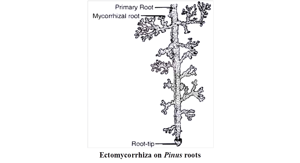
MYCORRHIZA (FUNGAL ROOTS)
The mutually beneficial or symbiotic association of a fungus with the roots of higher plants is termed mycorrhiza. Mycorrhizal roots differ in shape from normal roots and often show a wooly covering. These roots lack root cap and root hairs.
A fungus may get associated with roots of a number of plants and a particular plant may form association with a number of fungi. Depending upon the location of the fungus, the mycorrhiza is of two types, i.e., ectomycorrhiza and endomycorrhiza.
In ectomycorrhiza, the fungal hyphae are mainly external, forming a wooly covering on external surface of root and forms network of mycelium (Hartig net) in the intercellular spaces of the cortex. Fungal partner is commonly basidiomycetes, e.g., Pinus roots
In endomycorrhiza, the fungal hyphae enter the tissue of the root, spreading intercellularlly and intracellularly.
The fungus is able to break the cell wall in a limited way and is restricted to cortical region of the root. Some hyphae send small projections into cortical cells without destroying them.
Such fungi are termed VAM (Vesicular Arbuscular Mycorrhiza), e.g., Orchid roots.
Mycorrhizal association is a symbiotic relationship as both the partners are mutually beneficial to each other. The fungal partner obtains nourishment from the cortical cells of the root and depends upon the plant for shelter.
The root cells excrete sugars and other soluble gradients which are used by fungal hyphae spreading in intercellular spaces. The hyphae may get nourishment from the cells directly and also by sending small projections into cortical cells.
The fungus seems to be essential for the growth of the plant having mycorrhiza.
The plant also gets benefit from the association as the fungal hyphae spreading in soil substantially increases the surface area of absorption, thereby enabling the plant to get enhanced supply of water, nitrogen, phosphorus and other minerals from the soil.
Orchids seldom occur without mycorrhiza. Certain forest trees like pines, birches show stunted growth if their roots are not associated with fungus.
LICHENS
Lichens are dual (composite) organisms or entities which contain a permanent association of a fungal partner or mycobiont and an algal partner or phycobiont.
Mycobiont is dominant partner and mostly belongs to ascomycetes (Ascolichens -, e.g., Graphis, CIadonia, Parmelia, Usnea, etc.) or sometimes basidiomycetes (Basidiolichens - e.g., Corella, Cora, etc.).
Phycobiont is mostly a member of Chlorophyceae (e.g., Chlorella, Trebouxia, Protococcus, Palmella, etc.) or can be a BGA (e.g., Nostoc, Chlorococcus, Scytonema, etc.).
The term lichen was coined by Theophrastus (370 -285 B.C.), also called Father of Botany.
Lichens often grow in most inhospitable and uninhabited places like barren rocks (saxicolous), soil (terricolous), icy tundra or alpines, sand dunes, roofs, walls, wood (Iignicolous), tree bark (corticolous), leaves, etc.
They commonly live under humid and exposed conditions but can tolerate extreme desiccation. However, lichens, cannot tolerate air pollution, especially due to sulphur dioxide (so are considered indicators of SO2 pollution).
Lichens are perennial. Their growth is slow. Lichens have greyish, yellowish, greenish, orange, dark brown or blackish colouration.
Structure
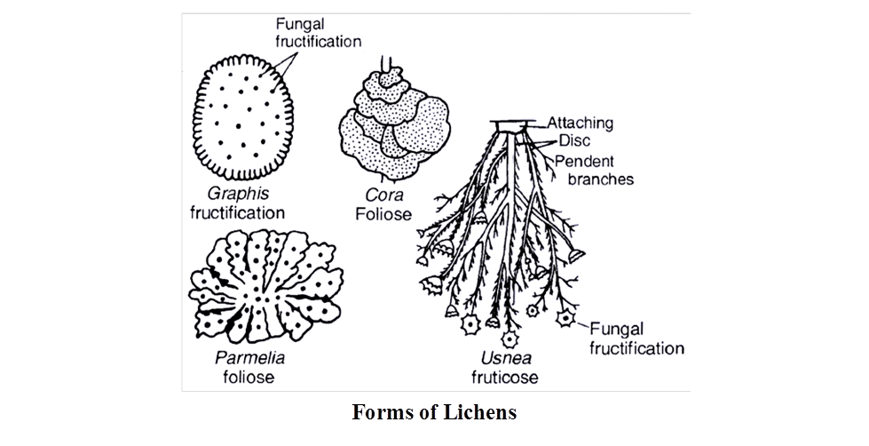
Based upon external morphology, the lichens are of three types :
(i) Crustose. Crust like, closely appressed to the substratum and attached to it at several places, e.g., Graphis, Lecanora, Rhizocarpon.
(ii) Foliose: The body of the lichen is flat, broad, lobed and leaf-like, which is attached to the substratum at one or a few places with the help of rhizoid like structures called rhizines, e.g., Parmelia, Peltigera.
(iii) Fruticose : The lichen is branched like a bush and attached to the substratum by means of a disc, e.g., CIadonia, Usnea, Evernia, Bryonia.
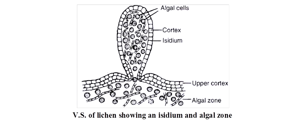
The bulk of lichen body is formed by fungal partner called mycobiont. It includes the surface, medulla (or interior) and rhizines (attaching devices).
The algal partner or phycobiont constitutes hardly 5% of the lichens body.
It is generally restricted to a narrow zone (algal zone) below the surface.
Relationship :
The fungus performs following functions:
Body structure and covering (ii) Anchoring (iii) Absorption of water and minerals. It can absorb water from wet air (atmosphere), dew and rain. Minerals are picked up both from substratum and atmosphere. Special chemicals are excreted by the fungal partner of the lichen to dissolve minerals from the substratum. (iv) Sex organs and fruitifications are of fungal origin.
The major function of alga is photosynthesis.
The cyanobacterial alga additionally takes part in nitrogen fixation. The alga picks up water and mineral salts from the fungus while the fungus obtains part of the food manufactured by the alga.
Therefore, in a lichen the association between alga and fungus is that of mutual benefit (mutualism) popularly called symbiosis.
However, at times the fungus is found to (i) send haustoria into algal cells (ii) induce alga to secrete organic substances and (iii) prevent alga to develop pectic covering. Therefore, some workers believe that the fungus is a controlled parasite over the alga. The phenomenon is called helotism.
Reproduction: Lichens multiply by four methods:
(i) Progressive death and decay resulting in the separation of a lichen thallus into two or more parts.
(ii) Fragmentation caused by mechanical injury, due to wind or animal bites.
(iii) Isidia are superficial outgrowths of the lichens which are primarily meant for increasing surface area and photosynthetic activity. At time, they are broken off. Each isidium is capable of forming a new lichen because it has a core of a few algal cells surrounded by a sheath of fungal hyphae.
(iv) Soredia. These are most efficient means of asexual reproduction. They are microscopic lichen propagules which are produced in large numbers inside sori called pustules. Soredia are dispersed by air currents. After falling on a suitable substratum each soredium gives rise to a lichen, because it has a few algal cells surrounded incompletely by a weft of fungus.
Concept Builder
- Special structures in the thallus of lichen:
(i) Cyphellae help in exchange of gases, present in lower cortex.
(ii) Cephalodia help to retain moisture and its algal partner fix nitrogen also.
(iii) Breathing pores for aeration, present in upper cortex of thallus.
Early Colonisers : Lichens are early or pioneer colonisers of barren rocks, cliffs, mountains and new terrains. During their growth, lichens stick to the rocks and cliffs by secreting acids. It produces minute crevices where organic matter accumulates. It paves the way for growth of mosses.
Food: In tundra, Cladonia rangifera (Reindeer Moss) constitutes the staple food of reindeer, caribou, musk ox, etc. Cetraria islandica (Iceland Moss) is used as a food article in Iceland, Sweden and Norway Lecanora esculenta is regarded as bread of heaven by Jews. Parmelia (Rock Flower) is also a table delicacy Dermatocarpon miniatum (Stone Mushroom) is a vegetable in Japan.
Dyes : Orchil is obtained from Rocella tinctoria. The latter was also the source of litmus (R.montagnei) before the advent of synthetic products. Litmus is a pH indicator.
Perfumes: Scented incense is got from species of Ramalina and Evernia.
Medicines: Usnic acid got from Usnea (Old Man's Beard) and Cladonia has antibiotic properties. It is used in preparation of ointment for burns and wounds.
Air Pollution : Decrease in lichen population of an area is indicative of SO2 pollution.
Fires: In hot season, Usnea may produce forest fires.
VIRUS
Term virus was coined by Pasteur.
Viruses are obligate intracellular parasites. They are intermediate between living and non living entities.
Non living nature of virus
Lacking protoplast.
Ability to get crystallized, e.g., TMV, poliomyelitis virus.
Inability to live independent of a living cell. (Lack functional autonomy)
High specific gravity which is found only in non living objects
Absence of respiration.
Absence of energy storing system.
Absence of growth and division.
Living nature of virus
Being formed of organic macromolecules.
Presence of genetic material.
Ability to multiply.
Occurrence of mutations.
Occurrence of certain enzymes like, neuraminidase (first discovered), transcriptase and lysozyme in certain viruses.
Infectivity and host specificity.
Viruses can be 'killed' by autoclaving and ultraviolet rays.
They take over biosynthetic machinery of the host cell and produce chemicals required for their multiplication.
Viruses are responsible for a number of infectious disease like common cold, epidemic influenza, chicken pox, mumps, poliomyelitis, rabies, herpes, AIDS, SARS etc.
Concept Builder
Mayer described Tobacco Mosaic disease in 1886.
Iwanowsky is credited with the discovery of virus in 1892. TMV was the first virus to be discovered.
Beijerinck called virus as "Contagium vivum fluidum " (living infectious fluid).
In 1935, Stanley crystallised TMV.
Twort and d'Herelle discovered bacteriophage.
Lwoff and Wollmann discovered temperate viruses.
Shafferman and Morris discovered cyanophage, e.g., LPP-1.
Bawden and Pirie studied the chemical nature (nucleoproteins) of TMV.
Sinsheimer discovered single stranded DNA in bacteriophage f × 174
Issac and Lindemann discovered interferon
Delbruck (1938), found viruses to undergo mutations.
Reverse transcription in Retroviruses was discovered by Temin and Baltimore, so the phenomenon is called teminism. The enzyme reverse transcriptase is RNA dependent DNA polymerase.
Structural Components of Viruses
(i) Envelope is the outer thin loose covering composed of proteins (from virus), lipids and carbohydrates (both from host). It has smaller subunits known as peplomers, e.g., Herpes virus, HIV, Vaccinia virus etc. If it is not present the virus is said to be naked.
(ii) Capsid : It is the outer protein coat made up of subunits called capsomeres, their number is virus specific. These possess antigenic properties.
(iii) Nucleoid : Viruses contain either DNA or RNA. No virus contains both DNA and RNA.
(a) DNA containing viruses are called deoxyviruses.
These are of two types:
(i) Double stranded DNA (dsDNA) virus, e.g., Pox virus, Cauliflower mosaic virus.
(ii) Single stranded DNA (ssDNA) virus, e.g., Coliphage ×174, M 13 phage.
(b) RNA containing viruses or riboviruses are of two types.
(i) Double stranded RNA (ds RNA) virus, e.g., Reo virus, Wound Tumour Virus.
(ii) Single stranded RNA (ss RNA) virus, e.g., TMV, Influenza virus, Foot and Mouth disease virus, Retroviruses (HIV).
Classification of Virus
Holmes (1948) has divided viruses into three groups on the basis of specific hosts.
(a) Phytophagineae are plant viruses. They generally have ssRNA, e.g., TMV, Potato Mosaic Virus, Yellow Vein Mosaic virus, Cauliflower Mosaic virus.
(b) Zoophagineae are animal viruses. They commonly have ssRNA or dsRNA or dsDNA. e.g., Poliomyelitis virus, Influenza viruses, Small pox virus, Mumps virus, Rabies virus.
(c) Phagineae attack lower organisms
(i) Bacteriophages are bacterial viruses and they usually possess dsDNA, e.g., T2, T4, lambda ![]() phage.
phage.
(ii) Coliphages are viruses of E.coli, e.g., Coliphage fd.
(iii) Cyanophages attack blue green algae, e.g., LPP-1, SM-1 .
(iv) Phycophages attack algae.
(v) Mycophages attack fungi.
(vi) Zymophages attack yeast.
Reproduction
It is of two main types: Phagic and Pinocytic
(a) Phagic Reproduction : It is further of two types :
(i) Lytic cycle : Occurs in virulent phages, e.g., T4 bacteriophages.
(ii) Lysogenic cycle : Occurs in temperate viruses such as phage.
(b) Pinocytic Reproduction: It is found in viruses like TMV, HIV, Hepatitis B etc., in which whole of virus particle enters host cell except envelope (if present).
Structural details of some viruses
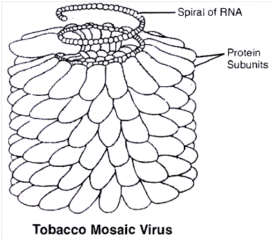
(1) Tobacco Mosaic Virus (TMV) is elongated rod like, 3000 Å long, 180 Å in diameter with molecular weight Protein 39.4 × 10⁶ dalton. 2130 capsomeres are arranged helically to form the capsid. RNA strand is helical. ssRNA consists of 6400 nucleotides. Thus, the ratio of nucleotides : capsomeres = 3 : 1
(2) Pox virus / variola is the causal agent of small pox. These are among the largest of animal viruses, are rectangular (brick shaped), 300 × 230 nm in size. Genome is dumbell shaped with central core of dsDNA. The core has two enzymes RNA polymerase and ATP phosphohydrolase.
(3) AIDS virus consists of single stranded RNA. It has 2 copies of ssRNA. Outer cover has 5 layers, i.e., outer most glycoprotein, followed by double lipid layer and the innermost has two protein layers.
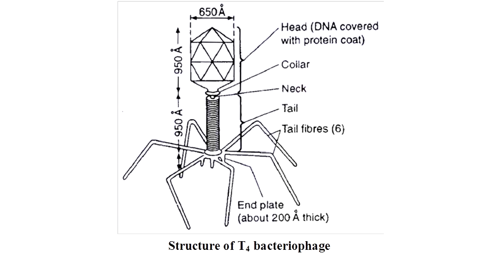
T4 Bacteriophage has a tadpole like structure with polyhedral head connected to a helical tail (binal). The head consists of nucleic acid surrounded by a protein coat or capsid. Nucleic acid is double stranded DNA. Tail is proteinaceous tube-like, core surrounded by sheath. At one end, tube is joined to the head by thin collar. At the other end, it has a hexagonal base plate with six small tail pins and six tail fibres which help in attachment of the phage to the host cell.
- Sub Viral Agents: These are viruses which lack one of the essential component, e.g., viroids, virusoids, prions
- Viroids (L. virus -poison, eidos -diminutive)
- They are the smallest self replicating particles which were discovered by Diener (1971). Viroids are infectious RNA particles which are devoid of protein coat.
- They are obligate parasites. Molecular weight of a viroid is low.
- The RNA is tightly folded to form circular or linear structures. Viroids are known to cause diseases (some 20) in plants only, e.g., Potato spindle tuber disease (PSTD), Chrysanthemum stunt and Citrus exocortis.
(2) Virusoids
- Discovered by Randle et. al., these are RNA viruses but inside the capsid of other larger virus. They replicate within the host and do not cause any infection.
(3) Prions
Discovered by Alper et al.
Proteinaceous infectious particles, causing certain diseases like
(i) Kuru disease (laughing death disease in humans)
(ii) Bovine spongiform encephalopathy (BSE or Mad cow disease)
(iii) Scrapie disease in sheep
(iv) Creutz Feldt Jakob disease
Table 5 : Viral diseases of Plants
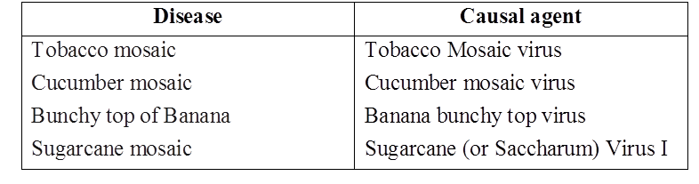
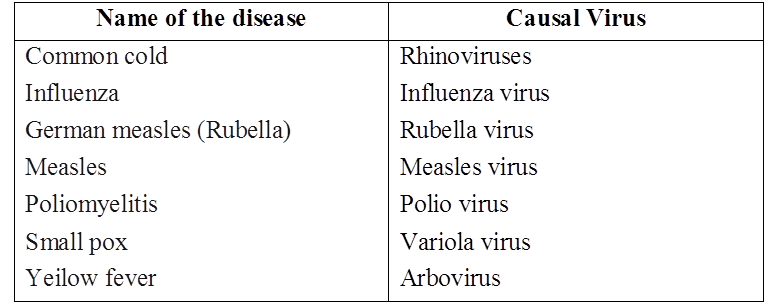
Table 6 : Viral diseases of Man
Animal viral diseases -Foot and mouth disease, Rinderpest, Ranikhet, Bird flu etc.
Concept Builder
Nomenclature of viruses :
International committee of virus nomenclature has given a system of naming the virus. The system consists of two parts. First part is common name of the virus and second part has the coded information about the virus. This is called as Cryptogram.
Kingdom Fungi
Fungi are eukaryotic organisms that include yeasts, molds, mushrooms as well as other microbes.Fungi are a distinct kingdom of heterotrophic creatures. They have a wide range of forms and environments. Fungi can often be observed on stale bread and rotten fruits. Toadstools and the common mushroom that we eat are both fungi. A parasitic fungus causes white patches on mustard leaves. Some unicellular fungi, such as yeast, are utilized in the production of bread and beer. Other fungi produce illnesses in plants and animals; Puccinia, which causes wheat rust, is a good example. Penicillium, for example, is a source of antibiotics. Fungi are widely distributed and can be found in the air, water, soil, and on animals and plants. They thrive in warm, humid environments.Mycology is the branch of biology concerned with the study of fungi.
Fungi are filamentous, with the exception of yeasts, which are unicellular. Their bodies are made up of hyphae, which are long, slender thread-like structures. Mycelium refers to the network of hyphae. Coenocytic hyphae are hyphae that are continuous tubes filled with multinucleated cytoplasm. In the hyphae of others, there are septae or cross walls. Fungi's cell walls are made up of chitin and carbohydrates.
Saprophytes are fungi that are heterotrophic and absorb soluble organic materials from dead substrates. Parasites are organisms that rely on live plants and animals for their survival. They can also live as symbionts, forming lichens with algae and mycorrhiza with the roots of higher plants.
Fungi reproduce via vegetative processes such as fragmentation, fission, and budding. Spores known as conidia, sporangiospores, and zoospores are instrumental in asexual reproduction, while oospores, ascospores, and basidiospores are responsible for sexual reproduction. Fruiting bodies are structures that produce different types of spores.
The three phases of the sexual cycle are as follows:
(i) Plasmogamy, which is the fusion of protoplasms between two motile or non-motile gametes.
(ii) Karyogamy, which is the fusion of two nuclei.
(iii) In a zygote, meiosis results in haploid spores.
Two haploid hyphae of compatible mating types come together and merge when a fungus reproduces sexually. In some fungi, the fusing of two haploid cells results in the formation of diploid cells right away (2n).
Kingdom fungi can be divided into three groups based on their nutritional needs.
(i)Saprophytic fungus get their sustenance from dead organic matter. Rhizopus, Penicillium, and Aspergillus are other examples.
(ii)Parasitic fungus get their food by living on other living organisms (plants or animals) and absorbing nutrients from them. Taphrina and Puccinia are two examples.
(iii)Symbiotic fungi are those that exist in an interdependent relationship with other species in which both parties benefit. Lichens and mycorrhizae are two examples. Lichens are the result of a symbiotic relationship between algae and fungi. Algae and fungi benefit from each other in this situation because fungi give protection to algae and algae provide carbohydrates for fungi.
The morphology of the mycelium, mode of sporeformation, and fruiting bodies form the basis for thedivision of the kingdom into various classes which are discussed below.
(a) Phycomycetes:
Phycomycetes can be found in aquatic areas and on decaying wood in moist and humid environments, as well as obligatory parasites on plants. Algal fungi are sometimes known as Phycomycetes. They have aseptate coenocytic hyphae that are distinctive. Asexual reproduction occurs via zoospores (motile) or aplanospores (asexual) (non-motile). In the sporangium, these spores are produced endogenously. A zygospore is created when two gametes fuse together. The morphology of these gametes is either similar (isogamous) or distinct (heterogamous) (anisogamous or oogamous). Mucor, Rhizopus (the bread mold), and Albugo(the parasitic fungi on mustard) are other typical examples.
(b) Ascomycetes:
The ascomycetes, sometimes known as sac-fungi, are typically multicellular (e.g., Penicillium) or occasionally unicellular (e.g. Saccharomyces or yeast). These fungi may be Saprophytic, decomposers, parasitic, or coprophilous(growing on dung). Mycelium is septate and branched. Conidia grown exogenously on special mycelium termed conidiophores are the asexual spores. Conidia create mycelium when they germinate. Ascospores are sexual spores that are produced endogenously in sacs similar to asci (singular ascus). These asci are grouped in ascocarps, which are distinct types of fruiting structures. Aspergillus, Claviceps, and Neurospora are some examples. Neurospora is widely utilized in biochemical and genetic research. Many of the members, such as morels and truffles, are edible and regarded as delicacies.
(c) Basidiomycetes:
Mushrooms, bracket fungi, and puffballs are examples of basidiomycetes. They can be found growing in soil, on logs and tree stumps, and in living things. Rusts and smuts, for example, are parasitic plant bodies. The mycelium is septate and branching. Although asexual spores are rare, vegetative reproduction by fragmentation is prevalent. Although there are no sex organs, plasmogamy is caused by the fusing of two vegetative or somatic cells of different strains or genotypes. The dikaryotic complex that results eventually gives rise to basidium. The basidium undergoes karyogamy and meiosis, resulting in four basidiospores. The basidiospores are formed exogenously on the basidium (pl.: basidia). Basidia are arranged in basidiocarps, which are fruiting bodies. Agaricus (mushroom), Ustilago (smut), and Puccinia are some of the most prevalent members.
(d) Deuteromycetes:
In this category, only asexual or vegetative phases of these fungi are known, therefore they are commonly referred to as imperfect fungi. When the sexual forms of these fungi were discovered, they were placed in the appropriate groups. It's also likely that the asexual and vegetative stages were given one name (and were classified as Deuteromycetes), while the sexual stage was given another one (and placed under another class). The fungi were accurately recognized and moved out of Deuteromycetes after the links were discovered. Deuteromycetes were frequently shifted to ascomycetes and basidiomycetes after their perfect (sexual) stages were discovered. Conidia are asexual spores that Deuteromycetes use to reproduce. The mycelium is branching and septate.Some individuals are saprophytes or parasites, whereas the majority are litter decomposers who aid in mineral cycling. Alternaria, Colletotrichum, and Trichoderma are other examples.