 ACME SMART PUBLICATION
Earthworm
The earthworm is a reddish-brown terrestrial invertebrate that lives in the moist soil's upper layer. They spend the day in tunnels created by burrowing and swallowing earth. Worm castings, or faecal deposits, are used to track them down in the gardens. Pheretima and Lumbricus are the most prevalent Indian earthworms.
(i) Morphology: Earthworms' bodies are long and cylindrical. The body is divided into over a hundred short, identical pieces (metameres about 100-120 in number). Along the longitudinal axis of the body, a dark median mid dorsal line (dorsal blood vessel) marks the dorsal surface. The presence of genital apertures distinguishes the ventral surface (pores).
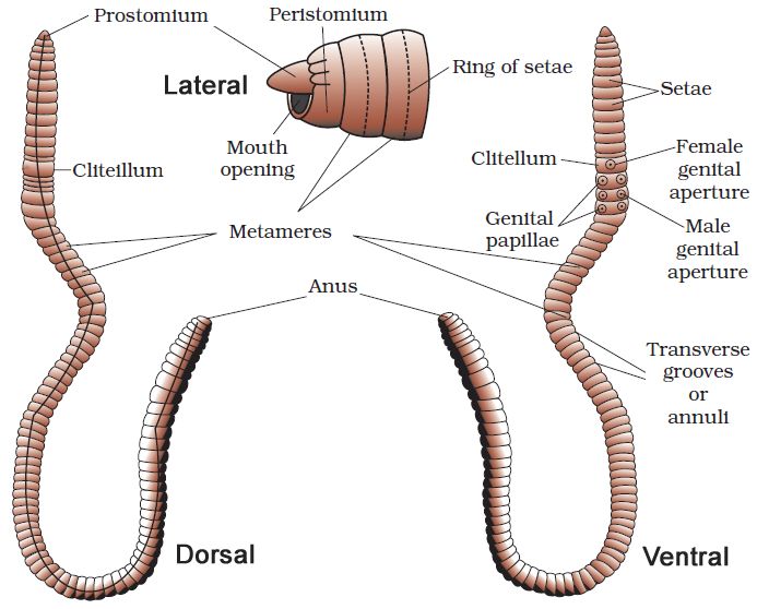
The mouth and the prostomium, a lobe that serves as a mouth cover and a wedge to force open soil crevices into which the earthworm may crawl, make up the anterior end. The role of the prostomium is sensory. The peristomium (buccal segment) is the first body segment and houses the mouth. A conspicuous dark band of glandular tissue termed clitellum covers segments 14-16 in a mature worm. As a result, the body is divided into three distinct regions: preclitellar, clitellar, and postclitellar. The ventrolateral sides of the intersegmental grooves, i.e. the 5th -9th segments, have four pairs of spermathecal openings. In the mid-ventral line of the 14th segment, there is a solitary female genital pore.On the ventrolateral sides of the 18th segment, there are two male genital pores. Nephridiopores are tiny pores that open on the surface of the body. Except for the first, last, and clitellum, each body segment has rows of S-shaped setae inserted in the epidermal pits in the middle. Setae can be retracted or expanded. Their primary function is movement.
(ii) Anatomy: A thin non-cellular cuticle covers the earthworm's body wall, which is followed by the epidermis, two muscle layers (circular and longitudinal), and an innermost coelomic epithelium. The epidermis is a single layer of columnar epithelial cells with secretory gland cells.
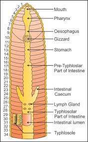
The alimentary canal is a straight tube that connects the first and last body segments. The buccal cavity (1-3 segments) leads into the muscular pharynx through a terminal mouth. The esophagus (5-7 segments) is a tiny, narrow tube that leads to the muscular gizzard (8-9 segments). It aids in the grinding of soil particles and rotting leaves, among other things. The stomach is divided into 9-14 parts. Earthworms eat decaying leaves and organic debris mixed with soil as nourishment.The humic acid in the humus is neutralized by calciferous glands in the stomach. The stomach begins at the 15th section and continues until the last part. On the 26th segment, a pair of short and conical intestinal caecae protrude from the gut. Except for the last 23rd-25th segments, the existence of an internal median fold of the dorsal wall called typhlosole is a distinguishing feature of the intestine after the 26th segment. This expands the absorption surface area in the gut. A little spherical opening called the anus connects the alimentary canal to the outside world. Digestive enzymes break down complicated meals into tiny absorbable components when the organic-rich soil moves through the digestive tract. These simpler molecules are absorbed and used through intestinal walls.
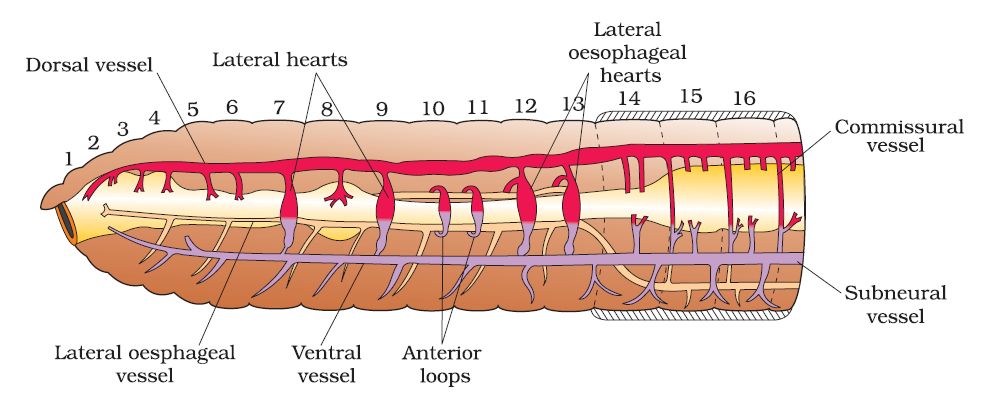
The blood vascular system of Pheretima is closed, consisting of blood arteries, capillaries, and the heart. Blood is restricted to the heart and blood vessels due to the closed circulatory system. Blood circulates in one direction due to contractions. The gut, nerve cord, and body wall are all supplied by smaller blood vessels. The 4th, 5th, and 6th segments all have blood glands. They manufacture blood cells as well as haemoglobin, which is dissolved in blood plasma. Blood cells are naturally phagocytic.
Earthworms do not have specific breathing apparatus. The wet body surface allows for respiratory exchange into the bloodstream.
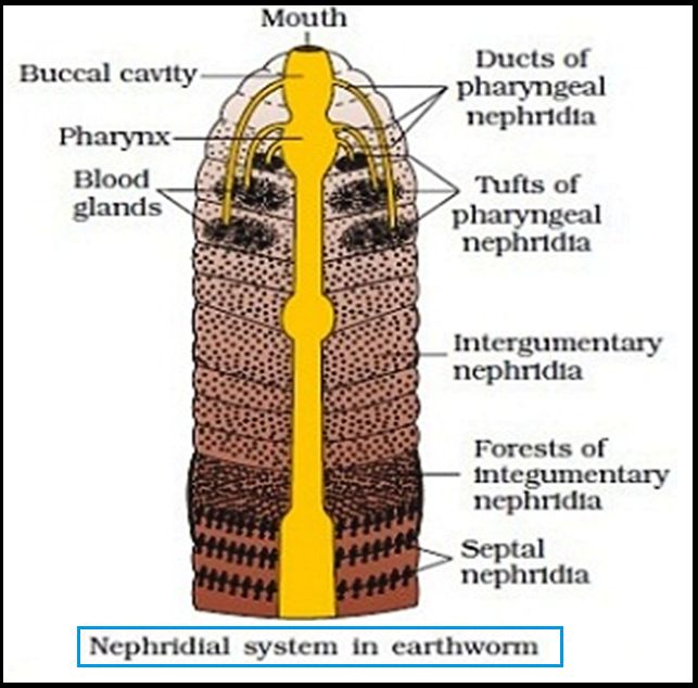
Nephridia are segmentally arranged coiled tubules that make up the excretory organs (sing.: nephridium). There are three types of nephridia:
(i) Septal nephridia, which are present on both sides of intersegmental septa from segment 15 to the last that open into the intestine,
(ii) Integumentary nephridia, which are attached to the lining of the body wall from segment 3 to the last that open on the body surface, and
(iii) Pharyngeal nephridia, present as a three-paired outfit in 4th, 5th and 6th segment. The structure of these many nephridia is remarkably similar. The volume and composition of the bodily fluids are regulated by nephridia. A nephridium begins as a funnel, collecting surplus fluid from the coelomic chamber.The funnel connects to a tubular component of the nephridium, which transports wastes to the digestive tube through a pore in the body wall.
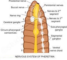
Ganglia are positioned segment-wise on the ventral paired nerve cord to represent the nervous system. In the front region (3rd and 4th segments), the nerve cord bifurcates, laterally enclosing the pharynx and dorsally joining the brain ganglia to form a nerve ring. The cerebral ganglia, along with other nerves in the ring, integrate sensory input and control bodily muscle responses.Although the sensory system lacks eyes, it does have light and touch-sensitive organs (receptor cells) that allow it to discern between light intensities and feel ground vibrations. Chemoreceptors (taste receptors) in worms are specialized to respond to chemical inputs. These sensory organs are found on the worm's front end.
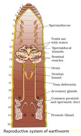
Earthworms are hermaphrodites (bisexual), which means they have both testes and ovaries. In the 10th and 11th segments, there are two pairs of testes. Their vasa deferentia connect to the prostatic duct at the 18th segment. In the 17th and 19th segments, there are two pairs of auxiliary glands, one pair in each.A pair of male genital holes on the ventrolateral side of the 18th segment access the outside the shared prostate and spermatic duct (vasa deferentia). In the 6th-9th segments, there are four pairs of spermathecae (one pair in each segment). During copulation, they receive and store spermatozoa. At the inter-segmental septum of the 12th and 13th segments, one pair of ovaries is attached. Underneath the ovaries are ovarian funnels that continue into the oviduct, unite, and open on the ventral side as a single median female genital hole on the 14th segment.
During the mating process, two worms exchange sperm. One worm must discover another worm, and they mate by exchanging spermatophores (packets of sperm) at opposite gonadal apertures.Cocoons made by clitellum gland cells contain mature sperm and egg cells, as well as nourishing fluid. Fertilization and development take place within the soil-deposited cocoons. The sperm cells within the cocoon fertilize the ova (eggs), which subsequently fall off the worm
and land in or on the earth. The worm embryos are kept in the cocoon. Each cocoon generates two to twenty baby worms on average after about three weeks. Earthworms develop directly, without the formation of a larva. Earthworms are known as "farmer's friends" because they dig burrows in the soil and make it porous, which aids in plant root penetration and respiration.Vermicomposting is the method of using earthworms to increase soil fertility. They're also utilized as game fishing bait.
