ACME SMART PUBLICATION
Digestive System
- Books Name
- ACME SMART COACHING Biology Book
- Publication
- ACME SMART PUBLICATION
- Course
- CBSE Class 11
- Subject
- Biology
DIGESTIVE SYSTEM
On the basis of the embryonic origin, the alimentary canal of vertebrates can be divided into three parts –
(A) Fore gut / Stomodaeum : Ectodermal. It includes buccal cavity / oral cavity.
(B) Mid gut / Mesodaeum : Endodermal. It includes pharynx, oesophagus, stomach, small intestine, and large intestine.
(C) Hind gut / Proctodaeum : Ectodermal. It includes anal canal and anus.
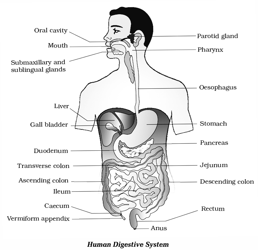
Digestive system of man consists of alimentary canal and some accessory digestive organs.
The Alimentary Canal:
It is a coiled muscular tube about 6 -9 metres long extending from mouth to anus.
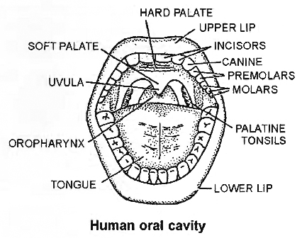
Mouth:
The mouth is an opening bounded by upper and lower lip.
Lips are attached on the inner side with the gums by thin transparent fold called Labial frenulum.
The space between lips and teeth is called vestibule.
The roof of buccal cavity is palate consisting of hard palate (maxilla, premaxilla and palatine bones) anteriorly and soft palate posteriorly.
Mucus epithelium has thick transverse folds called palatine rugae.
Terminal part of soft palate hangs in the throat called uvula. On sides of uvula tonsils are present which are made of lymphatic tissue.
The floor of buccal cavity is occupied by a muscular tongue attached at base by a fold called lingual frenulum.
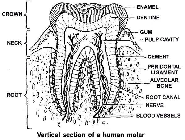
Teeth present on upper and lower jaws are
(i) Incisors: for cutting, have one root.
(ii) Canines: for tearing, have one root.
(iii) Premolars: for crushing, grinding and chewing, in upper premolar 2 roots and lower premolar 1 root.
(iv) Molars: for chewing, in upper molar 3 roots and lower molar 2 roots.
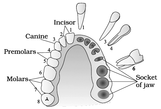
one side and the sockets on the other side
Dental formulae
In man, 20 teeth grow twice during life time i.e., diphyodont![]() ; (premolars and last molars absent In primary dentition) and 12 teeth appear only once i.e., Monophyodont.
; (premolars and last molars absent In primary dentition) and 12 teeth appear only once i.e., Monophyodont.![]()
Child =![]()
17 yr. old =![]()
Adult =![]()
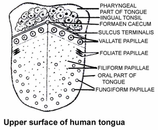
Tongue
The tongue is a voluntary muscular and glandular structure which occupies the floor of the mouth.
It is attached to the floor of the mouth by a fold called the frenulum of the tongue.
An inverted V-shaped furrow called the sulcus terminalis divides the upper surface of the tongue into anterior oral part and posterior pharyngeal part.
The apex of the sulcus terminalis projects backwards and is marked by a small median pit, named the foramen caecum.
The foramen caecum is an embryological remnant and marks the site of the upper end of the thyroglossal duct. Oral part of the tongue has papillae on its surface.
These are :
(i) Filiform papillae : smallest, most abundant and have no taste buds.
(ii) Fungiform papillae : appear as red dots on tongue and contain taste buds.
(iii) Foliate papillae: absent in man.
(iv) Circumvallate papillae: largest in size and knob like, also contain taste buds.
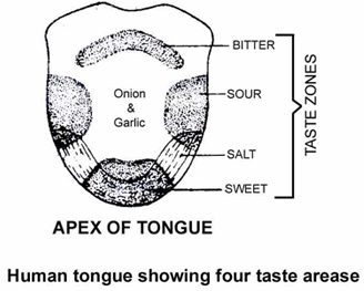
The different areas of tongue are demarcated as follows:
Tip – sweet
Tip and sides – salt
Sides – sour
Base – bitter
Sweat glands of dogs are present on tongue (panting of dog).
Salivary Glands
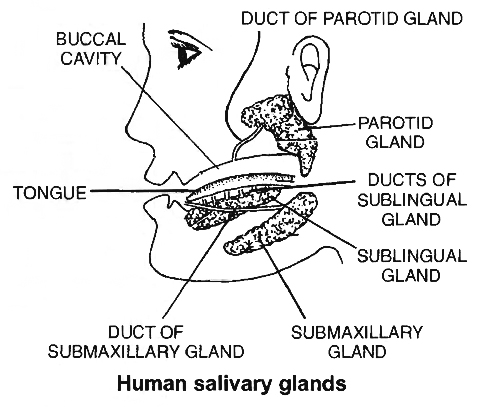
Four pairs of salivary glands open in the mouth cavity:
(i) Parotid: Largest -present below and in front of ears -Stenson's duct.
(ii) Submaxillary: Medium sized -present at the angles of jaw lower -Wharton's duct
(iii) Sublingual : Smallest -located below the tongue - Rivinus duct.
(iv) Infra orbital : Absent in man, otherwise present below eyes e.g. , in rabbit.
Daily Secretion of saliva is 1.5 litres, (pH of saliva is 6.7) and has salivary amylase (ptyalin), maltase and lysozyme.
Salivary glands are stimulated to secrete saliva by parasympathetic innervation while sympathetic nerves causes reduced secretion leading to drying of mouth.
Cl– are required for activation of salivary amylase. Mumps is viral infection of salivary glands (mainly Parotid).
Pharynx opens through gullet into the oesophagus and through glottis into the larynx.
An elastic cartilage plate, epiglottis, covers the glottis at the time of swallowing. Food mixed with saliva in buccal' cavity-Bolus.
Oesophagus
It is a long and thin tube, 25 cm long that pierces the diaphragm and enters the abdominal cavity.
Oesophagus is characterised by :
(i) Absence of visceral peritoneum. Its outermost fibrous (non-coelomic) covering is called tunica adventitia.
(ii) Absence of digestive glands. It has mucus-secreting goblet cells.
(iii) Presence of mucous membrane formed of non-keratinised stratified squamous epithelium some cells of which are ciliated.
(iv) Presence of voluntary (upper 1/3rd) and involuntary muscle fibres (lower 2/3rd).
Stomach
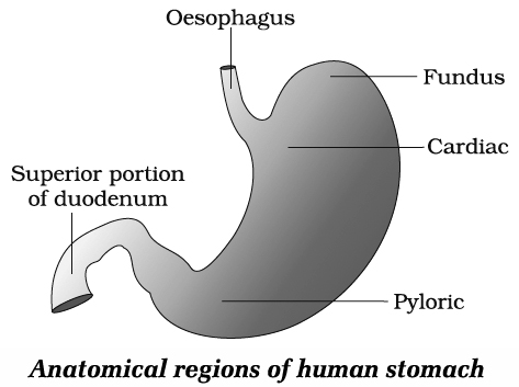
Stomach is oval and pouch like organ, divisible into cardiac, fundic, main body and pyloric parts.
Cardiac sphincter is present at the opening of oesophagus into stomach and prevents the regurgitation of food into oesophagus.
The pyloric part opens into small intestine and opening is guarded by pyloric sphincter.
The wall of stomach has three layers of muscles, outermost longitudinal layer, middle circular layer and innermost of oblique layer.
Mucosa has folds called rugae and cardiac, fundic and pyloric glands.
Only fundic glands secrete gastric juice.
These contain neck cells (secrete mucus and present in all three types of glands), oxyntic or parietal cells (secrete HCl and Castle's intrinsic factor for absorption of B12).
HCl of gastric juice converts Fe3+ into Fe2+ which makes the absorption of iron possible.
Non-secretion of HCl (achlorhydria) or gastrectomy can lead to iron-deficiency anaemia.
The peptic cells or chief cells or zymogenic cells release large quantity of pepsinogen and other enzymes.
Small Intestine
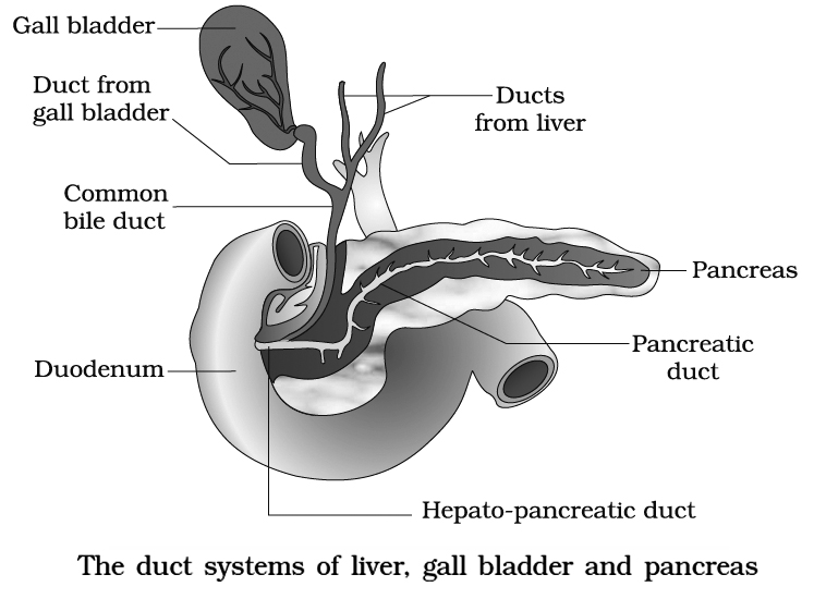
First part of small intestine is duodenum. It is 25 cm long, U or C-shaped in humans and receives the opening of hepatopancreatic duct (Bile duct + Pancreatic duct).
A small swelling is present at the opening of hepatopancreatic duct and is called 'Ampulla of Vater' or Hepatopancreatic ampulla and the opening is regulated by sphincter of oddi.
Other parts of small intestine are jejunum and ileum.
The wall of intestine has thin layers of longitudinal and circular muscles.
Mucosa has folds called plicae circulare (folds of Kerkrings or Valvulae conniventes) and villi towards lumen of the intestine.
Epithelial cells lining the villi have microvilli which further increase the absorptive area.
Intestinal glands or Crypts of Leiberkuhn have epithelial cells (secrete mucus), Paneth cells (secrete digestive enzymes) and argentaffin cells (probably secrete hormones).
In duodenum, Brunner's glands are also present (located in submucosa) which secrete mucus.
Diffused patches of lymphoid tissues are present throughout the small intestine and are aggregated in ileum to form Peyer's patches.
Large Intestine
It is about 1.5 m long and consists of three parts -Caecum, Colon and Rectum.
A blind pouch of caecum is vermiform appendix.
These parts help in digestion of cellulose in herbivores.
Wall of colon has sac like haustra.
Histologically, wall of colon has three bands of longitudinal muscles called taeniae coli.
Another characteristic of colon surface is the presence of small fat filled projections called epiploic appendages.
The colon is divisible into ascending, transverse, descending and sigmoid colon.
Ascending colon is the smallest part and lacks mesentry.
Last part of rectum is anal canal having a strong sphincter. It opens outside by anus.
DIGESTIVE GLANDS
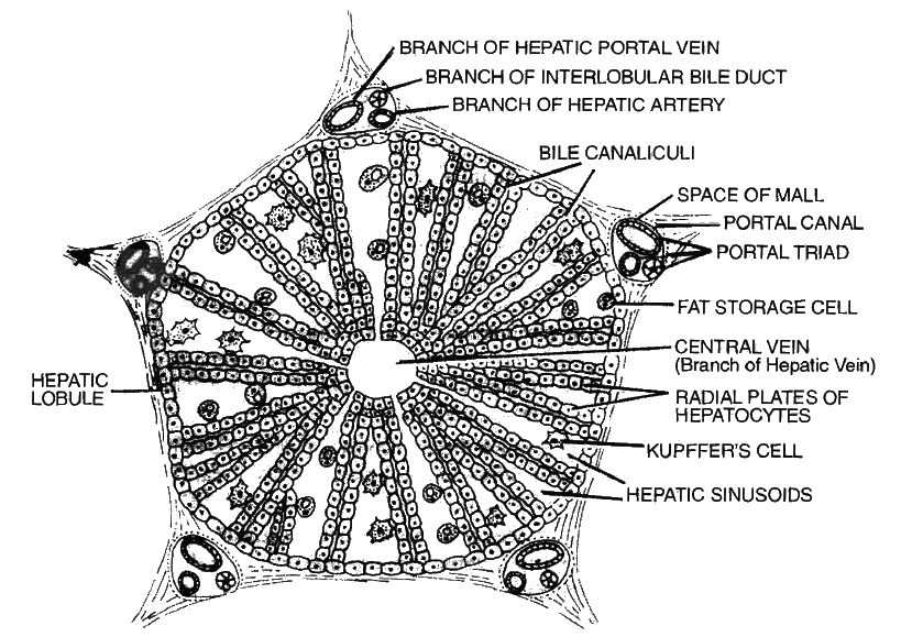
Liver
Largest digestive gland. It lies in the upper right side of the abdominal cavity just below diaphragm.
The liver is divided into two main lobes-right and left.
Between the right and left lobes, falciform ligament is present.
The right lobe is differentiated further into right lobe proper, a quadrate lobe and caudate lobe.
Liver is surrounded by Glisson's capsule, its trabeculae divide liver lobes into hexagonal lobules.
Polyhedral hepatocytes are arranged in cords around a central venule.
Portal triads contain hepatic artery, portal venule, bile ductule and lymphatics.
Blood sinusoids are present.
Kupffer cells are present in sinusoids and are phagocytic.
Gall bladder is situated on the inferior surface of right lobe. It is 8 cm long and 2 cm wide.
Bile is secreted by hepatocytes into the bile canaliculi, a series of narrow spaces between adjacent liver cells.
The canaliculil drain via bile ductules into bile ducts, which run in portal tracts; the bile duct themselves discharge into the right and left hepatic ducts which unite to form the common hepatic duct at the hilum of the liver.
Gall bladder has a capacity of 30 to 50 ml. It consists of smooth muscles lined by columnar epithelium.
It fills and empties via cystic duct which joins the common hepatic duct to form the bile duct; this in turn empties into the duodenum through the ampulla of Vater (hepatopancreatic ampulla).
At the point of its entry into the duodenum, the bile duct and adjacent pancreatic duct join each other.
The sphincter of Boyden surrounds the opening of bile duct.
Sphincter of oddi surrounds the ampulla of Vater.
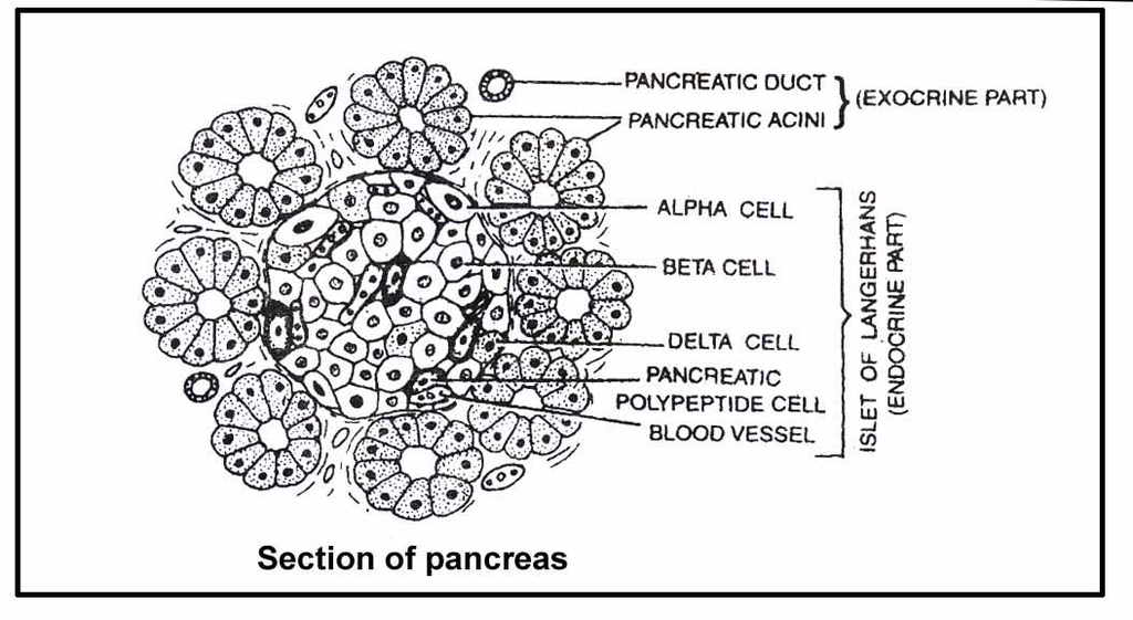
Pancreas
It is a racemosely branched gland, situated between stomach and duodenum.
Pancreas consists of acini (which secrete digestive enzymes) and islets of Langerhans (which secrete insulin and glucagon hormones).
Pancreas has two ducts within it.
The first is duct of Santorini which is accessory or nonfunctional, opening directly into duodenum and the other is duct of Wirsung which is functional and combines with bile duct to form common hepatopancreatic duct.
mechanism of food digestion
- Books Name
- ACME SMART COACHING Biology Book
- Publication
- ACME SMART PUBLICATION
- Course
- CBSE Class 11
- Subject
- Biology
Mechanism of Food DIGESTION
Digestion of Carbohydrates :
The diet of most of the animals including man consists of carbohydrates.
Depending upon the complexity, carbohydrates are of three types : polysaccharides, disaccharides and monosaccharides.
During the process of digestion, both poly and disaccharides are broken down to monosaccharides and in this form they can be absorbed into the body.
Some of these complex carbohydrates are starch and cellulose present in cereal grains, potato, fruits and tubers; sucrose present in cane sugar; lactose present in milk etc.
Enzymes that act on carbohydrates are collectively known as carbohydrases.
Pancreatic juice and intestinal juice also contain carbohydrate digesting enzymes.
Pancreatic juice contains pancreatic amylase that acts on starch to digest it into maltose, isomaltose and dextrin.
The secretions of the brush border cells constitute the intestinal juice or succus entericus.
This juice contains a variety of enzymes like disaccharidases (e.g., maltase), dipeptidases, lipases, nucleosidases etc.
Starch![]() Maltose + Isomaltose + Dextrin
Maltose + Isomaltose + Dextrin
Maltose + Isomaltose + Dextrin![]() Glucose
Glucose
Sucrose![]() Glucose + Fructose
Glucose + Fructose
Lactose![]() Glucose + Galactose
Glucose + Galactose
Digestion of Proteins:
Proteins are complex organic compounds made up of smaller units called amino acids. So in the process of digestion, all proteins are broken down to amino acids.
Enzymes that hydrolyse proteins are collectively known as proteases or peptidases.
Many of these enzymes are secreted in their inactive form or proenzymes as their active form would hydrolyse cellular and extracellular proteins of the organism itself.
Inactive enzymes are converted to their active form only at the site of action.
Saliva as such does not contain any protein digesting enzyme, but it can denature the uncooked natural proteins like the ones present in raw egg, unboiled milk or uncooked germinating seeds.
However, this is not a process of hydrolysis as in digestion.
Action of Gastric Juice:
The gastric glands of stomach produce a light coloured, thin and transparent gastric juice.
It contains water, hydrochloric acid (0.3%) and inactivated enzymes prorennin and pepsinogen.
The presence of HCl makes the medium highly acidic (pH = 1 or 2) so that pepsin can act on proteins to convert them into peptones and proteoses.
However, there is no pepsin in invertebrates.
Both prorennin and pepsinogen are converted to their active forms in the presence of HCl.
Pepsin and rennin can also do the same function once they are formed.
HCl also helps to kill bacteria and other harmful organisms that may be present along with the food.
Rennin acts on the casein protein of milk and converts it into paracasein which in the presence of calcium ions forms calcium paracaseinate (curdling of milk).
The function of rennin is then taken over by pepsin and other milk-coagulating enzymes. Pepsin then acts on it.
These reactions are summarized below:
Prorennin (inactive)![]() Rennin (active)
Rennin (active)
Pepsinogen (inactive)![]() Pepsin (active)
Pepsin (active)
Milk Casein![]() Paracasein
Paracasein
Paracasein + Ca++![]()
![]()
Calcium paracaseinate![]() Peptones and proteoses
Peptones and proteoses
Action of Pancreatic and Intestinal Juice:
Both pancreatic juice and intestinal juice (succus entericus) are poured into small intestine.
Pancreatic juice contains trypsinogen, chymotrypsinogen, procarboxypeptidases, lipases, amylases or amylopsin, DNAases and RNAases.
All these enzymes of pancreatic juice can act only in the alkaline medium.
This change in the medium of food, from acidic to alkaline, is done by the bile juice.
Therefore, bile juice acts on the food before the action of pancreatic juice.
All these actions are given below :
Trypsinogen (inactive)![]() Trypsin (active)
Trypsin (active)
Peptones and proteoses![]() Peptides
Peptides
Chymotrypsinogen (inactive) is activated to chymotrypsin by trypsin itself.
Chymotrypsin is another important milk coagulating enzyme and can hydrolyse casein into paracasein which then coagulates to form calcium paracaseinate.
However unlike rennin, it acts in the alkaline medium.
Chymotrypsin can act on other proteins also.
Digestion of Fats:
Fat digestion starts only when the food reaches the stomach.
Some amount of gastric lipase is present.
Gastric lipase is of little importance except in pancreatic insufficiency.
Most of the fat digestion begins in the duodenum, pancreatic lipase being one of the most important enzymes involved.
Bile juice contains bile salts that break down the bigger molecules of fat globules into smaller droplets by reducing the surface tension of fat droplets.
This process is known as emulsification of fats.
Lipase is the chief enzyme that acts on emulsified fats.
It is present both in the pancreatic juice and intestinal juice.
Pancreatic lipase (steapsin) is the principal enzyme involved in fat digestion.
Lipase converts emulsified fats into diglyceride and monoglycerides releaSing fatty acids at each step.
At the end of digestion, all fats are converted into fatty acids, glycerol and monoglycerides.
Absorption of Digested Products
- Books Name
- ACME SMART COACHING Biology Book
- Publication
- ACME SMART PUBLICATION
- Course
- CBSE Class 11
- Subject
- Biology
Absorption of digested products
Absorption is the process by which the end products of digestion pass through the intestinal mucosa into the blood or lymph.
It is carried out by passive, active or facilitated transport mechanisms.
The passage of various substances into the blood depends upon the concentration gradients.
However, some of the substances like fructose and some amino acids are absorbed with the help of the carrier proteins.
This mechanism is called the facilitated transport.
Transport of water depends upon the osmotic gradient.
Active transport occurs against the concentration gradient and hence requires energy.
Various nutrients like amino acids, monosaccharides like glucose, electrolytes like Na+ are absorbed into the blood by this mechanism.
Fatty acids and glycerol being insoluble, cannot be absorbed into the blood.
They are first incorporated into small droplets called micelles which move into the intestinal mucosa.
They are re-formed into very small protein coated fat globules called the chylomicrons which are transported into the lymph vessels (Iacteals) in the villi.
These lymph vessels ultimately release the absorbed substances into the blood stream.
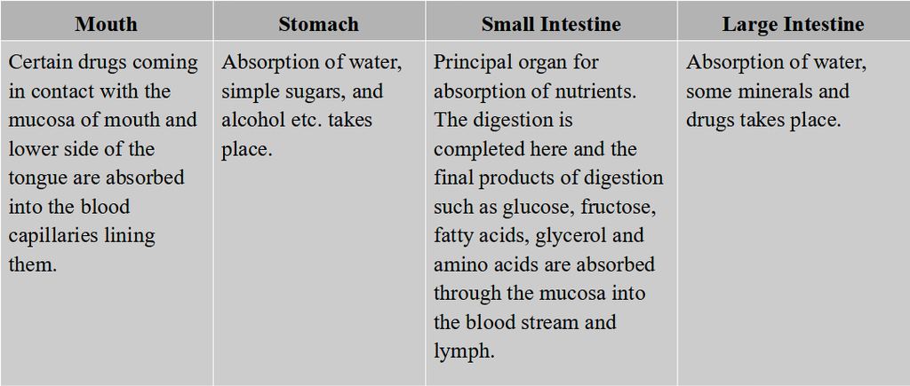
Absorption of substances takes place in different parts of the alimentary canal, like mouth, stomach, small intestine and large intestine.
However, maximum absorption occurs in the small intestine. A summary of absorption (sites of absorption and substances absorbed) is given in table.
Table: The Summary of Absorption in Different Parts of Digestive System
The absorbed substances finally reach the tissues which utilise them for their activities. This process is called assimilation'
The digestive wastes, solidified into coherent faeces in the rectum initiate a neural reflex causing an urge or desire for its removal.
The egestion of faeces to the outside through the anal opening (defaecation) is a voluntary process and is carried out by a mass peristaltic movement.
Disorders of Digestive System
- Books Name
- ACME SMART COACHING Biology Book
- Publication
- ACME SMART PUBLICATION
- Course
- CBSE Class 11
- Subject
- Biology
Disorders of Digestive System
The inflammation of the intestinal tract is the most common ailment due to bacterial or viral infections. The infections are also caused by the parasites of the intestine like tape worm, round worm, thread worm, hook worm, pin worm, etc.
Jaundice: The liver is affected, skin and eyes turn yellow due to the deposit of bile pigments.
Vomiting : It is the ejection of stomach contents through the mouth. This reflex action is controlled by the vomit centre in the medulla. A feeling of nausea precedes vomiting.
Diarrhoea: The abnormal frequency of bowel movement and increased liquidity of the faecal discharge is known as diarrhoea. It reduces the absorption of food.
Constipation: In constipation, the faeces are retained within the rectum as the bowel movements occur irregularly.
Indigestion: In this condition, the food is not properly digested leading to a feeling of fullness. The causes of indigestion are inadequate enzyme secretion, anxiety, food poisoning, over eating and spicy food.
NUTRITIONAL REQUIREMENTS OF HUMANS
Energy Yielding Nutrients
Carbohydrates are used primarily as sources of chemical energy, to be either metabolised immediately as glucose or stored as glycogen.
The synthesis of glycogen is called glycogenesis.
The liver can store enough glycogen to maintain blood glucose level for several hours. Under acute starved conditions, the liver cells begin to convert fatty acids and the glycerol (digestive products of fat molecules) into glucose.
Such production of new glucose is known as gluconeogenesis. Proteins are used as structural components of tissues, as channels, transporters, regulatory molecules and enzymes.
Proteins can also be utilised as energy sources, when broken down to amino acids.
Out of the 20 amino acids identified so far as the constituents of proteins, 8 (10 in children) cannot be synthesised in human body.
These must be provided in the diet from outside are designated as essential amino acids.
Lipid (fat) molecules are especially suitable as concentrated energy reserves.
The fat cell of adipose tissues are often called the fat depot of body.
Triglycerides are used as fuel.
Human body is able to synthesise most of the lipids in enough quantity, except three polyunsaturated fats, such as linoleic, linolenic and arachidonic acids.
These essential fatty acids must be provided to the human body through diets.
Minerals and Vitamins
Both minerals and vitamins occur as small molecules and mostly, do not require digestion.
Minerals are ingested as salts dissolved in water or as part of organic compounds (food).
Still, a few of the minerals are absorbed with the aid of digestive Juices (like bile) and gastric juices.
Of the twenty-one essential minerals required by man, some are important for maintaining fluid balance whereas others help to regulate metabolism by acting as a component of enzymes.
Vitamins are essential for normal metabolism, growth and sound health.
Humans can synthesise vitamin A (retinol) with the help of plant pigment, carotene, which is available in yellow and green leafy vegetables.
Vitamin A forms retinal pigment of human eyes, such as rhodopsin of rod cells and iodopsin of cone cells.
Humans can also synthesise vitamin D (calciferol) in their skin in presence of ultra-violet rays. Although most animals can synthesise vitamin C from glucose, humans cannot; hence, they require it in their diet.
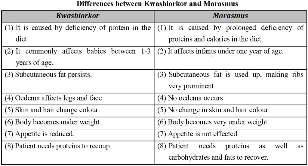
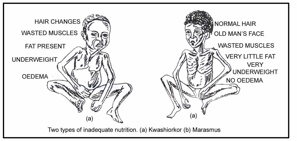
Nutritional Deficiencies and Disorders
Deficiencies of nutrients, like vitamins, minerals and proteins, in the food are related to specific disorders, diseases and abnormalities in humans.
Impairment of health due to improper intake of food or nutrients results in malnutrition.
Malnutrition is a term which covers problems of both undernutrition and overnutrition.
An individual or a group of individuals, may be undernourished due to non-availability of food, and hence, deficiency of minimum required food and nutrients.
In this situation of undernutrition, the intake of food is too insufficient to meet the needs for metabolic energy.
Consequently, the individual shall have to make up the shortfall by metabolising some molecules of its own body.
Excess intake of food and nutrients may cause a great deal of harm to the body.
The excess nutrients are stored as increased body mass. Such a situation is attributep as overnutrition.
Excess intake of saturated fats, like butter, ghee, vegetable oils, red meat, eggs, etc., often leads to hypercholesterolemia, a condition in which blood cholesterol content becomes abnormally high, ultimately leading to cardiac disorder.
Deposition of cholesterol on the walls of blood vessels stiffens the blood vessels and increases blood pressure.
Besides, excessive intake of calories (sugar, honey, ghee etc.) may produce overweight and obesity (excessive accumulation of fat in tissues), which is the most common form of overnutrition.
Very high intakes of minerals and fat-soluble vitamins (obtained from food sources alone) can be toxic.
This is because they are stored in the body.
With the exception of folic acid (women of child-bearing age), people who have well-balanced diet that supply enough energy, do not usually need to take dietary supplements.
But, if they do decide to take supplements, then they should follow the advice on the label to reduce the risk of an overdose.