 Maria Habib
- Books Name
- ACME SMART COACHING Biology Book
- Publication
- ACME SMART PUBLICATION
- Course
- CBSE Class 11
- Subject
- Biology
ANATOMY OF DlCOT AND MONOCOT PLANTS
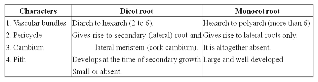
Anatomy of Root
Anatomically, three zones can be distinguished in a root. These are:
(i) Epidermis. It is single layered (uniseriate) and consists of tightly placed, thin walled, uncutinised cells. This epidermis layer is called as epiblema or rhizodermis. Epiblema in younger roots bears unicellular root hairs (water absorbing organs), and is also called piliferous layer.
(ii) Cortex. It consists of thin walled parenchymatous cells with intercellular spaces. In most monocots and some dicots, the cortex layer below epidermis becomes suberised to form protective tissue called exodermis. The cells of cortex store food material (e.g., carrot). The innermost layer of cortex develops into endodermis. It is made up of closely packed living cells characterised by the presence of band like thickenings made of lignin and suberin on their radial and tangential walls. These bands or strips are called Casparian bands or strips. Some cells of endodermis lying opposite to protoxylem remain thin walled and are called passage cells which allow radial diffusion of water.
(iii) Vascular bundles. Vascular bundles are radial and exarch. The centre of monocot root is occupied by parenchymatous cells called pith.
Differences between dicot and monocot root
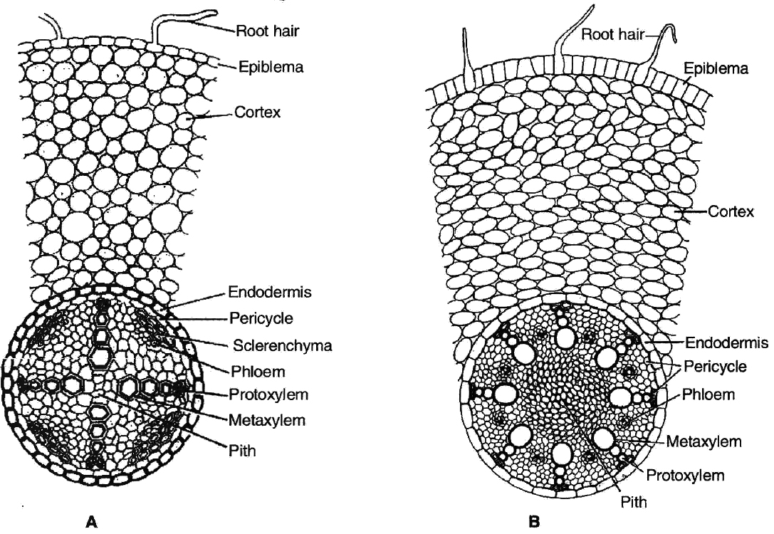
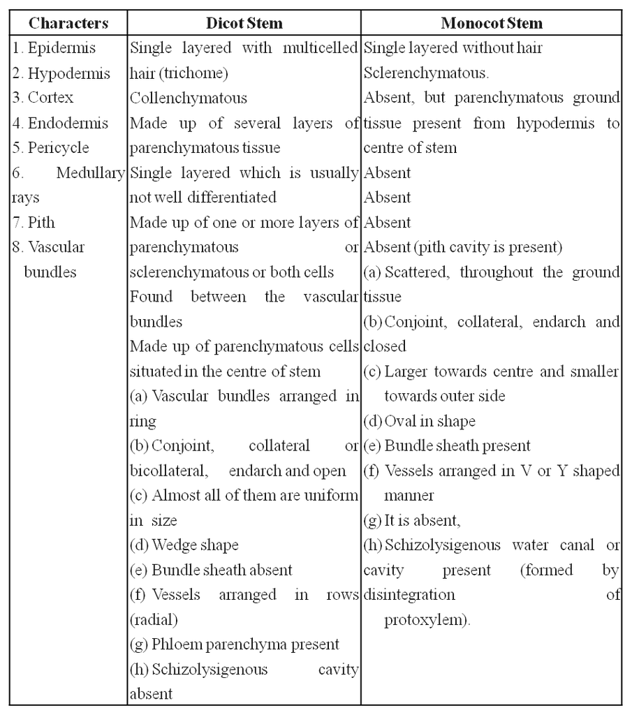
Anatomy of Stem
1. Primary structure of dicot stem
Dicot stem consists of following layers:
(i) Epidermis: It is the outermost layer consisting of single layer of closely arranged cells with cuticle (cutinized). It bears multicellular hairs.
(ii) Cortex: It is differentiated into hypodermis, general cortex and endodermis. Hypodermis is collenchymatous. General cortex is parenchymatous. Endodermis is wavy. It has starch grains hence it is called starch sheath or endodermoid.
(iii) Pericycle: It lies inner to endodermis. Pericycle is few layered thick. Above vascular bundle, it is sclerenchymatous and outside medullary rays it is parenchymatous.
(iv) Vascular bundles: These are in the form of a ring or eustele. They are conjoint, collateral and open. In the family Cucurbitaceae, the stem is wavy, having five ridges and five furrows and vascular bundles are present in ridges and furrows. Vascular bundles are bicollateral and open.
(v) Medullary or pith rays: These are radial strips of parenchyma present between adjacent vascular bundles. They help in radial conduction of food.
(vi) Pith : It is the central portion of stem consisting of parenchymatous cells with intercellular spaces. Narrow, radially elongated parenchymatous cells extend from pith toward the periphery are called medullary rays. The main function is food storage.
Part of transverse sections of stem: A-B. Dicot stem C-D. Monocot stem
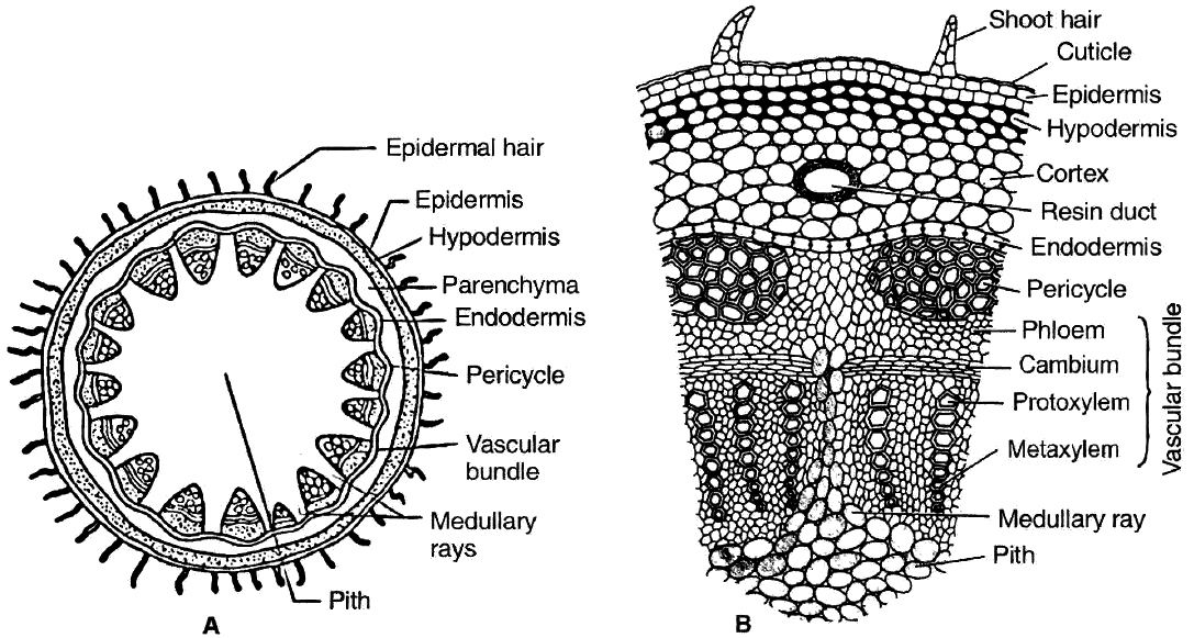
2. Primary structure of monocot stem
Monocot stem consists of following layers:
(i) Epidermis: It is the outermost layer and consists of compactly arranged parenchyma cells which are usually covered with cuticle.
(ii) Hypodermis: Cells of hypodermis are sclerenchymatous, providing mechanical strength to the stem.
(iii) Ground tissue: All the tissues inner to hypodermis represents the ground tissue. It is made up of parenchymatous cells rich in food reserve, like starch.
(iv) Vascular bundles: They lie scattered in the ground tissue. Each vascular bundle is surrounded by 2 or 3 layered sclerenchymatous sheath called as bundle sheath. The vascular bundles are conjoint, colateral, closed and endarch (Atactostele). Vessels are arranged in V shaped manner. Schizolysigenous water cavity or canals are present below protoxylem.
Differences between dicot and monocot stem anatomy
Anatomy of Leaf
1. Structure of dorsiventral leaf (dicot) :
In cross section of dicot leaf, following parts can be observed
(i) Epidermis.
The upper and lower surfaces are covered by the epidermis.
Cells of epidermis are parenchymatous and are closely packed together without any intercellular spaces.
Mostly the stomata are restricted to lower surface of leaf. Such leaves are called hypostomatic.
The outer walls of the epidermal cells are thickened and cutinized which prevents the loss of water.
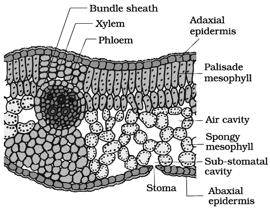
(ii) Mesophyll.
Between the two epidermal layers, there are numerous parenchyma cells which constitute the mesophyll.
In dicots, there are two distinct layers of mesophyll-the palisade (upper layer consisting of closely arranged column shaped cells containing abundant chloroplasts) and spongy tissue (the lower layer of irregularly shaped cells containing fewer chloroplasts).
(iii) Vascular bundles.
Vascular bundles in the leaf are located in the midrib and the veins.
Vascular bundles are conjoint, collateral and closed. Bundles are surrounded by a compact layer of parenchymatous cells which is called bundle sheath.
The xylem (protoxlem) is towards upper epidermis (adaxial) and the phloem on the lower epidermis (abaxial).
2. Structure of isobilateral leaf (monocot) :
Like the dicot leaf, it can also be differentiated into three types of tissues:
(i) Epidermis.
It consists of upper and lower epidermis, both of which may be interrupted by equal number of stomata.
Both the epidermal layers are cutinized. In some grasses e.g., Poa, Agropyron Maize, Psamma, epidermal cells are large with thin flexible walls which are called motor or bulliform cells.
These cells help in the rolling and unrolling of leaves.
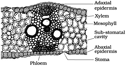
(ii) Mesophyll. Mesophyll cells are not differentiated into palisade and spongy parenchyma. Mesophyll cells are made up of parenchyma cells which have chloroplasts.
(iii) Vascular bundles. They are arranged in parallel manner. Vascular bundles are conjoint, collateral, closed and enclosed by a bundle sheath. The xylem is towards the upper side (adaxial surface) and phloem on the lower side (abaxial surface).
