 ACME SMART PUBLICATION
Anatomy of dicotyledonous and monocotyledonous plants
Transverse sections of the mature zones of roots, stems, and leaves are useful for better understanding the tissue organization of these organs.
A.Dicotyledonous Root: The interior tissue organization is as follows:
1. Epiblema is the outermost layer in the interior tissue architecture. Epiblema cells emerge as unicellular root hairs on many occasions.
2. The cortex is made up of many layers of parenchymal cells with thin walls and intercellular gaps.
3. Endodermis is the cortex's deepest layer. There are no intercellular gaps in the single layer of barrel-shaped cells.
4. Water-impermeable waxy substance suberin is deposited in the form of Casparian strips on the tangential as well as radial walls of endodermal cells.
5. A few layers of thick-walled parenchymatous cells called pericycle are found next to the endodermis. During secondary growth, these cells initiate the formation of lateral roots and vascular cambium.
6. The pith is small and undetectable.
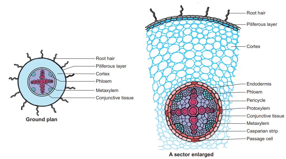
7. Conjuctive tissue is made up of parenchymatous cells that reside between the xylem and the phloem. Two to four xylem and phloem patches are typical. A cambium ring forms later between the xylem and the phloem.
8. The stele is made up of all tissues on the inner side of the endodermis, such as the pericycle, vascular bundles, and pith.
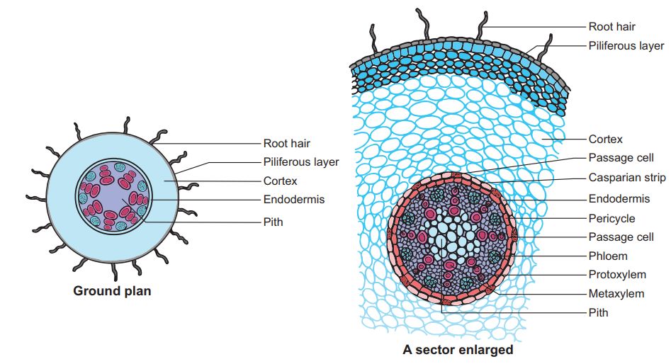
B. Monocotyledonous Root: In many ways, the monocot root's anatomy is identical to that of the dicot root.
1. Epidermis, cortex, endodermis, pericycle, vascular bundles, and pith are all present.
2. Unlike the dicot root, which has fewer xylem bundles, the monocot root frequently has more than six (polyarch) xylem bundles.
3. The pith is thick and developed.
4. There is no secondary development in monocotyledonous roots.
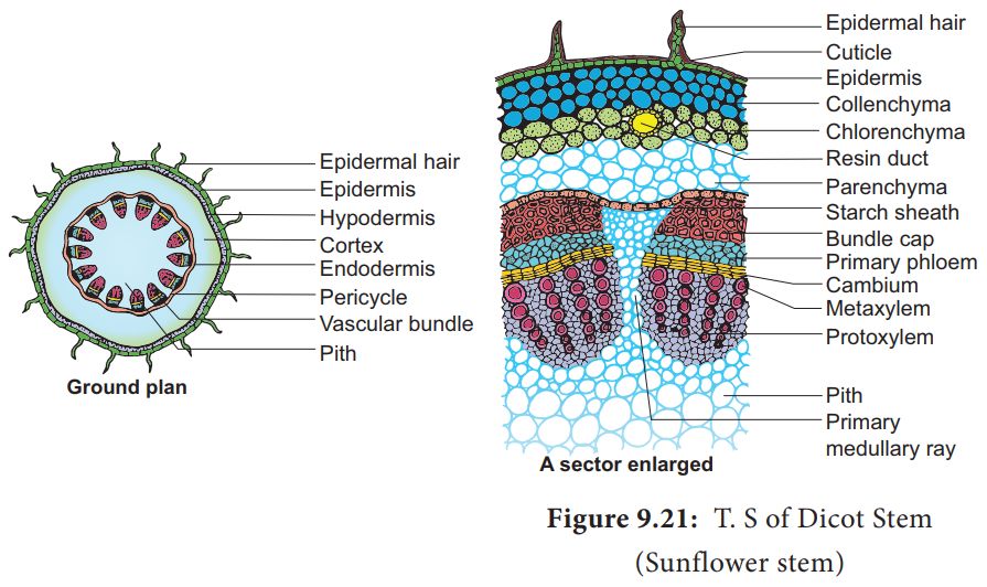
C. Dicotyledonous Stem:
1. The epidermis is the stem's outermost protective covering, as shown in this transverse section of a typical immature dicotyledonous stem.
2. It may have trichomes and a few stomata and is covered in a thin layer of cuticle.
3. The cortex is made up of cells organized in many layers between the epidermis and the pericycle. It is divided into three zones.
4. Just below the epidermis, the outer hypodermis consists of a few layers of collenchymatous cells that offer mechanical support to the embryonic stem.
5. The cortical layers underneath the hypodermis are made up of spherical parenchymatous cells with visible intercellular gaps.
6. The endodermis is the deepest layer andbecause the endodermis cells are densely packed with starch grains, the layer is also known as the starch sheath.
7. Pericycle appears as semi-lunar patches of sclerenchyma on the inner side of the endodermis and above the phloem.
8. Medullary rays are a few layers of radially arranged parenchymatous cells that lie between the vascular bundles.
9. A ring of vascular bundles surrounds the stem of a dicot. Each vascular bundle is joined, open, and has a protoxylem at the end.
10. The pith is made up of a large number of spherical, parenchymatous cells with extensive intercellular gaps that occupy the middle region of the stem.
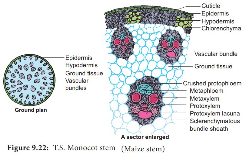
D. Monocotyledonous Stem:
1.A sclerenchymatous hypodermis, numerous distributed vascular bundles, each enclosed by a sclerenchymatous bundle sheath, and a broad, prominent parenchymatous ground tissue characterize the monocot stem.
2. The vascular bundles are joined and closed together. Vascular bundles at the periphery are typically smaller than those in the center.
3. The phloem parenchyma is missing, and there are water-filled holes inside the vascular bundles.
E. Dicotyledonous Leaf:
They are also called Dorsiventral Leaves.
1. The epidermis, mesophyll, and vascular system are visible in a vertical piece of a dorsiventral leaf through the lamina.
2. The epidermis of the leaf has a prominent cuticle that covers both the upper (adaxial epidermis) and bottom (abaxial epidermis).
3. In general, the abaxial epidermis has more stomata than the adaxial epidermis.
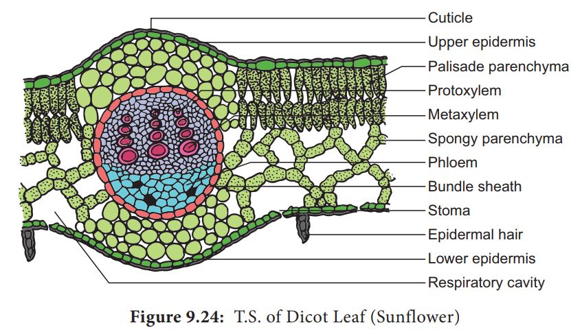
4. Stomata may even be absent in the upper epidermis. The mesophyll is the tissue that lies between the top and lower epidermis.
5. The mesophyll is made up of parenchyma, which contains chloroplasts and performs photosynthesis.
6. The palisade parenchyma and the spongy parenchyma are two types of cells. The elongated cells that make up the adaxial palisade hepatocytes are organized vertically and parallel to each other.
7. The spongy parenchyma, which is oval or spherical and loosely organized, is found underneath the palisade cells and reaches the lower epidermis. Between these cells are various huge gaps and air cavities.
8. Vascular bundles can be found in the veins and midrib of the vascular system. The size of the vascular bundles is determined by the vein size.
9. The veins in the dicot leaves' reticulate venation vary in thickness. A layer of thick-walled bundle sheath cells surrounds the vascular bundles.
F. Monocot Leaf:
They are also known as Isobilateral Leaves.In many ways, the anatomy of the isobilateral leaf is identical to that of the dorsiventral leaf. It demonstrates the following distinguishing characteristics.
1. Stomata are present on both epidermal surfaces in an isobilateral leaf, and the mesophyll is not divided into palisade and spongy parenchyma.
2. Certain adaxial epidermal cells along veins in grasses transform into huge, empty, colorless cells called Bulliform cells.
3. The leaf surface is exposed when the bulliform cells in the leaves have absorbed water and are turgid. They curl the leaves inwards to reduce water loss when they are flaccid due to water stress.
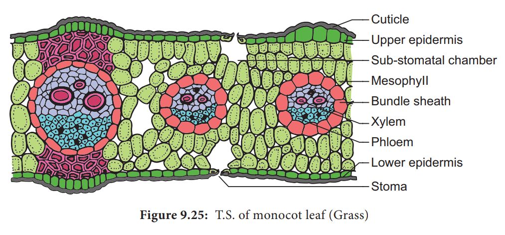
4. In vertical sections of monocot leaves, the parallel venation is represented in the near comparable diameters of vascular bundles.
